Showing posts with label EAE. Show all posts

Thursday, August 6, 2020

Is it possible to engineer Foxp3+ Tregs from primary T cells?

Here is the most recent paper that claims that they can do it. It was published in Science Translational Medicine. This is fairly respectable journal run by Science. This research group is so confident in their data that they even set up a new biotech company to commercialize their approach. I am going to examine how strong are their claims.

Here is a short description what they did: they used a combination of specific nuclease (TALEN) and virus (AAV6) to insert a new promoter into Foxp3 gene in an in vitro activated T cells. They called these Tregs edTregs.

insertion of MND promoter


The edTregs displayed very similar functionality known to occur in thymus-derived Tregs (tTregs) such as no or limited expression of IL-2 and other cytokines in an in vitro stimulation assays. 


The edTregs were suppressive towards effector T cells in a proliferation assay as should be expected from tTregs. Moreover this essential function required endogenous Foxp3 activity as edTregs from IPEX individuals with a defective Foxp3 gene did not show suppression.


proliferation assay


However, edTregs were significantly different from tTregs in Treg-specific demethylated region (TSDR). The point is it is now accepted that Treg identity is not established solely because of Foxp3 expression but requires specific and selective epigenetic modification within and outside of Foxp3 gene. Nonetheless, in in vitro assays, edTregs behaved as bona fide tTregs. 


What about in vivo? There the story gets a little bit murky. The authors used two models to assess edTregs in vivo. First, they co-transferred edTregs with effector T cells into immunodeficient mice to assess if edTregs could prevent graft versus host disease (GvHD). They do see reduction of mice mortality with edTregs.

However, there are some inconsistency between experiments describing GvHD model. In one set of experiments it produced 100% lethality by day 21 (see below, red line) while in other set of experiments it produced only 20% lethality (see above, red line). 


Such inconsistency casts doubts about edTregs ability to inhibit effector T cells in vivo and could explain why the authors did not see much difference in GvHD scores with or without edTregs (see below).


This could also explain why the authors did not see improvement in brain inflammation in mice EAE model when co-transferring antigen-specific edTregs with effector T cells.

In summary, this paper has done a lot of interesting in vitro work trying to convince us that their edTregs work as intended. However, in vivo work lacks consistency. It is not surprising. It has been known for some time now that Tregs behave differently in vitro vs. in vivo. Suppression in vivo appears to be strictly antigen-specific phenomenon unlike in vitro where it could be observed antigen-nonspecific manner (even though Treg activation in itself still require presence of cognate antigen). 


posted by David Usharauli


Tuesday, August 13, 2019

Do regulatory CD8+ T cells control autoreactive CD4+ T cells in the mouse model of human MS?

Recently journal Nature published a very thought-provoking study from Mark Davis' lab. In it, the authors have described the existence of a specialized population of CD8+ T cells that prevented auto-reactive CD4+ T cells from causing autoimmune brain inflammation (EAE) in mice, a laboratory model for human multiple sclerosis.

Let's analyze what the study shows. Both Fig. 1 and Fig. 2 are rather superfluous as they simply show either time kinetics of CD4+, CD8+ and γδ+ T cells responses in the blood or CNS following autoantigen immunization (Fig. 1) or frequency of TCR clonal  distribution based on TCR β or both γ and δ sequencing (Fig. 2). It is not clear what was the purpose of showing them within the paper itself.
    

Next, the authors tested the TCR specificities for expanded clones of CD4+ or CD8+ T cells. Four of these CD4+ TCRs expressed in human leukemia SKW αβ−/− cells yielded robust staining with a MOG35–55 I-Ab peptide–MHC tetramer. 
Curiously, out of nine TCRs from CD8+ T cell clones expressed in a mouse T cell hybridoma 58 αβ−/− cells, none of them get stimulated when co-cultured with bone-marrow-derived dendritic cells pulsed with myelin protein-derived peptides (total of 350 myelin peptides were tested). So, something else, besides myelin protein, was driving CD8+ T cell expansion.

To identify epitope specificity for TCRs from CD8+ T cells, the authors used H2-Db yeast-pMHC libraries. Six of the clonally expanded and one positive control CD8+ TCRs were used. Two, EAE6 and EAE7 TCRs showed robust tetramer staining but no matches were found in the mouse genome. They referred to these peptides identified in the peptide library screen the surrogate peptides (SPs).

Interestingly, the co-immunization of these SPs with myelin peptide inhibited the development of brain inflammation in mice. 

More importantly,  CD8+ T cells harvested from SPs-immunized mice but not control, naive mice, inhibited myelin-specific but not ovalbumin-specific CD4+  T cells in vitro. 


Moreover, only Ly49+ but not Ly49− fraction of CD8+CD44+CD122+ T cells from SPs-immunized donor mice showed inhibitory function both in vitro and in vivo. Of note, the application of the anti-Qa-1b antibody had no effect on the CD8 suppression of Myelin-specific CD4+ T cells. Here, the authors also tested CD8+ T cell reactivity to CFA (complete Freund’s adjuvant) or PTX (pertussis toxin) used in EAE immunization protocol and found that it did not increase Ly49+ fraction.


So, how can we summarize this paper? First, focus on γδ+ T cells here is extra and feels out of context. Second, no endogenous peptides were found that mimic SPs found in the library screen. Could it be microbiota-derived? The authors did not consider this possibility, it appears. Third, how SPs-specific Ly49+ CD8+ T cells inhibit myelin-specific CD4+ T cells? Not clear. It does appear highly specific to myelin-specific CD4+ T cells in the context of brain inflammation. But myelin-specific CD4+ T cells see myelin peptides but these CD8+ T cells do not seem to recognize them. Very confusing indeed. 
In general, the "memory-like" CD8+ T cells, such as Qa-1b-restricted population, has been known to inhibit immune response. This paper simply provides some new evidence in that direction. But it is not a novel idea or observation and without some novel mechanistic evidence I don't see how it could have landed in Nature's pages.  


posted by David Usharauli  


Thursday, February 18, 2016

pMHC tetramer-coated nanoparticles treat autoimmune conditions in mice

This week Nature published "figure-dense" paper showing that nanoparticles coated with auto-antigen+MHC complexes (pMHC) could treat spontaneous or experimentally-induced autoimmune diseases in mice. In fact, this study contains so many experimental results it should have been published in two part, in my view. I will try to condense its basic findings in few paragraphs and provide its interpretation below.

Non-obese diabetic (NOD) mice spontaneously develop [autoimmune] diabetes. Part of T cells that infiltrate and damage β cells in NOD mice express T-cell receptor (TCR) resembling diabetogenic BDC2.5-specific TCR that can be recognized by pMHC–2.5mi/IAg7 tetramers. When the authors injected nanoparticles (dextran-coated or pegylated iron oxide NPs) coated with 2.5mi/IAg7 tetramers (pMHC–NP) it induced expansion of cognate CD4+ T cells in blood and spleens of NOD mice. These cells had a memory-like (CD44hiCD62Llow) FOXP3− TR1-like phenotype.


These "activated/experienced" CD4+ TR1 cells from donor mice treated with pMHC–2.5mi/IAg7-NPs suppressed diabetes development in T-cell-reconstituted NOD-scid hosts upon adoptive transfer. This effect of TR1 cells was augmented by treating hosts with pMHC–NPs.


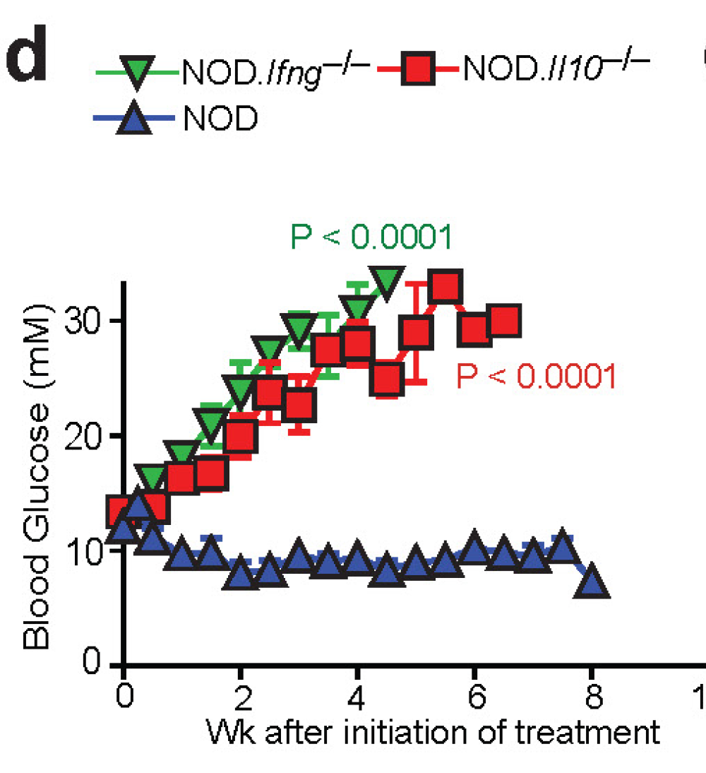
Moreover, 90–100% of the already diabetic mice that received nanoparticles coated with β cell antigens, 2.5mi/IAg7, IGRP4–22/IAg7 or IGRP128–145/IAg7, reverted to stable normoglycaemia.


Of note, treatment withdrawal resulted in loss of the normoglycaemic state in 25–60% of mice, in association with the loss of the tetramer+CD4+ T-cell pools. In other words, these data suggest that almost half of treated diabetic mice maintained normoglycaemic state after 5 weeks of treatment.


Beyond diabetes, the authors showed that nanoparticles coated with myelin oligodendrocyte glycoprotein, pMOG38–49/IAb–NP, dampended progression of experimental autoimmune encephalomyelitis (EAE, a model of multiple sclerosis), when given on day 14 after immunization and even restored motor function in paralytic mice when given on day 21.


Similar therapeutic effects were seen in 3rd autoimmune disease model, collagen-induced arthritis (CIA). HLA-DR4-IE-transgenic mice receiving nanoparticles displaying mouse collagen (mCII)259–273/DR4-IE showed reduced joint inflammation. In all these models, pMHC–NPs effect was antigen-specific.

Indeed, the effects of pMHC–NP therapy were not associated with impaired systemic immunity because pMHC–NP-treated mice showed unimpaired anti-viral immunity and mounted antibodies against an exogenous antigen as efficiently as control mice.

Interestingly, pMHC–NPs could not expand tetramer+ T cells in non-diabetic control mice which harbor mostly naive T cells. Further experiments confirmed that therapeutic effect of pMHC–NP required presence of "experienced" T cells. For example, whereas pMHC–NP therapy afforded 100% diabetes protection to T-cell-reconstituted NOD-scid hosts bearing memory BDC2.5 T cells, such therapy was inconsequential in hosts receiving naive BDC2.5 T cells.


The role of "experienced", but not naive T cells, in therapeutic effect of pMHC–NP was also supported by observation that whereas diabetic NOD G6pc2−/− mice (which lack IGRP antigen and corresponding memory/experienced T cells, but most likely harbor IGRP4–22 specific [aka, nonself-specific] naive T cells) responded to 2.5mi/IAg7–NPs like wild-type NOD mice, they did not respond to IGRP4–22/IAg7–NPs.


At molecular level, the authors showed blockade of IL-10, TGF-β and IL-21R (but not IFNγ) abrogated the anti-diabetogenic properties of 2.5mi/IAg7–NPs or IGRP4–22/IAg7–NPs in diabetic NOD mice.

However, to make things more complicated, the authors also showed that the development of the TR1 precursors and/ or TR1-like cells that expand in response to pMHC–NP therapy required IFNγ in addition to IL-10 in pre-diabetic NOD mice.

Finally, the authors found that at the cellular level, pMHC–NP treated NOD mice harbored increased numbers of so called regulatory B cells, BREG cells (but only in pancreatic lymph nodes, PLNs). These BREG cells were producing IL-10 in response to lipopolysaccharide (LPS). In vivo, these B cells suppressed diabetes development in T-cell-reconstituted NOD-scid hosts as compared to PLN B cells from control mice (and worked together with TR1 cells). The role of BREG cells was supported by observation that treatment of newly diabetic NOD mice with a B-cell depleting anti-CD20 mAb abrogated the anti-diabetogenic activity of 2.5mi/IAg7–NPs (see above).


In summary, this study suggests that when injected into mice these pMHC–nanoparticles expand antigen-specific and antigen-experienced T cells with regulatory properties. These TR1 work with BREG cells to block the chain of events that perpetuates autoimmune cycle. 

David Usharauli


Friday, February 5, 2016

New molecule, BHLHe40, links adjuvant activity of pertussis toxin to T cell pathogenicity in brain inflammation

Multiple sclerosis (MS) is a human neuro-inflammatory disease of autoimmune nature. Mouse model of MS is called experimental autoimmune encephalomyelitis (EAE) and it's induction in mice depends on dirty little secret: to induce EAE, mice are injected not just with peptide derived from myelin oligodendrocyte glycoprotein (MOG35-55) emulsified in CFA (to activate T cells) but mice are also injected with the co-adjuvant pertussis toxin (PTX), an ADP-ribosylating exotoxin derived from Bordetella pertussis (PTX is a whooping cough toxin that has been shown to be necessary for MS induction in this mouse model). No one really knows how or what way PTX primes mice for MS/EAE induction or why we even needed it in the first place (by the way, next time you hear that another drug failed in clinical trial for multiple sclerosis you know now it is because mouse model is completely artificial).

In this regard, new paper in Journal of Experinental Medicine is of interest. The authors report that transcription factor basic helix–loop–helix family member e40 (BHLHe40) was required for adjuvant activity of PTX for EAE development.

Using BHLHe40GFP mice the authors first showed that CD4 T cells expressing GFP (surrogate marker for BHLHe40) were enriched for effector T cells expressing cytokines implicated in EAE pathology (IFN-γ, IL-17, GM-CSF).

Next, the authors showed that ADP-ribosylating activity of PTX was necessary to augment GFP/BHLHe40 expression in T cells and to induce EAE.


Adoptive transfer of BHLHe40-deficient 2D2 T cells (that are specific for MOG peptide) confirmed that BHLHe40 expression was required for T cell pathogenicity in EAE.


In summary, this study showed that during EAE induction PTX sensitizes both innate cells (for IL-1 production) and adaptive immune T cells (for GM-CSF and IFN-γ production) via action of BHLHe40.

David Usharauli


Monday, December 21, 2015

Spontaneous death of myelin-producing oligodendrocytes could trigger delayed MS-like symptoms

I usually don't check Nature Neuroscience. It does not typically publish immunology-related studies [though MS studies are of interest]. So I was surprised to see such "heavily" packed immunology article there and decided to review it to understand how it ended up there.

It is immediately clear that is not written from immunologist point of view. In general, inter-disciplinary studies are encouraged but if it is not done properly it produces lesser quality research


To this end, the authors have used Plp1-Cre-ERT;ROSA26-eGFP-DTA mouse model where tamoxifen injection releases stop signal from diphtheria toxin A production in oligodendrocytes leading to their death. It appears that this is a rare model of oligodendrocyte deletion where mice actually survive long-term (but it appears this DTA model show late-onset [starting at weeks 26] "leakiness" in absence of tamoxifen injection. This knowledge in itself creates host of issues in data interpretation).  

So, the authors noticed that starting 40 weeks post tamoxifen injection [but not at 10 weeks], spleen and cervical lymph nodes of Plp1-Cre-ERT;ROSA26-eGFP-DTA mice contained MOG-specific effector T cells. These were accompanied with clinical symptoms of EAE.


Similar results were found with 2D2 transgenic CD4 T cells [specific for MOG] transferred into tamoxifen-treated Plp1-Cre-ERT;ROSA26-eGFP-DTA host.  

Now, next experiments were quite surprising. To clearly show the role of T cells in the development of late onset MS-like symptoms in tamoxifen-treated Plp1-Cre-ERT;ROSA26-eGFP-DTA mice the authors tried to cross this DTA model with RAG KO mice. Interestingly, these T/B cell-deficient DTA mice did not survive after tamoxifen injection, implying that recovery from acute oligodendrocyte deletion [following tamoxifen injection] somehow required presence of T or B cells [reminds of studies done by Michal Schwartz lab]. However, the authors neither tried to use CD3KO or B cell KO or simply Ab depletion to test these hypotheses.

Other set of experiments with adoptive transfer of T cells harvested from tamoxifen-treated Plp1-Cre-ERT;ROSA26-eGFP-DTA mice into RAG deficient mice (but not in WT host) produced MS-like symptoms.


In the remaining experiments the authors tried to show that injection of MOG peptide coupled to nanoparticles could tolerize self-reactive T cells.

So what we learned from this study? First, I am surprised that it even get into Nature Neuroscience [it does not belong there]. Now, this could mean few things: (1) this research was rejected from Nature Immunology and ended up in Nature Neuroscience; (2) It was directly sent to Nature Neuroscience but reviewed by non-immunologists; (3) standards for Nature Neuroscience is much lower compared to Nature Immunology, in general.

David Usharauli

Wednesday, November 4, 2015

MΦ-specific catecholamine biosynthesis sustains neuroinflammation

A few weeks ago I reviewed new Science paper describing role of Nr4a1-positive monocytes in patrolling melanoma metastasis in the lungs. Now, the same research group has published another paper in Nature Immunology describing role of Nr4a1-positive macrophages (MΦ) in mouse model of neuroinflammation [EAE] that mimics human relapsing-remitting multiple sclerosis (RRMS).

Here, the authors have used the same Nr4a1-GFP mice to monitor its cellular expression during donor [2D2]-T cells induced EAE. Flow cytometric analysis revealed that CNS infiltrating MΦ expressed high level of GFP (i.e. Nr4a1). Of note, in their Science paper the same people mentioned that "Nr4a1 expression in macrophages and Ly6C+ monocytes is relatively low suggesting limited Nr4a1 function". It appears the authors are contradicted themselves from one to another paper.


Next, the authors showed that myeloid-specific deletion of Nr4a1 exaggerated EAE clinical scores.


The authors found, that mechanistically, exaggerated neuroinflammation seen in Nr4a1-deficient mice was [partially] related to MΦ-specific catecholamine production (summary of α1 adrenergic receptor inhibition, catecholamine depletion by 6-OHDA, and MΦ-specific tyrosine hydroxylase (ThΔLysm) deficiency). 


In summary, these results suggest that Nr4a1 orphan receptor influences catecholamine biosynthesis in MΦ and that inhibition of catecholamine [epinephrine, norepinephrine, and dopamine] production could benefit RRMS patients. While results with ThΔLysm mice reached statistical significance, more complete deletion of Th enzyme in ThΔCsfr1 or ThΔNr4a1 mice [as in Figure 2] would have provided better and cleaner view of the role of MΦ-specific catecholamine production on neuroinflammation.

David Usharauli