Saturday, November 29, 2014

Serendipity in Science: Personal Observation

Re-published from niaIDEAlist's Report.
For a scientist reading review articles about his/her research topics is a quite boring task, since review articles usually contain summaries of what are already known. The reviews have no real scientific purpose except as a source of citations and cool looking figures.
There is one reason I found review articles to be very useful to read. Frequently, the authors of the review puts in the review unpublished data or discusses the results of experiment as an "unpublished observation" or a "personal communication". These are data not available elsewhere. These can be so-called "negative data" that current publishing policies do not favor, though I think there is no such thing as negative data in science, if experiments were done correctly, of course.
Many times negative data can be a foundation for the scientific breakthrough. Here is one example from my personal experience.
Few years ago, I was working on one project related to in vitro Foxp3 induction in naive mouse T cells. At that time, Foxp3+ T cells were very popular. Almost every second paper in immunology was somehow related to Foxp3+ T regs. For me, the interest in Tregs has to do with the fact that current theoretical frameworks on how immune system operates can not tolerate the presence of negative regulatory populations. It is a strange idea but true. Neither self-nonself nor PAMP/DAMP based immune system requires Tregs to operate, theoretically speaking, of course.
My boss then, Polly, had an idea that Tregs are T cell subset driving different type (or class) of immune response rather that inhibiting anything. Since scientists did not measure all type of responses, they saw a down-regulation of a particular known immune type response as an evidence of inhibition rather than considering that system was shifted to unknown type (cytokine) not measured in the assay (for example, IgA or IL-???).
Unlike many laboratories working on Foxp3 induction using anti-CD3/anti-CD28 antibodies, I decided to use more physiological approach of Foxp3+ T cell induction using peptide pulsed DCs and naive peptide-specific CD4 T cells. Experiments went as expected that we were able to generate peptide-specific Foxp3+ CD4 T cells. During this same time period, new T cell subset, called Th17, has been discovered and characterized. It turned out that while Foxp3 induction required TGF-beta, one could generate Th17 simply by adding IL-6 in addition to TGF-beta (TGF-beta + IL-6). It was already shown by then that presence of IL-6 drove Th17 induction and while inhibiting Foxp3+ induction.
Since I was doing an assay with Foxp3+ T cells, I have decided, for no apparent reason, to add IL-6 in my assay. I was expected to see a decrease in Foxp3 expression and increase IL-17. Strangely, when I added IL-6 to Foxp3 induction milieu (TGF-beta, peptide, DCs, CD4 T cells) I noticed that percentage of Foxp3+ T cells went up rather than go down. Th17 induction was expected. I repeated this assay multiple times and over and over I saw that IL-6 was promoting rather than inhibiting Foxp3 induction in my hands.
Now, these results went completely opposite to what everyone else were reporting in the literature. I asked my fellow postdoc to repeat the same experiments to make sure that I was not doing something strange with my culture. Again, we saw the same: IL-6 promoted Foxp3+ T cell induction.
Using IL-4 or IFN-gamma KO mice on the same background, we observed that IL-6 effect on Foxp3 induction was abolished on IFN-gamma KO but not on IL-4KO background. Indeed, using anti-IFN-gamma antibodies with wild-type CD4 T cell assay confirmed that when IFN-gamma was blocked IL-6 lost its ability to increase Foxp3 expression. Parallel experiments showed that exogenous IFN-gamma inhibited Foxp3 expression and that IL-6 inhibited IFN-gamma expression, as expected from published literature.
In the end, after 5 months of continues work, we come up of new hypothesis that could explain our observation. Our finding suggested that IL-6 promoted Foxp3 expression by suppressing IFN-gamma expression in activated peptide-specific CD4 T cells. We reasoned that other laboratories failed to see the same effect since they were using anti-CD3/anti-CD28 activation mode rather more physiological, peptide activation mode. Indeed, in our hand too, IL-6 inhibited Foxp3 expression when anti-CD3/anti-CD28 antibodies were used to activate CD4 T cells.
So, we were very confident that we found something worthy of publication in one of prestigious journals.
Then, suddenly, we started to receive reports that mice from our mouse colony started to die in higher numbers than normally observed. People had no clue what was going on. In the end, after thorough investigation that took few months, it was revealed that new type, not yet described Streptococcus was causing mice death (one of clinical signs was a cardiomyopathy). The mouse colony was shut down, newly re-derived and repopulated.
As you may have already suspected, when we repeated our experiments with IL-6 on T cells from "clean" mice, we saw that now IL-6 was inhibiting Foxp3 expression rather than promoting it and this effect was seen both times when peptide or anti-CD3/anti-CD28 were used.
These are the type of experiences that make someone like me to appreciate why we do science. As to new Streptococcus we discovered, I did not have a chance to study it but I hope someone will be doing his/her PhD by studying this unusual bacteria and its unusual effects on immune system.
David Usharauli

Tumor immunotherapy: 1 year at a time

Tumor immunotherapy is both an old and new idea. Whenever there is a new discovery in immunology, people usually are asking 3 questions: can it cure HIV, cancer or allergy?

Recently, a promise for successful immunotherapeutic approach to cancer treatment was boosted by clinical introduction of anti-CTLA4 and anti-PDL1 humanized antibodies. These antibodies target and exploit the natural inhibitory checkpoints in T cell activation program relevant for natural tolerance process to self or food antigens. There is another approach as well, called CAR-T technology, that I will review later.

Still, the actual, long-term benefits of current immunotherapeutic approaches are debatable. Here is one example. This is a Science paper. The senior author on this paper is Steven Rosenberg. This is a guy who actually has started clinical immunotherapy with IL-2 in 1980's.

This paper describes a single patient's personalized tumor immunotherapy protocol. Initially, the authors sequenced a metastatic tumor obtained from resected lung tissue and found 26 mutations. Next, they have prepared 3 different tandem minigene constructs (8-9 mutations each), transfected them in autologous antigen-presenting cells and run ex vivo TIL (tumor-infiltrated lymphocytes) activation assay. Only one construct, TMG-1, showed TIL reactivity.

Next, from this tandem minegene construct (that contained 9 mutant minigens), the authors synthesized 9 individual minigene constructs with each containing reversion of one mutation back to wild-type sequence, so to permit detection TIL specificity by antigen elimination. Interestingly, TIL's reactivity was abolished when erbb2 interacting protein was reverted to wild-type sequence
In addition, TIL's tumor reactivity was localized in CD4 T cell subset and was made up mostly of Vbeta22+ CD4 T cells

Thus this patient's TIL (CD4 T cells) specificity was directed towards one mutation in erbb2ip.

Next, the authors has expanded patient's TIL in vitro and injected back 42.4 billion TIL (25% Vbeta22+) into patient. This adoptive transfer of tumor specific TIL produced >1 year tumor burden stabilization and regression of metastatic tumor in liver and lung.
After 1.5 year from the initial TIL transfer, tumor re-emerged, so the patient was re-injected with in vitro expanded 100 billion tumor specific Vbeta22+ TIL and again such transfer produced additional >6 months stabilization and regression of tumor metastasis.
It appears that the difference between this graph and Fig. 2 D has to do with the fact that the authors measured only those tumor lesions which were present during both treatments.

What we have learned from this study? First, tumor in this case did express neo-antigen detected by CD4 T cells. Second, in vitro expansion of tumor-specific TILs and their adoptive transfer back into patient yielded >1 year (after single transfer) and in total >2 years (after 2nd transfer) of tumor burden stabilization and metastatic regression. Third, this approach, however, did not provide a long-lasting tumor-protective effect, despite presence of transferred tumor-specific CD4 T cells.

Does it mean that TIL approach is not viable in a long run? Why were billions of tumor-specific T cells not able to eliminate tumor? Because of inhibitory milieu at tumor sites? Were they terminally-differentiated T cells lacking self-renewal properties? It seems that tumor was able to escape CD4 T cell control despite the fact it still continued to express neo-antigen. How? 

David Usharauli


Wednesday, November 26, 2014

TLR5 ligand flagellin goes anti-viral

We live in an environment filled with all types of bacterial, viral, fungal and parasitic life forms. Some of those life forms actually live permanently with us, on our epithelial surfaces in a symbiotic relationships.

Broadly speaking, body's immune system is divided into two major subsystems: innate and adaptive. Since adaptive immune system (T and B cells) carried an essential receptor diversity sufficient to recognize myriad of antigens, it was assumed that the organisms deficient in adaptive immune system should easily succumb to infections.

However, more recently, the significance and the capability of innate immune system started to be recognized. So any paper that adds new evidence in this direction requires proper appreciation.

Such is this new paper published in journal Science. It focus on Rotavirus (RV) infection in mice. This virus causes diarrhea in children and it appears that neonatal mice show similar phenotype. So, at least this mouse model is clinically relevant. Paper shows how innate system activation via TLR5 agonist, flagellin, can prevent or cure viral infection in absence of antibodies and T cells.

A flagellin, TLR5 agonist ligand become a popular science reagent, especially among Emory immunology club members.

First, the authors found that in adult mice, flagellin injection totally prevented RV infection. Surprisingly, this effect was maintained in RAG KO mice which lack functional T and B cells (it would be interesting to know whether oral flagellin can have a protective effect as well). 
Second, flagellin was found to have a therapeutic effect too on established RV infection in RAG KO mice. 
Flagellin injection also reduced diarrhea intensity in neonatal mice.  
As expected, the authors found that TLR5KO, MyD88KO, NLRC4KO and TLR5KO/NLRC4KO abolished partially or fully flagellin's anti-RV effect (for some reason, the authors have used different Y scales to show flagellin's effect in different KO mice. I personally think it is confusing and totally unnecessary).
Additional experiments indicated that DCs played major role in flagellin's effect.

Furthermore, IL-22KO, IL-18KO, p40KO (lacks IL-12 and IL-23), RAG2KO/IL-2RgKO (lacks T, B, NK, ILC) but not anti-IL-17 injection or IL-1RKO mice showed reduced or abrogation of flagellin's anti-RV effect.
Surprisingly, the authors did not discuss data with p40KO mice.

Finally, the authors showed that injections of IL-22/IL-18 (instead of flagellin) into WT, RAG1-KO or neonatal mice eliminated and reduced clinical symptoms of RV infection.
In summary, this paper shows that flagellin signaling via TLR5 (and partially via NLRC4) induces IL-18 and IL-22 secretion that protects mice from RV infection. It appears that IL-22 is secreted by ILC but the source of IL-18 was not defined. In addition, role of IL-12 family (p40KO mice) is not discussed at all. Are they preparing another paper?

Of course, one major limitation of this study is that all these KO mice have different gut flora. It is important to know if flagellin's effect on RV is mediated through endogenous microbiota. Use of germ-free mice would be useful. 

David Usharauli


Sunday, November 23, 2014

Sensing tumor with a STING

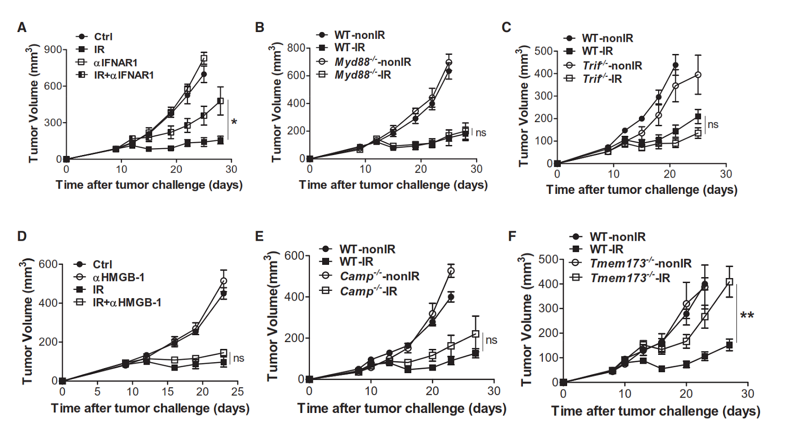
This is another paper that show that, in a mouse model, a local radiation therapy of established tumors activates immune system through DNA sensor STING.

First, the authors found that STING-KO mice or IFN-alphaR1 blockade, but not MyD88KO, TRIF-KO, CRAMP-KO (homolog to LL37) mice and anti-HMGB1 blockade, showed impaired ability to control tumor growth after local tumor irradiation.
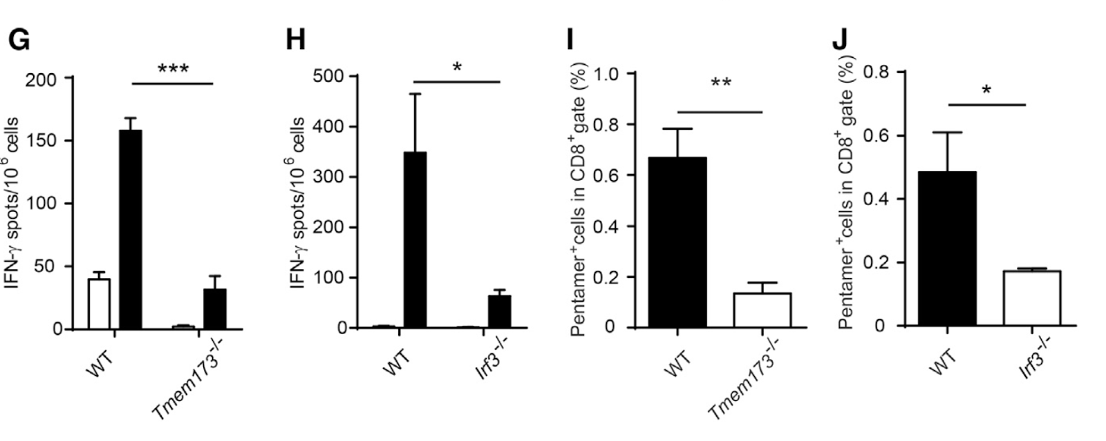
In vitro experiments showed that STING or IRF3 deficient BmDCs lacked an ability to cross-prime antigen-specific CD8 T cells. This deficiency was thought to be as a result of STING-KO BmDCs' inability to produce IFN-beta in response to irradiated tumor cells, since it was reversed by addition of exogenous IFN-beta.

Similar results were obtained with mice deficient in cGAS, a DNA  sensor molecule upstream of STING pathway. 

The authors also showed that radiation-induced tumor control was dependent on presence of CD8 T cells and autocrine IFNalphaR signalling in CD11c+ cells.
Finally, the authors showed that intra-tumoral injections of cGAS activators significantly improved tumor control in wild-type but not in STING-KO mice (for some reason, the authors did not show STING-KO control).  

In summary, the authors proposed the model wherein DNA (or DNA containing cellular fragments) released after tumor irradiation are captured by local DCs and cross-presented to CD8 T cells in IFN-alphaR dependent manner leading to T cell priming and tumor growth control.

Here too, the authors were unable to decipher the precise mechanisms for cytosolic uptake of tumor DNA by DCs.

David Usharauli

Saturday, November 22, 2014

STING-y nature of immune sensing of the tumors

Immune system is, in principle, capable of tumor recognition and rejection. So far, however, few mechanisms have been described which explain this process (ATP or HMGB1 release, for example).

This week two new, back to back papers in journal Immunity provided additional data regarding tumor detection by immune cells. I am going to review them individually.


Initially, the authors observed that mice deficient either in intracellular DNA sensing molecule STING or IRF3 failed to prime antigen-specific CD8 T cells against B16 melanoma cells expressing SIY antigen. In contrast, mice deficient in MyD88, TRIF, TLR4, TLR9, ATP receptor, or RNA-sensing MDA5/RIG-I showed normal CD8 T cell priming.   
In vivo tumor growth experiments show that the absence of STING and IRF3 significantly diminished mice ability to control both the growth of spontaneously, MCA-induced sarcoma as well as B16 melanoma expressing SIY antigen. In contrast, TRIF-deficient mice showed no impairment. For some reason, the authors presented tumor growth data from different experiments in different Y scale. Not the best practice.
To understand the mechanism behind tumor sensing, the authors exposed WT BmDCs to various B16 cells source (live, heat inactivated, irradiated, etc). Interestingly, only pure DNA from B16 cells induced IFN-beta secretion in BmDCs and that only in presence of membrane-opener lipofectamine.
However, this IFN-beta response was abolished in STING-deficient BmDCs.

Interestingly, both in vitro and in vivo, DNA derived from normal splenocytes could induce IFN-beta response in CD11+ dendritic cells, though not as strong as tumor DNA. This response in vivo was STING dependent.

Additional experiments show that STING deficiency affected CD8 T cell survival post cell division.
Mechanistically, failure of tumor-specific CD8 T cell accumulation was linked to deficient DCs activation in STING-deficient mice.
Finally, the authors showed that compared to WT mice, anti-tumor efficacy of clinically relevant anti-CTLA4/anti-PDL1 injections was reduced in absence of STING, though anti-CTLA4/anti-PDL1 injections still showed significant, almost 2-fold delay in tumor growth in STING-deficient mice.
The authors pointed out that they do not yet know how tumor DNA is able to gain access to DCs cytosols in vivo but not in vitro. In fact, in vitro, DNAs are notoriously hard to put inside cells, even with lipofectamine or with other transfection methods. So much so, that many labs and companies have moved into a RNA field as an alternative model for antigen delivery. But even with RNAs, we don't know much either. Such knowledge will be the basis for a rational design of tumor vaccines.   

David Usharauli
  

Thursday, November 20, 2014

NK cells need help to keep memory

NK cells are part of an innate immune system. It is thought to lack antigen-specificity and memory. Generally antigen-specific memory is defined as an augmented and faster response to antigen re-exposure. 

The data I have seen so far suggest that NK cells do have much wider antigen-specificity but I am still not entirely convinced that NK cells have a long-term memory as T cells do.

Here is a new article from JEM that points to this conclusion. Now, this study came from Lewis Lanier's lab. Initial studies of NK cells memory was based on experiments with Ly49H+ NK cells and mouse CMV antigen m157. In this new study, however, the authors has examined antigen-specificity and memory formation for Ly49D+ NK cells specific for alloantigen H-2Dd

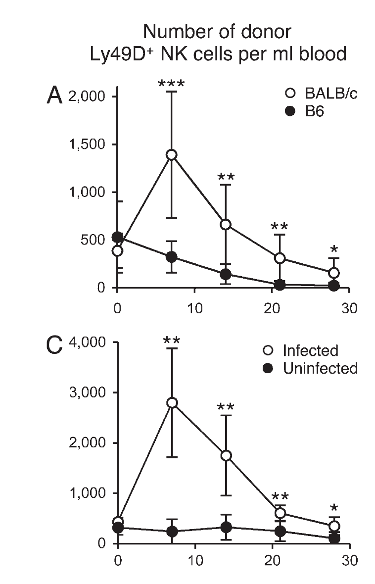
In this model system, purified Ly49D+ NK cells were transferred into Ly49D and Ly49H double deficient B6 host mice depleted of CD8 T cells (to prevent donor NK cell rejection). The host mice were then challenged with H-2Dd expressing splenocytes (from balb/c mouse). Donor NK cells showed no response to alloantigen alone. However, when host mice were exposed to  m157-deficient mouse CMV infection in conjunction with H-2Dd expressing splenocytes (to mimic conditions for an actual clinical tissue transplantation when human CMV becomes re-activated), the authors observed expansion of Ly49D+ NK cells (but at day 28 post infection, there was minimal difference between responding and non-responding NK cell populations).

This expansion of Ly49D+ NK cells was alloantigen-specific, required simultaneous infection with mouse CMV and was dependent of IL-12.


Interestingly, expansion of Ly49D+ NK cells was abolished when NK cells expressed another inhibitory molecule Ly49A. Both Ly49D and A recognize H-2Dd.

Surprisingly, at day 28 post infection, there was no difference between Ly49D+/A- and Ly49D+/A+ NK cell population and upon secondary challenge with alloantigen, Ly49D+/A- NK cell expanded at the same rate as during primary challenge (compare Fig.1 and Fig. 4).

An ex vivo analyses of effector functions of NK cells showed that presence of Ly49A inhibited NK cell's effector functions in both naive and primed NK cells (and only in presence of H2-Dd). Interestingly, in absence of Ly49A, difference between naive and primed NK cells disappeared.
To show that initial priming leads to antigen-specific enhancement during the secondary response (rather than general increase in NK cell responsiveness), the authors primed the host mice either with (a) m157-deficient mouse CMV infection in conjunction with H-2Dd expressing splenocytes, and then challenged with wild type mouse CMV, or (b) primed the host mice with wild type mouse CMV and then challenged with m157-deficient mouse CMV infection in conjunction with H-2Dd expressing splenocytes. Strangely, In both situations only NK cells primed during initial antigenic stimulation undergo secondary expansion but independent of secondary antigen-specificity. However, for some reason naive NK cells failed to expand in response to specific antigenic challenge in this setting (compare Fig. 1 and Fig. 6).  

Finally, the authors showed that primed NK cells showed slightly better protection against wild type mouse CMV in adoptive transfer experiment.
While I agree with the authors that NK cells have antigen-specificity, this study did not convince me that primed NK cells show enhanced secondary recall response (enhanced proliferative response) as typically observed for T cells.

David Usharauli

Tuesday, November 18, 2014

Flu detour

Influenza virus, commonly called Flu virus, has a volatile nature. There is, of course, Flu vaccines available in the market, but their efficacy are surprisingly barely 70%. 

Now, Flu virus infects upper and lower respiratory tissues, mainly epithelial cells. So clinical symptoms are usually proportional to the degree of tissue damage along the respiratory tract. However, in some individuals, Flu causes additional GI tract issues. Why is that?

New study in JEM may provide some new insight. It is important to point out at this stage that this study has examined a mouse model of Flu infection.

Initially, the authors observed that infection of mice with PR8, a mouse-adapted Flu strain, caused their weight loss and moderate diarrhea.


Interestingly, examination of GI tract for virus presence showed that unlike lung tissue, Flu virus did not infect GI tract directly. So why were the mice getting diarrhea?

IL-17 has been implicated for some time now in GI tract tissue damage. Indeed, infection of IL-17A-KO mice with Flu virus caused little or no intestinal tissue damage. Similar result was obtained when using anti-IL-17 antibody injections.

Since lung tissue damage was similar in both wild-type and IL-17-KO mice, the authors focused on IFN-gamma expression. Interestingly, IFN-gamma-KO mice also showed minimal intestinal damage.


Additional experiments showed Flu infection in mice induced high level expression of CCL25 in intestinal tissue and its blocking via anti-CCL25 treatment abolished intestine damage.
Other set of experiments indicated that Flu infection modified gut microflora composition by its effect on IFN-gamma expressing T cells which migrated from lung to intestinal tissue CCL25 dependent-manner. This changes in gut microflora was associated with local tissue damage in IL-15 and IL-17 manner.

It my opinion this study initially generated a clear data but as it progressed Flu model became more complex and at times confusing. For example, no data are presented of the level of lung tissue damage in IFN-gamma KO mice. It is not clear either whether IFN-gamma induces gut barrier damage and IL-17 production or vice versa.

David Usharauli

Friday, November 14, 2014

Toxic Entente: Aging, adipose tissue and TNF-alpha

The following study is a very interesting and neatly done research published in Journal of Experimental Medicine (JEM). Quite unusual results. It deals with aging, adipose tissue accumulation and susceptibility to cytokine storm.

A conceptual rationale for this study is a following: many cancers develop later in the life. Cancer treatments  now days incorporate immunotherapies. However, before immunotherapy is approved for human use, the scientists use it on young mice to test its efficacy. So the authors questioned what would happen if we were to try to test it on old mice? Exactly.

Now here is the surprise. This study, led by William Murphy from UC Davis, observed that unlike young mice, old mice (> 15 month) die rapidly when injected with a combination of anti-CD40 / IL-2 or CpG / IL-2. Why?


Usually, old mice of B6 mice become overweight with the aging. They accumulate a lot of adipose tissue (fat).


Interestingly, the authors found that when old B6 mice were artificially kept on a calorie-restricted diet, they became resistant to anti-CD40 / IL-2 injection.


Next, to validate the connection between adipose tissue and toxic response to anti-CD40 / IL-2 injection, the authors used leptin mutant ob/ob mice. This mice are very cute looking due to extreme overweight, even when young.


The authors found that similar to old B6 mice, ob/ob mice were highly susceptible to anti-CD40 / IL-2 injections.


Mechanistically, the authors found that a depletion of macrophages with Clodronate or neutralization of serum TNF-alpha with etanercept (FDA approved antibody) could rescue young ob/ob mice from toxic response to anti-CD40 / IL-2 injections.


Finally, the authors found that diet-induced obesity in young B6 mice also increased their susceptibility to anti-CD40 / IL-2 injections. However, unlike old B6 mice, only 50% of young obese B6 mice were sensitive to anti-CD40 / IL-2 injections.


In summary, the authors proposed that age-associated adipose tissue accumulation harbor macrophages that have heightened responsiveness to anti-CD40 / IL-2 injections leading to uncontrolled TNF-alpha release and death. This study also shows that calorie restriction during aging can have a beneficial, life-saving effect. 

It would be interesting to know if a gut microbiota played any role in the observed difference between young and old B6 mice. The fact that young obese B6 mice were not as susceptible to anti-CD40 / IL-2 injections as old B6 mice were suggests that age-associated susceptibility to cytokine storm is more complex than simple adipose tissue accumulation (still, in my opinion, this article should have been published in Nature Medicine even though it did not show a "perfect" correlation between adipose tissue and cytokine storm in young obese B6 mice). 

One drawback is that it is not completely clear whether anti-CD40 / IL-2 or CpG / IL-2 injection regimen is a typical immunotherapy regimen approved for human use. 

David Usharauli


Wednesday, November 12, 2014

Pulling RAG out of NK cells

Sometimes experimental results are just too unexpected to take at face value.

Take for example a new study from journal Cell about RAG gene and NK cell fitness. This research came from Joseph Sun's lab, a well known expert in NK cell biology.

Almost everyone in immunology knows that RAG genes (RAG1 and RAG2) are involved in TCR and BCR rearrangement. Simply put, RAG1 or RAG2 KO mice lack T and B cells but have everything else. So it seemed that way until now.  

This new study focused on NK cell biology. Initially, using RAG fate-mapping mice where any cells expressing RAG gene will be permanently marked with red fluorescence protein, RFP, the authors showed that a substantial proportion of NK cells expressed RAG gene at some point during their development. 


Interestingly, direct comparison of NK cells with or without history of RAG gene activity showed that NK cells with no RAG gene activity (RFP- NK cells) have more activated phenotype and are more cytotoxic compared to RFP+ NK cells.


More importantly, an adoptive transfer of equal number of RFP+ and RFP- NK cells into Ly49h-/- recipient, followed by MCMV infection which expands viral antigen specific NK cells, revealed that compared to RFP+ NK cells, RFP- NK cells showed diminished expansion in response to MCMV infection and accelerated contraction so much so that at day 35 post-infection, there were hardly any RFP- NK cells left in recipient mice. Such unusual behavior of RAG KO NK cells were cell-intrinsic since RAG KO NK cells from WT:RAG KO mixed bone marrow chimeric mice or even from natural mutant scid mice showed similar deficiency. Furthermore, NK cells from athymic mice, AID KO, MHC II KO and TCRbeta KO mice showed no such behavior. Even adding back of WT T and B cell to RAG KO mice could not rescue RAG KO NK cells' survival deficiency.


Additional experiments showed that RAG KO NK cells failure to survive had to do with higher rate of  cell death (high caspase activity).


Finally, the authors also showed that OVA-specific CD8 OT-I cells on RAG background were also significantly impaired in their survival as compared to WT OT-I cells.


In summary, this study shows that RAG gene expression at some point during the ontogeny of NK cells confers unexpected fitness to NK cells later in their fate. Since RAG gene is not expressed in fully developed NK cells (based on RAG-YFP knock-in reporter mice), this effect of RAG gene on NK cells is quite long-lasting (the authors speculate that this effect of RAG gene has to do with DNA break repair mechanisms).

What about all the studies using RAG KO NK cells or transgenic TCR mice on RAG KO background? Does it mean it we need to re-evaluate those results? Or maybe NK cells with no or RAG gene activity are different NK cell subsets each with special functions? If T cells and B cells have functional subsets, NK cells may have too.
     
David Usharauli

Tuesday, November 11, 2014

True blood is not telling the whole truth

Some studies are just very tedious to do but necessary. This is especially true for studies of descriptive nature. These studies do not usually have any hypothesis to follow. Their task is simply to generate a lot of data and to try to apply system analysis approach to find any correlations. Sometimes they can yield an important information, but mostly they have values as foundation blocks for other studies.

The following new paper from journal Cell is such a study. It deals with  a research topic that interests me so I have decided to write short opinion about it.

In this study, research group led by Donna Farber have analyzed human T cell subset distribution in various human tissues across different age group. They have obtained human tissues from organ donors (total of 56 donors).


They observed that there were significant differences between some of T cell subset in lymphoid and non-lymphoid tissues. For example, the frequency of central memory T cells (TCM), for both CD4 and CD8 T cells, were not significantly different among tissues. But the frequency of naive and effector memory (TEM) T cells were significantly different between blood and non-lymphoid tissues. In addition, blood and interestingly lung tissue contained significantly more RA+ terminally differentiated CD8 effector memory T cells (TEMRA) compared to non-lymphoid tissue (intestinal tissue).


More importantly, analysis of T cell subset distribution across age group revealed that the frequency of naive CD4 and CD8 T cells in the blood undergo a significant age-related reduction.

    
I would be interested to know how these differences of the frequency of T cell subsets between various tissues translates into functional differences in an in vitro assays. 

Recent push for more translational research implies that in the future lot more studies on cells from human tissues are anticipated. Human blood is, of course, the most widely accessible human tissue. However, focus on human blood-derived immune cells, without proper comparison with other tissue-derived immune cells, may undermine long-term goals. Comprehensive studies like this one in journal Cell play an important role in putting to diverse observations into simple, easily understandable concepts.

In addition, properly constructed human cell experiments would save lots of laboratory animals from unnecessary experimentation and many times from their pointless and unjustified sacrifice for the name of science. I think it is time for NIH to really apply 3Rs to animal protocol approvals.        

For more information about this study, please check the link in this post.
 
David Usharauli