Tuesday, December 20, 2016

Lactobacillus reuteri extends lifespan of FOXP3-deficient scurfy mouse via microbiota–inosine–A2A receptor axis

Scurfy mice harbor natural mutation in FOXP3 gene that clinically resembles FOXP3 deficiency. Both scurfy mice and genetically modified FOXP3-KO mice die prematurely within first month of life. In humans, clinically observed FOXP3 deficiencies do not seem to be as lethal as in mice though one could argue that scurfy mice lifespan could be extended if these mice were given human-like medical attention (for example, i.v. feeding, anti-inflammatory medication, so on). 

In this regard, a new paper in Journal of Experimental Medicine is of great interest. It showed that a member of gut microflora, Lactobacillus reuteri, a probiotic microbe, when given orally could drastically extend lifespan of scurfy mice (30d vs. >125d) via microbiota–inosine–A2A receptor axis.


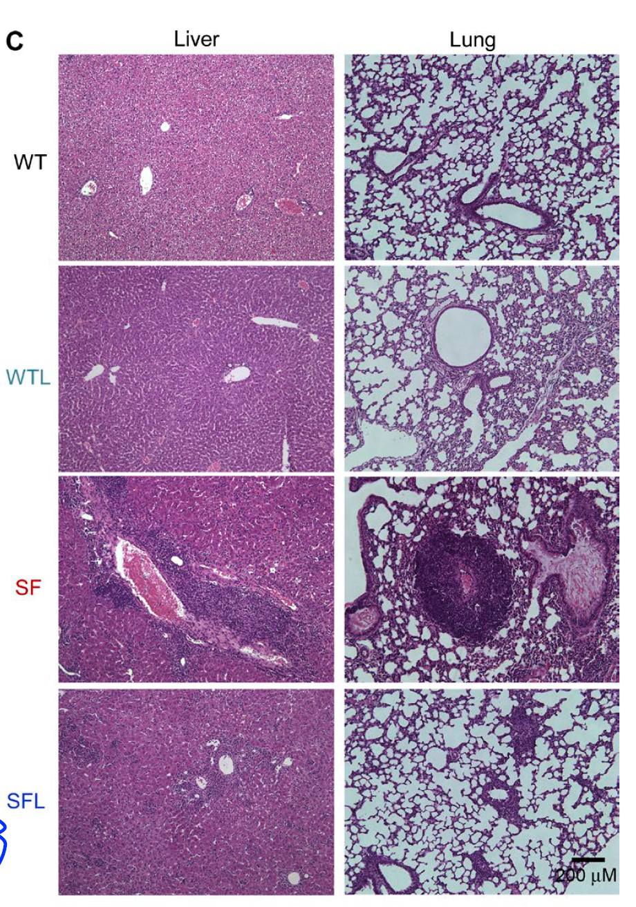
Morphologically, oral Lactobacillus reuteri significantly reduced tissue inflammation in scurfy mice.


Blood test showed that one molecule Lactobacillus reuteri could restore to WT level in scurfy mice was a purine metabolite inosine.


Indeed, oral inosine was able to recapitulate Lactobacillus reuteri effect on scurfy mice lifespan (and both Lactobacillus reuteri and inosine effects were specifically mediated via adenosine A2A receptor).


In summary, this study revealed that purine metabolites, inosine or adenosine could protect scurfy mice from tissue immunopathology and drastically prolong their lifespan. It is quite rare to see that one molecule could produce such effect. It would be interesting to see how caffeine consumption affects immunopathologies in humans as it acts as a natural antagonist to adenosine A2A receptor.

David Usharauli


Tuesday, December 13, 2016

Protozoa-enabled non-genetic colitis after T cell transfer in immune-deficient mice

Adoptive transfer of naive T cells into T-cell deficient host mice has been used as a colitis (gut inflammation) model that led to discovery of FOXP3+ T regulatory cells which when co-transferred with naive T cells prevented gut immunopathology.

A new study in Journal of Experimental Medicine, however, provided evidence to show that there is a limit how much FOXP3+ T regs could do. This study found that adoptive transfer of naive T cells into Rip2−/−Rag1−/− mice (RIP2 is an essential signaling adapter molecule downstream of both NOD1 and NOD2) led to protozoa, Tritrichomonas muris-enabled dominant colitis that could not be prevented by FOXP3+ T regs.

Initially, the authors observed in non-littermates that naive T cell transfer led to severe gut inflammation in  Rip2−/−Rag1−/− host as compared to just Rag1−/− mice [though it is strange that in their mouse facility Rag1−/− host did not show weight loss after T cell transfer].


Interestingly, colitis in Rip2−/−Rag1−/− hosts could not be prevented by co-transfer of FOXP3+ T regs.


Co-housing and littermate control control experiments confirmed that a non-genetic factor was responsible for colitis development in Rip2−/−Rag1−/− hosts and that factor could be transferred between mice when co-housed together.


Fecal matter analysis showed that Rip2−/−Rag1−/− hosts were selectively enriched with protozoa Tritrichomonas muris, and that its transfer to other mouse accelerated T cell-mediated colitis.


In summary, this study shows that Rip2−/−Rag1−/− double deficient hosts harbor protozoa Tritrichomonas muris that by itself or through modulation of gut microbiota establishes a dominant colitogenic gut ecosystem that is even transferable to genetically non-related mice.  

David Usharauli