Wednesday, April 29, 2015

Reciprocal T subset-specific tumor protection by RNA vaccine encoding mutant MHC class I and II binding epitopes

Efficacy of solid cancer immunotherapy mostly depends on effector activity of T cells. Initially, CD8 T cells were thought to mediate primary anti-tumor activity. However, for the past 15 years, growing evidence pointed to a stand-alone CD4 T cell role in cancer protection.      

This new paper in journal Nature provided another example of CD4 T cell specific tumor protection. Strangely, the authors' data suggest that in silico generated MHC class I and II binding mutant epitopes reciprocally activated CD4 and CD8 T cell tumor responses, respectively.

Using three different mouse tumor models, the authors showed that surprisingly mice immunized either with MHC class I binding cancer-specific mutant 27-mer peptide + polyI:C or with MHC class I binding mutant epitope-encoding RNAs, generated predominantly CD4 T cell immunogenic response.   


One of the cancer epitope (B16-M30) encoding RNA even induced fully CD4 T cell-dependent 80% protection of cancer bearing mice.


Strangely, when the authors designed RNAs encoding several MHC class II binding mutant epitopes in combination with one class I binding epitope (synthetic RNA pentatope), anti-tumor protection was CD8 T cell-dependent.


Even more strangely, when the authors designed RNA pentatopes with only MHC class II binding epitopes (based on in silico algorithm and expression level), anti-tumor protection was also CD8 T cell-dependent.


In summary, the authors showed that tumors carry multiple (sometimes hundreds) of mutations that can specifically bind to MHC class II molecules. However, why were epitopes selected based on prediction to bind class I molecules induced CD4 T cell-dependent anti-tumor response (and vice versa for class II epitopes and CD8 T cells) are not clear.

David Usharauli    

Thursday, April 23, 2015

Research and editorial dissonance in action: beware of 1 year long review process

Here is an example of the paper that should not be published in journal such as Immunity. Its title and abstract sounded interesting. I was always curious how dendritic cells (DCs) could mediate both immunity and tolerance, especially that in both situations DCs should undergo some type of maturation to efficiently interact with T or B cells.

In this article, the authors described mouse model with CD11c-specific inactivation of NF-Kb pathway (IkbkbΔitgax). Afterwards, the authors proceeded to analyze cellular and morphological features of this model. Only relevant figures are Fig. 1 and Fig. 7, however even these figures do not support the authors conclusion. To claim that DC-selective deficiency of NF-Kb pathway leads to autoimmune condition, showing spleen and lymph node enlargement or showing antigen specific CD8 T cells expansion and lack of iTreg conversion are insufficient. In fact, the most important finding that antigen-specific CD8 T cells in this model induce skin inflammation are mentioned as data not shown.

I have no idea why editors had allowed publication of this paper. It appears that there was some doubts initially in editors' mind because it took almost full 1 year before the article was accepted for publication. Usually, such a long review process is a strong indication of major weakness in research article. I guess eventually the editors caved in and allowed its publication. Editorial board of journal Immunity needs people with a little bit more integrity and scientific instincts.

David Usharauli

Tuesday, April 21, 2015

CD8 T effector cells monitor liver tissue with the help of platelet scouts

Liver is a body's chemical detox factory. Several viruses including HBV can infect liver cells called hepatocytes. What is interesting about HBV that it is essentially a non-cytopathic virus in immunocompetent hosts, meaning that it can infect hepatocytes and does no "damage" to it (unlike, for example, influenza virus infection of airway epithelial cells). More likely, due to continuing immunosurveillance of liver tissue by innate immune cells, like NKT and NK cells, liver cells showing obvious abnormalities are quickly eliminated.


The authors showed that contrary to conventional thinking, initial docking of CD8 Tcells on liver endothelial cells was antigen-independent in HBV infected hosts.


Next, the authors showed that this particular docking of CD8 Tcells on liver endothelial cells was independent of known mechanisms involving integrins, selectins or chemokines.


Unexpectedly, initial docking of CD8 Tcells on liver endothelial cells was reduced by platelet depletion or when adoptively transferred platelets lacked CD44 expression (CD44 interacts with hyaluronan).


The authors showed that after initial Ag-independent docking of CD8 Tcells on liver endothelial cells via platelets, CD8 Tcells subsequent liver tissue monitoring and effector function (crawling cessation and IFN-γ secretion) was Ag-dependent events.


In summary, this study described novel mechanism of cell-cell cooperation between CD8 Tcells and platelets. It is not clear what signals promotes adhesion of CD8 Tcells to platelets. It is not even clear why platelets are adhering to liver sinusoidal endothelial cells in the first place. Are platelets sensing subtle changes in liver tissue due to non-cytopathic HBV infection?

David Usharauli

Thursday, April 16, 2015

Extracellular matrix (ECM) degradation helps CAR-T cells to control solid tumors

Chimeric antigen receptor-transduced T cells (CAR-T) represent a new and powerful mode of cancer immunotherapy. Results generated so far clearly indicate that CAR-T cells are very good against fluid tumors such as B cell-derived lymphomas. It is not surprising. Anyone working in immunology knows that B cells are one of the best targets for cytotoxic assays. But what about solid tumors? Not so. Why?

New study in Nature Medicine may shed light on this issue. The authors reported that conventional CAR-T cells lack enzyme, heparanase, necessary to degrade ECM that coats solid tumors.

This is a short study, just 4 figures, but the results are very impressive. Initially, the authors showed that long-term culture of ex vivo-expanded (LTE) CAR-T cells down-regulates heparanase activity. Heparanase cleaves heparan sulfate proteoglycans, a part of ECM.


Next, the authors showed that heparanase-transduced CAR-T cells maintain enzyme activity long-term and show improved invasion activity in Matrigel assay. [Of note, the authors results indicate that baseline Matrigel-invasion activity of long-term cultured CAR-T cells is too variable (compare Fig. 1a vs. Fig. 2d; 8% ± 6% vs. 29% ± 18% for the same long-term cultured CAR-T cells)].


Still, in an in vivo assays, heparanse-transduced CAR-T cells showed far superior anti-tumor activity against solid tumors (against neuroblastoma and melanoma) compared to control CAR-T cells. As expected, CAR-T activity against B cell malignancy was not improved by heparanase activity.


In summary, these results suggest that optimization of CAR-T cell therapy against solid tumors would require improving ECM-degradation capacity of CAR-T cells.

David Usharauli

Sunday, April 12, 2015

TNF-α sensing by brain is required for early mobilization of adaptive immune system

Very few studies have been done with the focus on understanding the relationship between brain and adaptive immune system. In principle, rapid sensing of peripheral insults by neurons and information analysis by brain could facilitate efficient recruitment of adaptive immune cells.


Initially, the authors showed that direct injection of 10pg of TNF-α into mouse hypothalamus induced rapid mobilization of T and B cells in the spleen and adipose tissue (similar effect was seen with an intravenous L.M infection). 


Next, the authors showed that such mobilization of adaptive immune cells during an intravenous L.M infection was reduced when TNF-α antagonist was injected into hypothalamus


Similarly, such mobilization of adaptive immune cells in the spleen and adipose tissue during an intravenous L.M infection was reduced when TNF-α signaling was inhibited in hypothalamus by shRNA.


Conversely, when TNF-R deficient mice were injected in the hypothalamus with lentivirus construct encoding TNFR1, mobilization of adaptive immune cells in the spleen and adipose tissue was restored during an intravenous L.M infection.


Furthermore, sympathetic denervation of adipose tissue also abolished mobilization of adaptive immune cells in the spleen and adipose tissue in response to hypothalamic TNF-α injection.


Next, the authors showed that mobilization of adaptive immune cells in the spleen and adipose tissue in response to an intravenous L.M infection or hypothalamic TNF-α injection was reversed by lypolysis inhibitor, implying that brain signaled the adaptive immune system via lipid metabolites.


Finally, the authors made observation that diet-induced obesity rendered mice insensitive to hypothalamic injection of TNF-α, provided indirect evidence of diminished immune activity in obesity.


In summary, these results points to a novel inter-talk mechanism, based on lypolysis products, between brain and adaptive immune system at an early stage of infection. Right now it is not clear whether this brain-assisted rapid mobilization of adaptive immune cells could translate into efficient immune response against infection later on

David Usharauli

Saturday, April 11, 2015

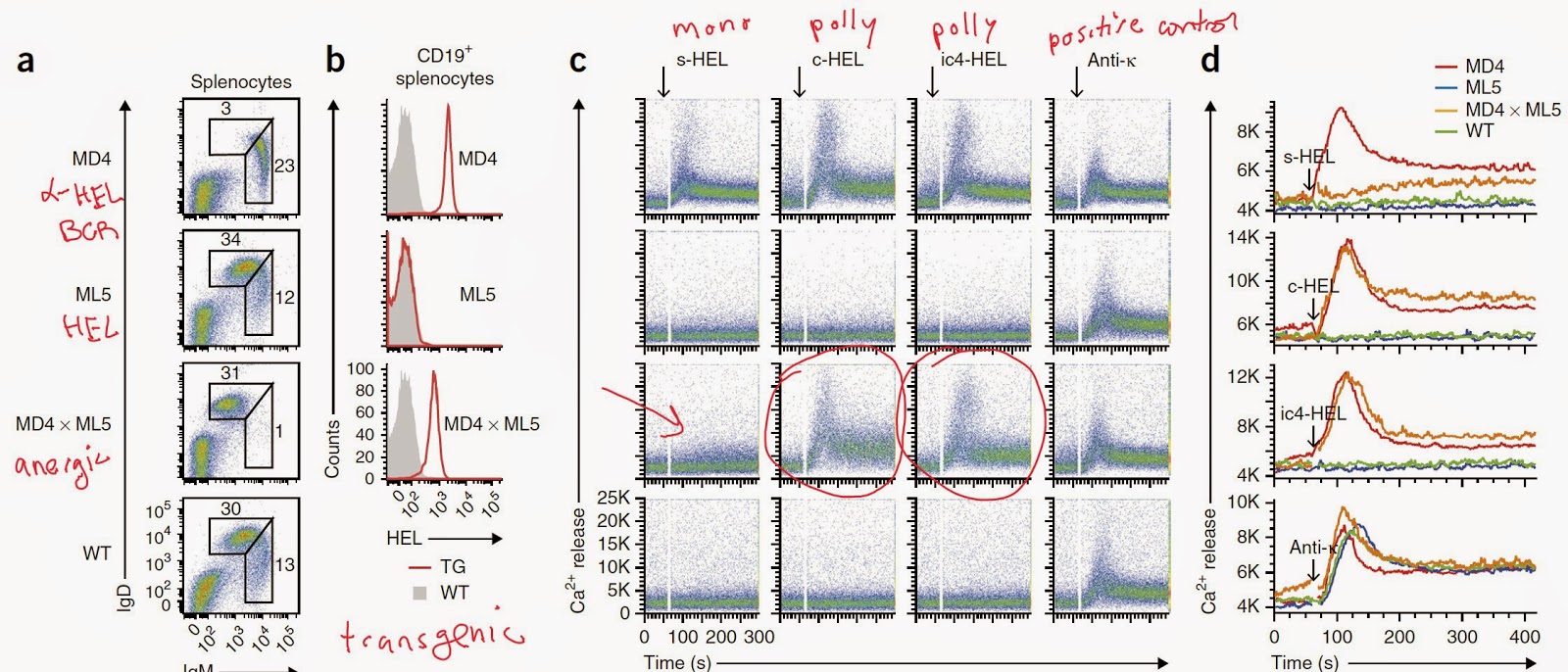
Anergic B cells respond to polyvalent antigens via IgD receptor

Naive B cells express IgM and IgD receptors. Both receptors share the same unique immunoglobulin variable region but differ in immunoglobulin constant regions. IgM is secreted in response to antigenic signaling but not much is known regarding the role of IgD in immune response.

New study in Nature Immunology provided very interesting data about the function of IgD. It turned out that unlike IgM, IgD receptors only respond to polyvalent antigens.

The authors, led by Hassan Jumaa at the Institute of Immunology (Ulm, Germany), first showed that unlike IgM receptor, cell line expressing IgD receptors specific for hapten (NIP) or antigen (HEL) responded only to polyvalent antigenic forms.


Next, the authors find that these difference in response between IgM and IgD was related to the difference in hinge region (that connects variable and constant regions). IgD with no hinge region (IgDΔhinge) responded as if IgM and IgM with IgD hinge region responded as if IgD.


The authors observed that anergic B cells obtained from antigen (HEL)-specific B cell double transgenic mouse expressing soluble HEL (s-HEL) could still respond to polyvalent HEL antigen.


The authors showed that monovalent antigen could competitively inhibit IgD signaling in response to polyvalent antigens.


Next, the authors observed that functionally, absence of IgM receptor could compromise innate B-1 cells scavenging response (natural antibody response) to a soluble auto-antigen phosphatidylcholine (PtC).


Indeed, IgD receptor itself were unable to respond to a soluble phosphatidylcholine (PtC).


Finally, the authors also showed that the absence of IgM signaling compromised IgG scavenging functions against multiple auto-antigens (oxidized LDL, etc) as well.


In summary, this study revealed that anergic B cells (IgMlowIgDhigh) can respond to antigen when stimulated with polyvalent antigens. Probably the role of IgD receptor is to prevent improper activation of naive B cells in response to soluble antigens. I wonder what is the (a) phenotype of IgD KO mice or (b) whether auto-antigens targeted by natural antibodies are mono or polyvalent in nature?

David Usharauli


Monday, April 6, 2015

Melanoma dendritic cell vaccine is safe but is it effective?


There is no doubt that cancerous tissue can yield multiple neo-antigens derived from non-synonymous mutations. Hypothetically and practically (as this new study and others as well have shown), immune system can and is able to detect these minute differences in the mutated proteins. It appears that by combining current in silico algorithms such as NETMHC-3.4 with epitope presentation assays (in vitro assays) provide quite accurate list of potential immunogenic tumor peptides.

The authors in this new paper, for example, were able to identify and confirm in a complementary in vitro assays (T2 cell peptide binding assay and tandem minigene constructs expression assay in DM6 cell line) the presence of several neo-antigens in melanoma tissue derived from 3 patients.


Re-injection of CD40L+TLR ligand matured, melanoma-peptide pulsed autologous dendritic cells back into patients yielded antigen-specific CD8 T cells expansion.


This was a small Phase I clinical study to determine the safety of the DCs vaccine. Since science fundamentals are strong behind this trial (especially considering the authors focus on IL-12p70 producing DCs as a source of cellular vaccine), the results were expected. Of note, these 3 patients were treated with ipilimumab (humanized α-CTLA4 antibody) prior to the experiments described in this paper. It is not clear how this could have skewed the [positive] outcome of DC vaccine here. Anyway, successful tumor treatment would require simultaneous approach from several directions (DC vaccine, checkpoint inhibitors, small drug tyrosine inhibitors).

David Usharauli      

Saturday, April 4, 2015

in vitro model to study commensal microbe-host interaction

Segmented filamentous bacteria (SFB) is a commensal gut microbe in laboratory mouse. Importantly, few years ago SFB was identified as microbe that could specifically drive TH17 development in the mouse gut as well as induce general gut immune system maturation.

It is anaerobic microbe with no known in vitro growth conditions. However, to reduce laboratory animal suffering and unnecessary in vivo experimentation, it would be advantageous to be able to study SFB-host interactions in vitro.

This is exactly what the authors of the new paper published in journal Nature had tried to accomplish (this paper was under review for 1 year). The authors were able to develop in vitro SFB-mammalian cell co-culture that yielded functional colonies of SFB.

The authors showed that in vitro culture of SFB required presence of live cells (both human (caco-2/TC7 line) or mouse cells (mICcl2 line) were tested) and iron.


Importantly, in vitro derived SFB could colonize germ-free mice and induce secretory IgA and  TH17 development.


The authors showed that in vitro derived SFB induced gene expression profile in host cells in an in vitro assay that mimicked in vivo observations.


In summary, I hope this type of research advances would contribute not to just general increase of our knowledge of commensal-host interaction, but also would indirectly improve laboratory animal welfare by reducing their use in the lethal, terminal experimentation.

David Usharauli
  

Thursday, April 2, 2015

Mutation in human interferon regulatory factor-7 (IRF7) predisposes to severe flu infection

Many times, unexpected reactions to infections (as in severe influenza) or innocuous agents (as in allergy) are based on unsuspected mutations in proteins relevant in immune regulation


The authors showed that the patient inherited one mutated copy of IRF7 from each parent. Functionally, the patient's derived IRF7 lacked the ability to induce IFN-α in a reporter assay (parents were heterozygous and had normal IRF7 functions).


In ex vivo experiments the authors showed that patient's derived plasmacytoid dendritic cells (IFN-producing cells) did not respond to H1N1 infection by up-regulating IFN-α system (though small amount of IFN-β was produced).


Furthermore, the authors showed that the patient's derived fibroblast were highly susceptible in a viral replication assay compared to control fibroblast samples, suggesting that high viral titre in this patient could have produced clinical signs of severe flu.


In summary, this short and simple study provides molecular basis in understanding the disease outcome. In the future, when every newborn will have their DNA sequenced, the parents will be in a better position to carry out necessary prophylaxis and avoid any complications from infections or allergy.

David Usharauli