Tuesday, August 27, 2019

The auto-reactive CD4+ T cells provide IL-2 to proto-Tregs in the thymus

The T cells expressing the transcription factor Foxp3 called regulatory T cells, abbreviated as Tregs, are the most important cell type in the immune system. Without them, the whole immune system goes haywire. As a result, the body simply dies in a very short time.

The Tregs develop in the thymus and require two things: TCR signaling and IL-2. The thymus expresses a very diverse set of epitopes including that from peripheral tissues such as the pancreas or prostate. The high-affinity interaction between TCR and epitope/MHC II makes proto-Treg sensitive to local IL-2, a necessary step to complete a Treg formation loop.

But what cell provides that crucial IL-2 to proto-Tregs? There hasn't been any consensus with this regard but a new paper in the Journal of Experimental Medicine from Sasha Rudensky's lab indicates that it is mature CD4+ T cells and CD25+Foxp3- CD4+ single-positive (SP) T cells that are the main source of thymic IL-2 required for Treg development.

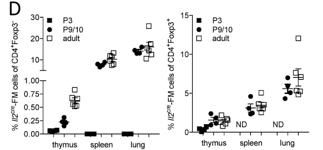
For this study, they used an IL-2 reporter mouse wherein cells expressing or having a history of the expression of IL-2 are genetically labeled and analyzed. They found that IL-2 expression was restricted to TCRbeta expressing CD4+ population.


Out of CD4+ T cells, the most IL-2 was made by mature CD4 SP and CD25+Foxp3- CD4+ T cell population. Of note, CD25+Foxp3- T cell population contains proto-Tregs.


Interestingly, the authors also detected mature Tregs with the history of IL-2 expression. It implies that bifurcation between Tregs versus IL-2 producer is a stochastic process.


As expected, TCR signaling together with IL-2 was essential for Treg formation. A "bystander" effect on Foxp3 upregulation on antigen-independent proto-Tregs (Vbeta 8- T cells) could be explained by the fact that these T cells were likely TCR activated in vivo before harvesting for ex vivo experimentation.


Based on these data, the authors suggested the following model: among mature SP CD4 T cells, a small pool produces IL-2 that in the context of high-affinity TCR/epitope interaction and CD25 upregulation promotes Foxp3+ Treg formation either autocrine or paracrine manner. Since the thymus is expressing self epitopes we can conclude that those IL-2 producing T cells are auto-reactive T cells.


The following questions remain unanswered:

1. What determines Treg, IL-2-producer or deletion pathways? All three options are open for high-affinity TCR+ CD4 SP cells.

2. Do TCR specificity overlaps between Tregs and IL-2 producers?

3. What cells provide IL-2 to Tregs in the periphery?

4. Is IL-2 delivery TCR/epitope-specific or non-specific event?


We have recently published a new model, called SPIRAL, that provides answers to these questions. The SPIRAL is based on the principle of epitope cross-reactivity.


Shared TCR epitope cross-reactivity could permit dyads of Foxp3+ regulatory and IL-2-producing T cell precursors to escape thymic purge 


posted by David Usharauli


Tuesday, August 13, 2019

Do regulatory CD8+ T cells control autoreactive CD4+ T cells in the mouse model of human MS?

Recently journal Nature published a very thought-provoking study from Mark Davis' lab. In it, the authors have described the existence of a specialized population of CD8+ T cells that prevented auto-reactive CD4+ T cells from causing autoimmune brain inflammation (EAE) in mice, a laboratory model for human multiple sclerosis.

Let's analyze what the study shows. Both Fig. 1 and Fig. 2 are rather superfluous as they simply show either time kinetics of CD4+, CD8+ and γδ+ T cells responses in the blood or CNS following autoantigen immunization (Fig. 1) or frequency of TCR clonal  distribution based on TCR β or both γ and δ sequencing (Fig. 2). It is not clear what was the purpose of showing them within the paper itself.
    

Next, the authors tested the TCR specificities for expanded clones of CD4+ or CD8+ T cells. Four of these CD4+ TCRs expressed in human leukemia SKW αβ−/− cells yielded robust staining with a MOG35–55 I-Ab peptide–MHC tetramer. 
Curiously, out of nine TCRs from CD8+ T cell clones expressed in a mouse T cell hybridoma 58 αβ−/− cells, none of them get stimulated when co-cultured with bone-marrow-derived dendritic cells pulsed with myelin protein-derived peptides (total of 350 myelin peptides were tested). So, something else, besides myelin protein, was driving CD8+ T cell expansion.

To identify epitope specificity for TCRs from CD8+ T cells, the authors used H2-Db yeast-pMHC libraries. Six of the clonally expanded and one positive control CD8+ TCRs were used. Two, EAE6 and EAE7 TCRs showed robust tetramer staining but no matches were found in the mouse genome. They referred to these peptides identified in the peptide library screen the surrogate peptides (SPs).

Interestingly, the co-immunization of these SPs with myelin peptide inhibited the development of brain inflammation in mice. 

More importantly,  CD8+ T cells harvested from SPs-immunized mice but not control, naive mice, inhibited myelin-specific but not ovalbumin-specific CD4+  T cells in vitro. 


Moreover, only Ly49+ but not Ly49− fraction of CD8+CD44+CD122+ T cells from SPs-immunized donor mice showed inhibitory function both in vitro and in vivo. Of note, the application of the anti-Qa-1b antibody had no effect on the CD8 suppression of Myelin-specific CD4+ T cells. Here, the authors also tested CD8+ T cell reactivity to CFA (complete Freund’s adjuvant) or PTX (pertussis toxin) used in EAE immunization protocol and found that it did not increase Ly49+ fraction.


So, how can we summarize this paper? First, focus on γδ+ T cells here is extra and feels out of context. Second, no endogenous peptides were found that mimic SPs found in the library screen. Could it be microbiota-derived? The authors did not consider this possibility, it appears. Third, how SPs-specific Ly49+ CD8+ T cells inhibit myelin-specific CD4+ T cells? Not clear. It does appear highly specific to myelin-specific CD4+ T cells in the context of brain inflammation. But myelin-specific CD4+ T cells see myelin peptides but these CD8+ T cells do not seem to recognize them. Very confusing indeed. 
In general, the "memory-like" CD8+ T cells, such as Qa-1b-restricted population, has been known to inhibit immune response. This paper simply provides some new evidence in that direction. But it is not a novel idea or observation and without some novel mechanistic evidence I don't see how it could have landed in Nature's pages.  


posted by David Usharauli