Wednesday, September 28, 2016

Wiskott-Aldrich syndrome protein (WASp) deficient FOXP3+ Tregs fail to control IgE mediated allergies

The inherited immunodeficiencies are frequently characterized with dysregulated Th2 responses, atopy, and elevated IgE levels. Mutations in Foxp3, STAT3, DOCK8, PGM3, LAT, ZAP70, or RAG result in hyper IgE phenotypes. Mechanism is unclear.


For this study the researchers analyzed "the overall burden of clinical food allergy within a cohort of 25 patients with mutations in the WAS gene" and found that individuals with WAS mutations were more likely to demonstrate serum sensitization to peanut, milk, and egg (compared to the general population).


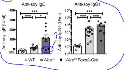
Lab mice deficient for WASp also display elevated IgE (and IgG1) antibody levels to components of their chow food (even on germ-free background).


Since WASp is expressed in different cell types, the authors tested mice selectively deficient for WASp in B cells, DCs or FOXP3+ Tregs. Out of these gene-modified mice, only Wasfl/fl Foxp3-Cre mice showed deviation to Th2 phenotype and development of IgE to chow food.


In vitro T cell stimulation confirmed that total T cells from Wasfl/fl Foxp3-Cre mice showed selective enhancement in Th2 cytokines.


In sum, these results indicate that mice with selective deficiency of WASp in FOXP3+ Tregs display excess in Th2 subsets. It is possible that absence of WASp destabilizes FOXP3+ Tregs and this somehow drives their de-differentiation into Th2-like cells (and not into Th1 or Th17, for instance).

David Usharauli


Saturday, September 24, 2016

Genomic alterations in IFN-γ pathway underlie resistance to anti-CTLA-4 (Yervoy) therapy

This week journal Cell published a short "reverse translational" study conducted on small cohort of patients non-responsive to immunotherapy with anti-CTLA-4 antibody (ipilimumab, Yervoy) that  showed that genomic alterations in IFN-γ pathway in non-responder patients could underlie their resistance to immunotherapy.  

For this study the authors compared 12 patients who did not respond to ipilimumab therapy (non-responders) and 4 patients who did respond to ipilimumab therapy (responders). They found that "tumor samples from non-responders were found to have significantly more somatic mutations, including copy-number alterations (CNAs) and single-nucleotide variants (SNVs) of the IFN-γ pathway genes".


Next, the authors showed that primary melanoma cell lines derived from anti-CTLA-4 responder or non-responder patients could be differentiated based on their in vitro sensitivity to IFN-γ.


Next, they showed quite bizarre experiment. By knocking down IFN-γ receptor in mouse B16 melanoma cells the authors showed that these cell line became less sensitive to IFN-γ in vitro. Not sure about logic behind these experiment.  

Finally, the authors showed that B16 melanoma cells deficient for IFN-γ signaling and transplanted into WT mice were less sensitive to anti-CTLA-4 therapy.


In summary, this small cohort study suggests that screening of melanoma patients for genomic alterations in IFN-γ pathway could be useful in better predicting therapeutic outcome for this checkpoint inhibitor therapy.

David Usharauli


Tuesday, September 20, 2016

Fusobacteria, a gut commensal, contributes to autoimmune type I diabetes in mice

Initiation of autoimmune disease is still an immunological mystery. Some forms of autoimmune diseases are results of genuine genetic defects in signaling molecules within immune system. Other forms show strong linkage to certain HLA haplotypes that present antigenic epitopes. More recently scientists focused on the role of gut commensals in autoimmune diseases.

A new study in Jounral of Experimental Medicine showed that cross-reactivity at the epitope level between gut commensal Fusobacteria-derived magnesium transporter and β islet-specific glucose-6-phosphatase catalytic subunit–related protein (IGRP) contributed in autoimmune diabetes development in IGRP-specific CD8 T cell transgenic, CD8+ TCR NY8.3 NOD mice.

Initially, the authors observed that unlike MyD88KO NOD mice, MyD88KO CD8+ TCR NY8.3 transgenic NOD mice showed accelerated diabetes development (though unlike the authors, I don't find this surprising).


Interestingly, when co-housed with WT NOD mice, MyD88KO TCR NY8.3 transgenic NOD mice showed enhanced protection against diabetes, suggesting dominant role of fecal bacteria present in WT NOD mice in providing this protection.


Since it is known that NOD mice susceptibility to diabetes is commensal-dependent, the authors sequenced fecal microbiome in MyD88KO NY8.3 NOD mice to determine its composition. Not surprising, certain families of commensals underwent changes on MyD88KO background.


When the authors compared the IGRP206–214 peptide sequence against bacterial protein sequences in the nonredundant protein sequence database, they found several hits shared strong homology with IGRP206–214 peptid, the native autoantigen detected by NY8.3 CD8+ T cells. One such peptide, W15944, was derived from L. goodfellowii, a member of the phylum Fusobacteria (gram-negative anaerobe), a human and NOD mouse oral commensal.


Indeed, W15944 stimulated NY8.3 CD8+ T cells could transfer diabetes in NOD mice.


Finally, introduction of L. goodfellowii into WT NY8.3 NOD mice accelerated diabetes development, further suggesting role of L. goodfellowii in this process (while in general, this paper is of high quality, it lacks some of the crucial experiments such as, for example, (1) introduction of L. goodfellowii into NY8.3 NOD mice on germ-free background and (2) experiments with L. goodfellowii lacking cross-reactive W15944 peptide).


In summary, the author showed that in this artificially constructed NOD mice, IGRP-specific CD8 T cells could be activated by commensal-derived cross-reactive peptide and initiate autoimmune destruction of  β cells (it is remains to be seen why Foxp3+ regulatory T cells are incapable of preventing such T cell attack).

David Usharauli


Wednesday, September 7, 2016

Eye-specific autoimmunity in AIRE/Lyn double deficient animal

AIRE protein in the thymus directs expression of tissue-specific antigens that on one hand reduces number of auto-antigen specific T cell clones and on the other hand leads to generation of auto-antigen specific Foxp3+ regulatory T cells. However relationship between between central tolerance and its peripheral counterpart is not fully understood.

New study in Journal of Clinical Investigation examined relationship between hypomorphic AIRE function on Lyn-/- background. The authors found that such combination selectively accentuated eye-specific autoimmunity.  

First, the authors observed that mice double deficient for AIREGW/+/Lyn-/- functions (but not single deficient) developed spontaneous eye inflammation.


These AIREGW/+/Lyn-/- mice showed expansion of eye-protein specific CD4 T cells and autoantibodies.


These auto-antigen specific T cells could infiltrate eye tissue causing inflammation.


The authors found that selective absence of Lyn in CD11c+ dendritic cells (AireGW/+CD11c-Cre Lynfl/fl) were sufficient to recapitulate this eye immunopathology.


Finally, the authors found that auto-antigen uptake by Lyn-/- DCs in eye draining lymph nodes contributed to eye inflammation.


In summary, this study indicates that some of the clinically relevant autoimmune phenotypes could be linked to failure to establish both central and peripheral tolerance.

David Usharauli


Tuesday, September 6, 2016

Foxp3+ Tregs control CD8 T cells, but not CD4 T cells, by IL-2 deprivation

This week Nature Immunology published another interesting study from Rudensky's lab done in collaboration with Jason Fontenot (who apparently moved from Biogen to Juno Therapeutics). So, if you are a fan of Tregs, below is my short scientific overview of that paper.

This study tries to answer what role IL-2 signaling plays in already established Foxp3+ Tregs. For such study mice with germline deficiency in IL-2 signaling cascade would have been impractical since IL-2's effect on thymocytes and etc. Instead, the authors went to already well established path of using mice lacking "molecule of interest" specifically in Tregs. In this case several Foxp3-cre mice were used, such as: l2rbfl/flFoxp3Cre, Il2rafl/flFoxp3Cre, Stat5afl/flStat5bfl/flFoxp3Cre, Rosa26Stat5bCAIl2rbfl/flFoxp3Cre and Rosa26Stat5bCAIl2rafl/flFoxp3Cre, Rosa26Stat5bCAFoxp3Cre–ERT2. All these gene-modified mice allows specific targeting of Foxp3+ Tregs.

Unsurprisingly, mice lacking IL-2Rβ or IL-2Rα or STAT5 signaling specifically in Tregs developed autoimmunity.


Surprisingly, however, while constitutive expression of STAT5 in Tregs lacking IL-2 signaling receptors (IL-2Rβ or IL-2Rα) could rescue mice from "CD4 effector phenotype" and early death, these mice still developed immunopathology later due to massive expansion of CD8 effector/memory cells, suggesting that sensing of IL-2 by Tregs, so called "IL-2 sink", was necessary to specifically control CD8 T cells, but not CD4 T cells.


In summary, this study suggests that Tregs are using distinct mechanisms to control CD4 and CD8 T cells. It kind of makes sense because MHC class-II restricted CD4+ Tregs cannot interact the same way with MHC class-I restricted CD8+ T cells as they could with conventional MHC class-II restricted conventional CD4+ T cells.

David Usharauli