Tuesday, March 31, 2015

Tumor converts neutrophils into metastasis-promoting cells via γδ T cell-derived IL-17

Immune system supposed to defend the body from infectious agents and genetically transformed cells (tumors). Usually, at the end of each immune response, that can be quite damaging (immune phase), immune cells will be involved in tissue healing, regeneration or remodeling (adaptation phase). However, sometimes infectious agents or tumors will circumvent these steps and jump directly to adaptation phase and will recruit immune cells to carry out their "agenda" at the expense of the host.

For example, we can speculate that tumor's "agenda" would be to grow and expand (metastasize). However, neither of these possible without local and distant tissue remodeling and its readiness to accommodate (accept) tumor cells. Here is where local immune cells become involved.

New study in journal Nature points to one of those possibilities. The authors showed that in a mouse model of mammary tumor metastasis, neutrophils promotes tumor lung metastasis via IL-17 produced by circulating γδ-T cells.

I would like to point out that the authors have used tumor transplantation model (to have shorter experiments) that is obviously very different from spontaneously arising tumors. Nevertheless, they found that lung or lymph node metastasis of skin transplanted tumor was reduced with neutrophil depletion using α-Ly6G antibody (this antibody supposedly selectively depletes neutrophils since they express it at high level).


Interestingly, tumor metastasis were also reduced in the recipients devoid of adaptive immune system and correlated with reduction of IL-17 and G-CSF (granulocyte colony-stimulating factor) serum levels.


Finally, the authors showed that γδ-T cell depletion or genetic deficiency reduced lung and lymph node metastasis of transplanted tumor.


In summary, these results suggest that tumor cells exploit not yet identified pathways within immune system (γδ-T cells / IL-17 / neutrophils axis) to prepare distant tissues to accommodate tumor colonies coming from original tumor niche. Only by understanding how immune cells interact with normal tissues during or after immune response, could we design ways to block such metastasis.

In general this paper is OK, especially if one considers other papers (two recent papers in JEM) corroborating the idea of IL-17's involvement in tumor initiation and metastasis. However, some data are not clear or not well explained, so not a Nature caliber paper, in my view. For example, in Fig. 3a, treatment with α-IL-17A did not modify IL-17 level in the serum. Also, the authors did not explain why they thought CD8 T cells were protective against metastasis in this model when tumor-bearing RAG KO hosts, which lack CD8 T cells, did not show increased metastasis (Fig. 2d versus. Fig. 3g)?


David Usharauli
 

Sunday, March 29, 2015

Tyrosine kinase hyper-activation rather than its inhibition overcomes acute lymphoblastic leukaemia cells resistance

Acute lymphoblastic leukaemia (ALL) in pre-B cells is driven by constitutive activation of tyrosine kinases, such as BCR-ABL1 (Philadelphia chromosome positive) that mimic BCR signalling (that normally serves as a survival signal). For that reason, treatment of ALL usually involves use of tyrosine kinase inhibitors (TKI) such as Gleevec (imatinib). However, frequently ALL cells develop resistance to TKI. 


The authors showed that in vitro assay ALL cells transfected with constitutively active Syk kinase undergo rapid cell death that was actually reversed by imatinib treatment.


Next, the authors showed that BCR-ABL1 transformed ALL cells deficient of signaling molecules with ITIM tail (pecam1, CD300, Lair1) that normally inhibit Syk kinase, undergo rapid cell death.


Further experiments revealed that inhibitory phosphatases, SHP1 (ptpn6) and SHIP1 (inpp5d), downstream of ITIM signalling contributed to ALL cell survival via Syk inhibition.


Finally, the authors showed that use of small molecule inhibitor of SHIP1, 3AC, could lead to rapid ALL cell death due to Syk hyper-activation


In summary, these results showed that even ALL cell still retain sensitivity to BCR negative selection when over-activated. Initially, mutation in BCR-ABL1 make pre-B cells to receive constitutive survival signal. However, additional activation of Syk kinase would push overall signalling within ALL to the threshold of negative selection and to the ultimate ALL cell death. Thus, combining of Gleevec with 3AC-like drugs would provide additional safeguards to prevent the emergence of resistant ALL clones and dramatically improve survival of ALL patients.   

David Usharauli


Friday, March 27, 2015

First-born thymic Foxp3+ regulatory T cells protect against tissue-selective AIRE-dependent autoimmune syndrome

Adaptive immune system should be able to distinguish between self and non-self antigens. This is a very fine process since at the molecular level both self and non-self antigens look the same. So how it is done?


AIRE is a gene that controls tissue-specific protein expression in the thymus. The authors observed that conditionally T reg-depleted neonatal mice supplemented with T regs from AIRE KO donor, but not from WT donor, developed tissue-specific autoimmune syndrome.


Further experiments with adoptive transfer of T reg population tagged either during neonatal stage (day 0-10) or after weaning (day 35-45) revealed that only neonatally tagged T regs were enriched in Foxp3+ population able to prevent the development of AIRE-dependent tissue-selective autoimmune syndrome.


The results from this study suggest that the role and functionality among Foxp3+ T regs varies depending, at least, on time of their generation. It appears that first-born T regs generated during a neonatal stage provide specialized protection against development of autoimmune syndrome (though tagging per se did not specifically identify AIRE-driven Foxp3+ T regs in newborn mice in this study).

Interestingly, the authors reported that in NOD.Foxp3-DTR mice T reg depletion after weaning did not result in death of the experimental animals. This is in contrast to earlier reports from Rudensky's lab where adult Foxp3-DTR mice die after T reg depletion. Wonder what could have made such difference.   

David Usharauli


Monday, March 23, 2015

IL-3 producing innate B cells drive sepsis susceptibility

Sepsis is a life-threatening condition characterized by over-production of first line defense cytokines such as IL-1β, IL-6, TNF-α (so called cytokine storm) and leading to death due to failure of vital organs (heart, kidney, liver) and coagulation / anti-coagulation systems.

It is not clear whether immune system can defend against sepsis or contrary make it worse. From an evolutionary point of view, sepsis (as infectious overload) would represent dead-end for the host.

However, if we could understand sepsis pathophysiology we can try to manage it. In this regard, new paper in journal Science is of great interest. This study shows that a little known cytokine IL-3 drives host's susceptibility to sepsis.

The authors have studied mouse model of septic shock. They observed that IL-3 KO mice were highly resistant to septic shock


IL-3 KO mice were able to control bacterial counts in the blood.


Reconstitution experiments confirmed that IL-3 was driving sepsis susceptibility.


Additional experiments showed that B cells were the major source of IL-3 during sepsis in this mouse model.

Indeed, adoptive transfer of wild-type B cells into IL-3 KO hosts could increase sepsis susceptibility of the host (though the authors failed to present host survival data in this setting, as in Fig. 1A).


Finally, the authors showed that in humans, survival of sepsis patients correlated with the level of serum IL-3.


In summary, this simple study points to a new therapy for sepsis patients. In would be interesting to know whether reduced innate response in IL-3 KO mice (a) allowed the host to clear the bacteria from the blood using alternative mechanisms (complement, C-reactive protein system, etc) or whether (b) it was easier for IL-3 KO hosts to repair gut wall damage and prevent further leak of gut flora into blood.    

David Usharauli


Friday, March 20, 2015

CCL3 and memory CD4 T cells improve tumor antigen RNA loaded dendritic cell vaccine performance

Dendritic cells represent the most potent antigen-presenting cells capable of priming naive T cells against infectious or tumor antigens. However, so far, clinical application of antigen-loaded autologous (patient's own) dendritic cell vaccines produced few encouraging results.

Unlike regular vaccines that contain protein conjugates emulsified in adjuvants, dendritic cell vaccine is a biological vaccine containing live cells pulsed with target antigen. The rules that govern efficacy of live cell vaccines would depend on several conditions such as immunological condition of vaccine injection site, migration and viability of injected live vaccine cells and etc. 


The authors have tested their model both in glioblastoma cancer patients and in mouse model of skin cancer. I would like to point out the this paper was under Nature's review for more than a year, implying that it's data are not as strong as the authors would like to argue.

For one thing, clinical protocol the authors have used is not entirely clear. Initially, the authors showed that immunization of patients with tetanus toxoid (Td vaccine) prior to DC vaccine injection extended glioblastoma patients survival almost two-fold compared to un-pulsed DCs. However, I do not understand why Td was compared to un-pulsed DCs. Even after careful reading of methods section, I still was not able to clearly understand the authors reasoning.


Next, using mouse model, the authors showed that presence of activated memory CD4 T cells contributed to the enhanced migration of antigen-loaded DCs to the local lymph nodes.


Interestingly, Td- or memory CD4 T cell-mediated enhancement of DCs migration was abolished in CCL3-KO host.


In addition, exogenous CCL3 was effective only in presence of activated memory CD4 T cells, implying that both memory CD4 T cells and CCL3 are required for enhancement of DCs migration to local lymph nodes.


In summary, these results revealed that locally Td-activated memory CD4 T cells creates a favorable environment for antigen-loaded DCs migration to local lymph node in a CCL3-dependent manner. Source of Td-driven CCL3 from the injection site is not clear.

If you ask me, only reason this paper ended up in Nature has to do with the fact that somehow the authors' protocol (Td + DC pulsed with pp65 antigen RNA) was able to substantially extend glioblastoma patients' survival. However, it is not clear why would Td-activated skin-resident memory CD4 T cells improve migration of DCs loaded with non-related antigen, CMV pp65? If we consider the fact that glioblastoma patients should already have CMV pp65-specific memory CD4 T cells capable of doing the same function as Td did, then I don't see how Td could have provided any additional benefits over simple CMV pp65 RNA-loaded DCs? This paper raises more questions than provides answers.

David Usharauli
Wednesday, March 18, 2015

IL-33 links innate lymphoid cells group 2 to lean body mass control

There is one aspect of immune system that I did not get at all. It is related to the role of immune system in maintaining a lean body mass by controlling browning [beiging] of white adipose tissue and increasing energy expenditure.

Previous studies have implicated immune cells, such as eosinophils, type 2 macrophages and IL-4rα signalling in body energy expenditure and lean body mass control. New study in journal Nature has provided evidence suggesting that type 2 innate lymphoid cells, ILC2 and IL-33 play an independent and non-redundant role in lean body mass control and energy expenditure

The authors showed that in both human and mouse, white adipose tissue contain LinIL-25IL-127+population expressing IL-33R typical for ILC2. The frequency and number of ILC2 population in white adipose tissue were inversely correlated with obesity or high fat diet.


Experiments with IL-33KO mice or recombinant mouse IL-33 showed that ILC2 population in white adipose tissue retracted or expanded depending on presence of IL-33.


Adipose tissue analysis revealed that IL-33 induced UCP1 expression in white adipose tissue. UCP1 is responsible for high energy expenditure in beige fat.


Adoptive transfer of ILC2 into ILC2-deficient hosts showed that IL-33 worked through ILC2. Additional experiments revealed that IL-33/ILC2 axis worked independently from eosinophils, regulatory T cells or IL-4rα signalling.


Somewhat similar results were presented in another paper published in journal Cell earlier this year. However, in that paper the authors observed that eosinophils and IL-4rα signalling were also involved alongside with IL-33/ILC2. It would be interesting to know what is the reason for this discrepancy.

In summary, these results points to a novel function of innate immune system. Though It may even nothing to do with immune function per se but rather linking ancient metabolic pathways to innate immune cells. Even if IL-33 can help to reduce body fat, it does not necessarily means that we could start injecting obese people with IL-33. IL-33 has been implicated in pathological responses as well, like asthma or allergy. Only global view of cytokine function could tell us the real usefulness of any finding. Discrete, individual disease models are insufficient for this task and could lead to wrong conclusions. 

David Usharauli


Monday, March 16, 2015

Antibody targeting IL-23(p19 subunit) beats psoriasis

Psoriasis is a common skin inflammation of autoimmune nature. If there is any disease that benefited from the fundamental immunological research is Psoriasis.

Initially thought to be a purely skin disorder, now it is firmly established that immune system's attack on it own skin tissue plays a major role in driving clinical signs of psoriasis. Pioneering studies with α-p40 antibodies suggested that IL-12 might have been involved in psoriasis. However discovery of IL-23 (that consisted of p40 and p19 subunits) in 2001 opened up new era in our understanding of molecular mechanisms behind psoriasis pathology.

New study in Nature provided additional data to clearly establish that selective targeting of IL-23 drastically improves psoriasis's skin pathology.

The authors showed that three, monthly intravenous injections of humanized antibody targeting p19 subunit of IL-23 (Tildrakizumab) could drastically reduce psoriatic skin pathologies in absolute majority of patients that lasted for at least 1 year.


α-p19 antibody injections reduced hyper-keratinization and hyper-proliferation in psoratic skin lesions.


In summary, this study suggests that IL-23 system that includes IL-17 are intimately involved in psoriatic skin pathology and its targeting (maybe simultaneously with α-IL-17 ) could finally bring the needed relief to people suffering from this skin disorder.

 David Usharauli


Sunday, March 15, 2015

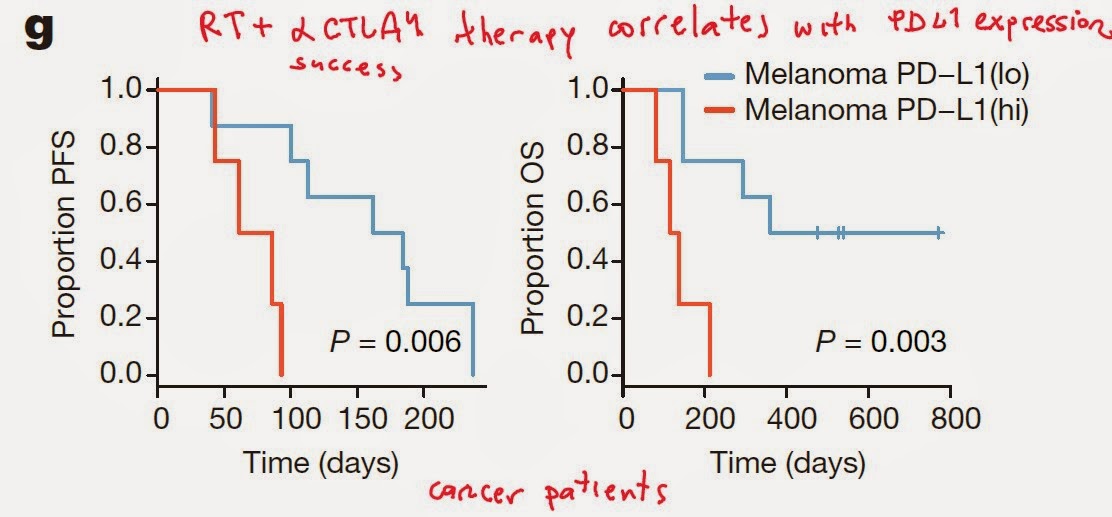
Synergy of checkpoint inhibitors for tumor immunotherapy

Introduction of checkpoint inhibitors, α-CTLA4 and α-PD1/PD-L1 humanized antibodies, injected new hope in cancer immunotherapy. Still, these clinical advances are at early stage and the significant benefits are achieved in small proportion of patients (~ 20%, for solid tumor patients). 


The concept is similar to treatment of antibiotic resistant pathogenic microorganisms. Single mechanism of action usually is not sufficient for successful treatment since microorganisms are quick to adapt and develop resistance to it.

Tumors, too, frequently behave as the separate macro-organisms within the body. When coming under immune pressure exerted by α-CTLA4 antibody, tumors will try to escape immune surveillance by up-regulating parallel inhibitory pathways such as PD1/PD-L1 system. 


Identification and simultaneous targeting of such inhibitory and/or escape strategies will finally provide clearly measurable clinical benefits in majority of tumor patients.

David Usharauli


Thursday, March 12, 2015

Nociceptor analog, MRGPRX2, mediates pseudo-allergic response to pharmacological compounds

Classical allergic response is mediated via IgE. However, some allergic responses to small peptides, pharmacological or chemical compounds are independent of IgE. 


The authors showed that mast cells from mice deficient for Mrgprb2 expression failed to respond to classical mast cell activator (secretagogue), compound 48/80, while retaining IgE mediated responsiveness.


Additional experiments confirmed that Mrgprb2 mediated injection site reactions (ISR) and mast cell histamine release in response to drugs such as Icatibant (bradykinin-receptor antagonist), tubocurarine (skeletal muscle relaxant) and ciprofloxacin (fluoroquinolone antibiotic).


Knowledge of chemical motifs (THIQ) responsible for Mrgprb2 activation will help to prevent and treat pseudo-allergic reactions to life-saving medical drugs.

David Usharauli

Wednesday, March 11, 2015

NK cell activation initiates type II diabetes

Type II diabetes is a complex syndrome involving endocrine, immune and metabolic abnormalities. It is well known that obesity can lead to type II diabetes. But how?

New study in Nature Immunology suggests that obesity-induced adipocyte stress activates NK cells  via NCR1 receptor driving IFN-γ mediated insulin insensitivity characteristic to type II diabetes.

The authors observed that NK cell depletion in mice fed high fat diet (HFD) ameliorated insulin insensitivity and glucose intolerance.


Ex vivo examination of visceral adipose tissue (VAT), a target tissue of type II diabetes, revealed that HFD induced expression of NK cell ligand in VAT detected by NCR1 (NKp46 in humans).


Interestingly, VAT but not subcutaneous (Sc fat) adipose tissue from HFD fed mice could stimulate NK cells.

In vivo experiments confirmed that NCR1 deficiency improved insulin sensitivity.


Finally, the authors showed that IFN-γ derived from NCR1 activated NK cells promotes inflammatory macrophages in VAT leading to glucose intolerance, that can be ameliorated with NCR1 blockade.


In summary, these results showed that HFD induces visceral adipose tissue stress that activates local NK cells via NCR1 ligand leading to inflammatory macrophage polarization and reduced insulin sensitivity. Targeting NK cell activation may interrupt this disease cycle and improve type II diabetes management.

I was always wondered why diet-induced obesity leading to VAT stress should activate inflammatory, M1 type macrophages? What is an evolutionary advantage for such response, in general? No idea.  

David Usharauli


Saturday, March 7, 2015

Persistence of immune complexes (ICs) impair Fc-receptor effector functions

Chronic viral infections represent incompletely understood immune condition. Previously it has been shown CD8 T cells functions were impaired during chronic viral infections. 

Now, two new back-to-back papers in journal Immunity provided evidence suggesting that chronic viral infection impairs Fc-receptor effector functions through excessive production and persistence of immune complexes (ICs).

First paper came from David Brooks lab at the University of California, Los Angeles (UCLA). 

The authors (First author Douglas H. Yamada) observed that antibody-mediated depletion of target cell population was severely impaired in chronic LCMV-infected mice (LCMV-Clone 13).


Similar impairment was observed with LCMV-Clone 13 infected mice that receive human CD20 expressing tumor cell line (hCD20-EG7) and depleting α-CD20 antibody (Rituximab).


The authors observed that impairment of antibody-depleting function were selectively impaired in mice infected with LCMV virus producing persistent infection (LCMV-Clone 13), but not with LCMV virus producing acute infection (LCMV-Armstrong).


Additionally, the authors found that the presence of endogenous antibodies were necessary to impair depleting function of exogenously administered antibody.


Adoptive transfer showed that target cells in chronically infected hosts remained susceptible to antibody depletion.  


Additionally, presence of endogenous immune complexes (ICs) but not of free antibodies correlated with depletion deficiency.


Mechanistically, the authors showed that deficiency of macrophage population could recapitulate antibody depleting functional impairment.    


Furthermore, the presence of excess ICs in chronically infected hosts impaired priming of antigen-specific CD8 T cells via defective dendritic cell acquired antigen-antibody complexes.


In summary, these results suggest that excessive production of circulatory ICs during persistent viral infections (HBV, HBC, HIV, etc) impairs antibody-dependent Fc-receptor function (clearance of infected target cells, priming of antigen-specific CD8 T cells). It appears that macrophage Fc-receptors are overwhelmed in the presence of excess circulatory ICs (almost identical research results were presented in the 2nd paper by Rafi Ahmed's lab).

This knowledge could help to better understand the limitations of vaccine efficacy in human populations with chronic viral infections.

David Usharauli