Wednesday, January 11, 2023
If T cell clones are so diverse, what prevents anti-tumor immune response?
Friday, September 18, 2020
A specific bacteria-infecting virus, bacteriophage, found in gut microflora, augments anti-tumor T cell immunity
Molecular mimicry between microbial and host's antigens could contribute to autoimmunity but also to the protection against tumors through epitope cross-reactivity. A new study in journal Science indicates that those cross-reactive epitopes could come from viruses that infect endogenous microbial species.
In this study the authors made a surprising observation that only certain Enterococcus hirae microbial strains (E. hirae 13144 or IGR11) augmented anti-cancer effect in experimental cancer model.
Next, the authors showed that this biological activity was linked to one dominant epitope, TSLARFANI, derived from TMP protein that originated in 39.2-kb prophage only in those specific E. hirae strains. Mice immunized with heat-inactivated E. hirae 13144 strain, or peptide TSLARFANI, or irrelevant E.coli engineered to express TMP, all augmented anti-cancer effect.
Mechanistically, the authors showed that epitope, GSLARFRNI, derived from cancer cells used in these experiments, was recognized by the same CD8 T cells which labeled with TSLARFANI epitope tetramers confirming cross-reactivity between these 2 epitopes.
In summary, this study suggests that microbiota and bacteriophages they carry represent new modality in fight against cancer. In this study overall anti-tumor effect is modest but we need to take into account that this is an effect of just one cross-reactive epitope in one type of MHC inbred mice, and it is likely that many other epitopes will be involved in outbred species such as humans. However, it is still extremely hard to do such analysis in humans in real world scenario due to lack of exact knowledge about human microbiota strains and poor reliability and performances of available bioinformatics approaches. However, once the mechanistic principles underlying anti-cancer effects are uncovered and accepted, then it is much easier to move the field forward.
posted by David Usharauli
Thursday, August 6, 2020
Is it possible to engineer Foxp3+ Tregs from primary T cells?
![]() |
![]() |
This
could also explain why the authors did not see improvement in brain
inflammation in mice EAE model when co-transferring antigen-specific edTregs with
effector T cells. ![]() |
Wednesday, April 15, 2020
Do Tregs inhibit or promote allergic responses? How to avoid data misinterpretation
Tuesday, January 28, 2020
Microbiota-derived peptide and autoimmune heart inflammation: a tale of missing data
The authors even introduced into germ-free TCR transgenic mice Bacteroides thetaiotaomicron (B. theta) lacking the β-gal. Up to now, it feels that the authors have checked all the boxes necessary for high-quality research. But then for some reason, they do not show survival data comparing Bacteroides thetaiotaomicron (B. theta) with and without the β-gal gene as in figure 1. They just showed how a lack of β-gal Bacteroides thetaiotaomicron modifies MYH6-T cells accumulation in the heart tissue.
Saturday, November 30, 2019
Tracking deletion of autoreactive clones versus Treg generation for thymically expressed epitopes
Similar results were obtained when thymus tissue was analyzed.
Saturday, October 26, 2019
Tumor elimination requires simultaneous expression of both class I and II neo-epitopes
Tuesday, August 27, 2019
The auto-reactive CD4+ T cells provide IL-2 to proto-Tregs in the thymus
The Tregs develop in the thymus and require two things: TCR signaling and IL-2. The thymus expresses a very diverse set of epitopes including that from peripheral tissues such as the pancreas or prostate. The high-affinity interaction between TCR and epitope/MHC II makes proto-Treg sensitive to local IL-2, a necessary step to complete a Treg formation loop.
But what cell provides that crucial IL-2 to proto-Tregs? There hasn't been any consensus with this regard but a new paper in the Journal of Experimental Medicine from Sasha Rudensky's lab indicates that it is mature CD4+ T cells and CD25+Foxp3- CD4+ single-positive (SP) T cells that are the main source of thymic IL-2 required for Treg development.
For this study, they used an IL-2 reporter mouse wherein cells expressing or having a history of the expression of IL-2 are genetically labeled and analyzed. They found that IL-2 expression was restricted to TCRbeta expressing CD4+ population.
Out of CD4+ T cells, the most IL-2 was made by mature CD4 SP and CD25+Foxp3- CD4+ T cell population. Of note, CD25+Foxp3- T cell population contains proto-Tregs.
Interestingly, the authors also detected mature Tregs with the history of IL-2 expression. It implies that bifurcation between Tregs versus IL-2 producer is a stochastic process.
As expected, TCR signaling together with IL-2 was essential for Treg formation. A "bystander" effect on Foxp3 upregulation on antigen-independent proto-Tregs (Vbeta 8- T cells) could be explained by the fact that these T cells were likely TCR activated in vivo before harvesting for ex vivo experimentation.
Based on these data, the authors suggested the following model: among mature SP CD4 T cells, a small pool produces IL-2 that in the context of high-affinity TCR/epitope interaction and CD25 upregulation promotes Foxp3+ Treg formation either autocrine or paracrine manner. Since the thymus is expressing self epitopes we can conclude that those IL-2 producing T cells are auto-reactive T cells.
1. What determines Treg, IL-2-producer or deletion pathways? All three options are open for high-affinity TCR+ CD4 SP cells.
2. Do TCR specificity overlaps between Tregs and IL-2 producers?
3. What cells provide IL-2 to Tregs in the periphery?
4. Is IL-2 delivery TCR/epitope-specific or non-specific event?
We have recently published a new model, called SPIRAL, that provides answers to these questions. The SPIRAL is based on the principle of epitope cross-reactivity.
Shared TCR epitope cross-reactivity could permit dyads of Foxp3+ regulatory and IL-2-producing T cell precursors to escape thymic purge
posted by David Usharauli
Tuesday, August 13, 2019
Do regulatory CD8+ T cells control autoreactive CD4+ T cells in the mouse model of human MS?
Let's analyze what the study shows. Both Fig. 1 and Fig. 2 are rather superfluous as they simply show either time kinetics of CD4+, CD8+ and γδ+ T cells responses in the blood or CNS following autoantigen immunization (Fig. 1) or frequency of TCR clonal distribution based on TCR β or both γ and δ sequencing (Fig. 2). It is not clear what was the purpose of showing them within the paper itself.
Next, the authors tested the TCR specificities for expanded clones of CD4+ or CD8+ T cells. Four of these CD4+ TCRs expressed in human leukemia SKW αβ−/− cells yielded robust staining with a MOG35–55 I-Ab peptide–MHC tetramer.
Interestingly, the co-immunization of these SPs with myelin peptide inhibited the development of brain inflammation in mice.
More importantly, CD8+ T cells harvested from SPs-immunized mice but not control, naive mice, inhibited myelin-specific but not ovalbumin-specific CD4+ T cells in vitro.
Moreover, only Ly49+ but not Ly49− fraction of CD8+CD44+CD122+ T cells from SPs-immunized donor mice showed inhibitory function both in vitro and in vivo. Of note, the application of the anti-Qa-1b antibody had no effect on the CD8 suppression of Myelin-specific CD4+ T cells. Here, the authors also tested CD8+ T cell reactivity to CFA (complete Freund’s adjuvant) or PTX (pertussis toxin) used in EAE immunization protocol and found that it did not increase Ly49+ fraction.
So, how can we summarize this paper? First, focus on γδ+ T cells here is extra and feels out of context. Second, no endogenous peptides were found that mimic SPs found in the library screen. Could it be microbiota-derived? The authors did not consider this possibility, it appears. Third, how SPs-specific Ly49+ CD8+ T cells inhibit myelin-specific CD4+ T cells? Not clear. It does appear highly specific to myelin-specific CD4+ T cells in the context of brain inflammation. But myelin-specific CD4+ T cells see myelin peptides but these CD8+ T cells do not seem to recognize them. Very confusing indeed.
posted by David Usharauli
Saturday, July 13, 2019
Pathological changes in the gut could initiate autoimmune diabetes microbiota-specific manner
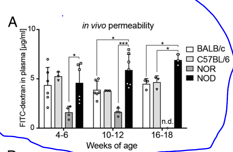
A new study in PNAS did some experiments on BDC2.5XNOD mice to understand the initiation of autoimmunity. First, they showed that the NOD background showed increased gut wall barrier permeability by measuring the FITC-dextran level in the blood after oral application (though they did not show the same permeability test for BDC2.5XNOD mice).
Interestingly, BDC2.5XNOD mice do not develop spontaneous autoimmune diabetes. However, oral application of low-dose dextran-sulfate sodium (DSS) activates T cells and initiates diabetes.
Diabetes, however, does not develop if DSS-treated BDC2.5XNOD mice are depleted of endogenous microbiota or if naive BDC2.5XNOD mice received DSS-modified microbiota.
In summary, this study showed that both gut inflammation and microbiota are necessary for the initiation of autoimmune diabetes in BDC2.5XNOD mice. The most likely scenario is that changes introduced by DSS allow endogenous microbiota to activate islet-specific T cells via cross-reactive antigens. DSS modifies both gut wall permeability and microbiota. Both of these phenomena have been observed by the authors. They conclude that "restoration of a healthy gut barrier through microbiota and diet modulation in diabetes-prone individuals could ultimately reduce intestinal activation of islet-reactive T cells and prevent T1D occurrence".
Others, in news/views for this article, even suggested using antibiotics to deplete endogenous microbiota, but in my opinion, this is a premature suggestion because the authors did not show that microbiota depletion after diabetes has already developed could stop it.
posted by David Usharauli
Saturday, June 29, 2019
Select microbiota species provides protection against food allergy via RORγt+ Tregs
In a separate set of experiments the authors noticed that Il4raF709 mice or mice specifically deficient for RORγt+ Tregs subset displayed similar phenotype in response to allergic challenge. They thought there could be a connection.
Indeed, Il4raF709 mice deficient for RORγt+ Tregs subset lost an ability to resist allergic reaction when colonized with defined mix of Clostridiales and Bacteriodales.
Finally, the authors attributed the loss of protection to loss of MyD88 adaptor signaling in Tregs because Il4raF709 mice deficient for MyD88 signaling in Tregs also showed loss of protection against allergic reaction when colonized with defined mix of Clostridiales and Bacteriodales (Note, oral short chain fatty acid (SCFA) therapy failed to protect Il4raF709 mice against allergic response) .
In summary, we could conclude based on this and other studies that RORγt+ Tregs do play a decisive role in protection against unwanted inflammatory response (Note, however, that allergic sensitization protocol employed here is not exactly "translational" approach).
One major drawback of this study is that the authors failed to examine why it is that Clostridiales and Bacteriodales but not Proteobacteria or other species could signal via MyD88 to provide protection against allergic response. In my view it is not a difference in innate signaling molecules that distinguishes protective versus non-protective microbiota species but rather their antigenic composition that provides epitopes to RORγt+ Tregs to keep them active and in a good functioning condition (MyD88 could be just necessary to keep such antigen-specific Tregs active due to its role in metabolic pathways).
posted by David Usharauli
Tuesday, June 18, 2019
A neonatal temporal window for thymic epitope-specific Foxp3+ Treg formation
Curiously, they noticed that thymic development of Padi492–105 specific Tregs was time restricted and their formation rapidly went down after 3 weeks post birth.
More importantly, specific antigen expression was primarily responsible for both initial Treg formation and later its reduction.
However, even if there was initially an age-related decline in the frequency of Padi492–105 specific Tregs both in the thymus and periphery, their absolute numbers were maintained at a constant level in the periphery afterward. This is important to highlight.
Notably, this age-related antigen-dependent Treg reduction could be reversed in chimera where only thymic stromal but not bone-marrow derived cells expressed specific epitope. It could mean dose effect or specialized antigen-presentation pathway contributes to age-related decline in Padi492–105 specific Tregs formation.
Furthermore, out of several Padi492–105 specific TCRs with different antigen response potency, only moderate potency responders were enriched in Tregs in the periphery (the highest potency T cells were lost in the thymus and the lowest potency T cells ended up in Tconv spleen pool). In my view, this is conveniently too clean to my liking.
Also, the authors found Treg formation best correlated with the TCR:self-MHC half-life (t1/2).
In summary, this study identified several self epitopes that drive mouse Treg formation in the thymus and this process is restricted to a few weeks post birth. It is not clear why or how the cessation of Treg formation is happening in the thymus here. As absolute numbers of such epitope-specific Tregs that seeded the periphery stayed constant it could indicate that a saturation feedback loop may exist between periphery and thymus that adjust Treg numbers. Additionally, the authors suggest that Treg formation could be predicted based solely on TCR:self-MHC dwell half-life (t1/2). However, dwell time cannot explain their own observation about the age-related decline of Treg formation. What has changed in 8-week versus 3-week thymus to upend dwell time so dramatically? Besides, this paper did not address a mechanism of bifurcation that determines deletion versus Treg formation at the single thymocyte level that has been shown to occur independently of TCR affinity.
Of note, these results could explain why some CD4+ TCR transgenic mice don't show thymic Foxp3+ Treg formation but still harbor them in the periphery, for example, marilyn CD4+ TCR transgenic mouse. As such mice are ordinarily examined when they are adults (>8 weeks) it will miss the thymic phase.
posted by David Usharauli