Foxp3+ regulatory T cells, Tregs, prevent autoimmunity and their total or partial defects can cause lethal or variable autoimmunity. For most part, endogenous ligands recognized by Tregs are not known.
Based on data derived from artificial transgenic models it is suggested that development and maintenance of naturally derived Tregs could be antigen-specific, though it is not known how diverse Treg TCR repertoire should be to keep auto-reactive T cells in check.
Based on data derived from artificial transgenic models it is suggested that development and maintenance of naturally derived Tregs could be antigen-specific, though it is not known how diverse Treg TCR repertoire should be to keep auto-reactive T cells in check.
To answer this question, Rudensky's research group conducted series of experiment on transgenic mice (G113TgFoxp3YFP-CreTcraFL/FL mice) which harbor normal repertoire of conventional T cells but where "all" naturally developed Treg cells would express single TCR specificity (derived from Vβ6+Vα2+ G113 TCR).
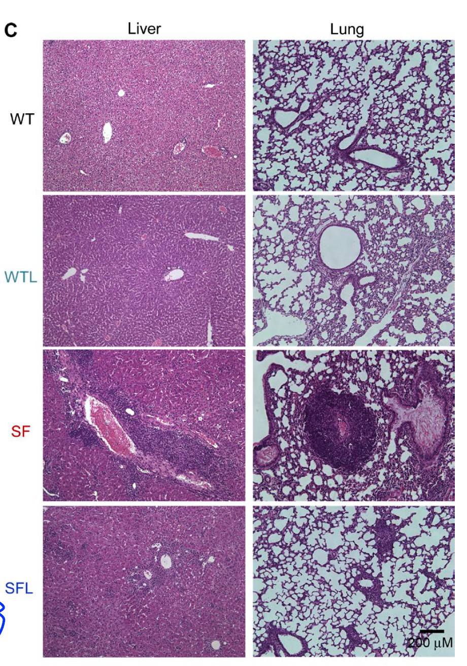
Surprisingly, unlike control mice (which did not survive past 4 wk of age), 50% of G113TgFoxp3YFP-CreTcraFL/FL mice survived to 4 mo of age (the end point of the experiment) though all of them displayed "visible evidence of significant autoimmunity".

So what does this result tell us? These are quite unexpected results. It would be highly unusual to imagine that a single antigen and single Treg specificity could be sufficient to prevent lethal autoimmunity in 50% of mice. We don't even know the source of the endogenous antigen G113 TCR supposed to recognize.
I personally would rather focus on technical aspect of this result: how robust is TCR modification in G113TgFoxp3YFP-CreTcraFL/FL mice? Maybe there is still "leakage" of endogenous TCRalpha chains? I wonder about this because of "50% data". Maybe in those 50% surviving mice Tregs were able to assemble TCR with endogenous TCRalphas?
David Usharauli
Surprisingly, unlike control mice (which did not survive past 4 wk of age), 50% of G113TgFoxp3YFP-CreTcraFL/FL mice survived to 4 mo of age (the end point of the experiment) though all of them displayed "visible evidence of significant autoimmunity".

So what does this result tell us? These are quite unexpected results. It would be highly unusual to imagine that a single antigen and single Treg specificity could be sufficient to prevent lethal autoimmunity in 50% of mice. We don't even know the source of the endogenous antigen G113 TCR supposed to recognize.
I personally would rather focus on technical aspect of this result: how robust is TCR modification in G113TgFoxp3YFP-CreTcraFL/FL mice? Maybe there is still "leakage" of endogenous TCRalpha chains? I wonder about this because of "50% data". Maybe in those 50% surviving mice Tregs were able to assemble TCR with endogenous TCRalphas?
David Usharauli