Tuesday, February 10, 2015

Mitochondrial DNA abnormality activates cytosolic type I IFN system

Mitochondria is a cell's energy hub. It is of a prokaryotic (simple bacterial) origin. It has its own DNA. However, prokaryotic DNA can activate innate immune system. So, what would happen if our mtDNA undergo misfolding stress?

This is exactly what the new Nature paper tried to uncover. This study led by Gerald Shadel at the Yale School of Medicine, has examined the outcome of mitochondrial DNA (mtDNA) packaging abnormality by studying mouse model heterozygous for mitochondrial transcription factor A (TFAM-/+), a protein responsible for mtDNA higher-order nucleoid organization.

Gene expression profiling revealed that cells in TFAM-/+ mice showed enrichment in IFN-stimulated pathway genes.


The authors showed that this type I IFN response was significantly abrogated in cGAS, STING, IRF3 and TBK1 deficient backgrounds indicating that in TFAM-/+ mice aberrant mtDNA released into cytosol activates cGAS-STING-IRF3 pathway leading to type IFN production.

Additional experiments showed that such heightened type I IFN response in TFAM-/+ mice reduced viral cellular burden.

Finally, the authors showed that viruses from herpesviridae family could induce mtDNA misfolding (by TFAM depletion via viral-encoded UL12 M185 molecule) and mtDNA dependent type I IFN response in wild type mice, mimicking TFAM-/+ mice, suggesting physiological function of mtDNA stress response.


In summary, these results highlight how mitochondrial DNA stress could activate innate immune system and cause inflammation (in human disorders such as systemic lupus erythematosus, cancer, multiple sclerosis, etc).

This study provides additional support for recently described immunological function of mtDNA and its detection by cGAS-STING-IRF3-type I IFN pathway. It is of note that many cellular abnormalities analysed from immunological point of view are converging on type I IFN response.

David Usharauli


Saturday, February 7, 2015

antigen-specific IgG1 isotype protects against type III allergy (hypersensitivity)

IgG antibody family consists of four isotypes: IgG1, IgG2a, IgG2b and IgG3 (IgG1-4, in humans). They differ in their Fc portions. Current knowledge suggests that IgG isotypes have different function depending on a degree of their interactions with complement and FcRs.  

New study in journal Nature, however, provided evidence for complement- and FcR-independent function of IgG1 isotype.

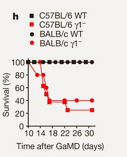
This research led by Fred Finkelman at the University of Cincinnati College of Medicine, have analysed immune response of mice deficient for IgG1 isotype (Igγ1mice). Strangely, immunization with goat anti-mouse IgD anti-serum (GaMD, protein immunogen) led to the death of Igγ1- mice, but not wild-type mice.


The authors showed that this death of Igγ1mice was caused by kidney insufficiency.


Analysis of immune response to GaMD in Igγ1mice revealed selective increase in antigen-specific IgG3 isotype and IgM in serum and their deposition in kidney's glomerular capillaries. Interestingly, kidney damage in Igγ1mice was still present in FcγR1- or FcγR1- and complement C3 double deficient mice.


The authors showed that kidney damage could occur even in wild-type mice by passive injection of antigen (TNP)-specific IgG3 and antigen (TNP-protein). Other IgG isotypes did not induce renal disease.


Importantly, concomitant injection of antigen specific IgG1 and disease-inducing IgG3 could prevent renal disease development independent of complement C3 or inhibitory FcγRIIb.


In summary, these results indicate that induction of antigen-specific IgG1 response during protein immunization prevents the development of type III hypersensitivity. Mechanistically, IgG1 competes for antigen binding with antigen-specific IgG3, thus inhibiting large antigen-antibody complex formations typical for IgG3.

This new knowledge could help to design new treatments for human cryoglobulinemia.

David Usharauli


Thursday, February 5, 2015

Tissue-specific regulatory T cells control asthma development

Foxp3+ CD4 regulatory T cells (T regs) maintain immune tolerance to self. Exactly how do they do it, no one really knows. Studies have described so many different mechanisms of T regs function that one can only wonder whether these cells really belong to one family.

Most likely, T regs may function in a tissue-specific manner as proposed by P. Matzinger and T. Kamala. New study published in Nature Immunology may support this hypothesis.

This study is a combined effort by French and German scientists. Here, the authors have examined the role of protein kinases in T regs function. Initial experiments showed that protein kinase, CK2 (casein kinase 2), was the most up-regulated kinase in activated T regs compared to effector T cells.


Next, the authors generated a mouse model carrying Foxp3-specific deletion of CK2 in T regs by crossing Foxp3-cre mice with CK2 fl/fl mice. 


Interestingly, analysis of different tissues in Foxp3-cre CK2  fl/fl mice revealed that only lung tissue was affected by Foxp3-specific CK2 deletion in T regs.


Further analyses revealed that Foxp3-specific deletion of CK2 in T regs resulted in selective increase in type 2 immune signature in lung draining lymph nodes and in serum.


In fact, spontaneous lung inflammation in naive Foxp3-cre CK2  fl/fl mice was comparable to allergic lung inflammation (asthma) in antigen sensitized control mice.


However, surprisingly, T regs homing into lung tissue was not affected by Foxp3-specific CK2 deletion in T regs. More detailed analysis of T regs from Foxp3-cre CK2  fl/fl mice revealed selective up-regulation of inhibitory receptor ILT3.


The authors showed that ILT3 expression in CK2-deficient T regs could dampen TCR signalling in T regs.


Finally, the authors showed that CK2 deletion in T regs led to selective increase in numbers of type 2 response promoting IRF4+ PD-L2+ dendritic cells (DCs).


In summary, these results indicate that CK2 deletion in T regs induces ILT3 up-regulation and attenuation of TCR signaling in lung-tissue T regs, leading IRF4+ PD-L2+ dendritic cells (DCs) expansion and type II inflammation in the lung

How these results advance our knowledge in Foxp3+ T cells? No data are provided to explain why lung-tissue T regs were so selectively affected by CK2 deletion or how IRF4+ PD-L2+ DCs are amplified. Very interesting and very strange.

Let me know what do you think about this study.

David Usharauli