Wednesday, January 28, 2015

What would studying twins could tell us about immune system? Nothing much.

Few weeks ago journal Cell published a study that was widely publicized by online media. The research article came from Mark Davis lab at the Stanford University School of Medicine.


The authors found between 58%-77% of analyzed immune markers are mostly or solely determined by non-heritable, environmental factors. These data suggest that one cannot predict the magnitude and class of the immune response (TH1,TH2,...etc) based on inherited gene analyses. In other words, these results would make studying immune-related therapies, like vaccine efficacy or anti-tumor immunology, more complicated and unpredictable.
However, these results were expected, anyway. The point is that immune system and especially the antigen-specific repertoires of adaptive immune system (TCR and BCR) are generated stochastic fashion, even in MZ twins. Unique adaptive immune system affects innate immune system as well. Basically, no humans would have the same immune system, everThese implies that neither genes nor environment could truly influence or determine the true direction of immune response. It will be random and individualized response. Only immune system and neuronal system have this additional, random, person-specific level of variation.

Interestingly, the authors themselves acknowledge this point in the discussion, however they dismissed it, as not sufficiently significant.

David Usharauli


Tuesday, January 27, 2015

Orphan interferon-lambda vetoes commensal microbes backdoor dealings

This is a second paper in IFN-lambda series. This new paper published in journal Science provided additional confirmation to the earlier study suggesting the role of commensal microbes in norovirus infection


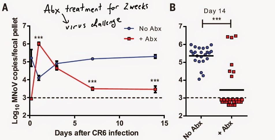
This resistance to intestine norovirus infection was reversed by normal fecal supplementation implying the role of gut microbes in supporting norovirus infection.


The authors showed that resistance to chronic norovirus infection upon antibiotic treatment were dependent on viral dose and IFN-lambda signaling.


These data suggest that murine norovirus employs endogenous gut microbial flora to establish chronic intestinal infection. It is not clear how microbiota alters IFN-lambda signaling to permit norovirus persistence in the gut.

David Usharauli 

Monday, January 26, 2015

Interferon-lambda can do it but suffers from amnesia

Ordinarily when we consider anti-pathogen response we think about adaptive immune response with antibodies or T cells. However, the more we study innate immune system the more we uncover about its hidden potential

This new study in journal Science from Herbert Virgin's lab at the Washington University School of Medicine, showed new function for interferon molecule called IFN-λ. IFN-λ is a member of family called type III IFNs. 


First, the authors observed that mice deficient for IFN-lambda receptor 1 expression (IFN-λR1) (but not IFNγR or IFNαR1) showed high viral burden in the gut. 


Indeed, exogenous application of IFN-lambda could clear chronic murine norovirus infection from the gut in control wild-type and IFNalphaR1-KO mice but not from IFN-lambdaR1-KO mice.


Surprisingly, IFN-lambda was active against chronic murine norovirus infection even in RAG-KO mice lacking adaptive immune system.


Interestingly, IFN-lambda treated mice that clear first norovirus infection remained susceptible to second norovirus infection, implying the absence of immune memory.


In summary, the authors showed that for certain viral infection innate system can provide "sterile" immunity, though it lacked memory component.

Chronic viral infection exist because they are able to find ways to co-exist with both innate and adaptive immune system. Here, for example, the authors showed that norovirus (CR6 strain) was able to establish chronic gut infection in wild-type, IFN-lambda sufficient environment, because it did not activate IFN-lambda system. This knowledge may help to design optimal natural anti-viral treatments for human noroviral infections.   

David Usharauli


Sunday, January 25, 2015

Between Scylla and Charybdis: how helminth control in wild animals affects their susceptibility to pathogenic microbes

When I read scientific articles I focus on how results fit into their conclusions. Especially I am very skeptical to research data or their interpretation which become headlines news all over the web.

In my opinion, when one writes about science news it would help if before publishing anything on open forum to read and analyse it in order to understand what novelty research brings and/or its limitation.

As an example, lets analyse research article recently published in journal Science. In this study, the authors has treated free roaming wild African buffalos with anti-helminth drugs to reduce acquisition or shedding of worms (reduce TH2 immunity) and increase their TH1 immunity to pathogenic microbe, mycobacterium bovis, a bovine tuberculosis (a deadly disease in buffalos).

All buffalos (total of 216), both in control (108) or treated (108) groups were mycobacterium bovis-free at the beginning of experiment. The authors claim that while anti-helminth treated group shed fewer worms or worm eggs during the course of observation, there was no much different in infection with mycobacterium bovis between treated (33 out of 100 = 33%) or control (36 out of 101 = 36%) groups. However the graph itself shows that by the end of observation period (~4 years), ~80% of analysed buffalos in control group acquired mycobacterium bovis infection compared to ~45% in treated group.


Next, the authors showed that anti-helminth treatment had a major impact on survival of infected buffalos: while ~36% of mycobacterium bovis-infected buffalos died in control group, mortality rate of mycobacterium bovis-infected buffalos in treated group was ~7%.


Using mathematical model for disease transmission, the authors estimated that due to increase in survival of mycobacterium bovis-infected buffalosmycobacterium bovis would spread more in buffalo population.   

In summary, the authors claim that anti-helminth treatment reduces the mortality from mycobacterium bovis in buffalos, but at the same time has no effect on rate of acquisition of mycobacterium bovis infection (???), but rather it increases spread of mycobacterium bovis in buffalo population

Here is what do not understand: (1) aren't the rate of acquisition of infection and its spread the same? How can be one different from another? (2) Fig. 2b shows that control buffalos were infected in higher rates compared to treated buffalos. What about that?

David Usharauli


Friday, January 23, 2015

Gene signature of tumors under immune pressure

Tumor is a tissue undergoing homeostasis-resistance turnover. Animal studies suggested that immune system can detect and sometimes eliminate tumor. In humans, however, the data are more complex and there is no clear answer, especially for solid tumors.

Since experimentation on human subjects ad libitum is obviously not an option, the scientists are constantly developing indirect, ex vivo approaches to study human tumor immunology. Advances in omics science made it possible to analyze large data set to find a clinically-relevant correlations between tumor-specific signatures.

This is exactly what this new paper published in journal Cell tried to do. This group led by Nir Hacohen at the Massachusetts General Hospital, has analyzed human tumor samples for gene signatures looking for evidence for their adaptation to local anti-tumor immune response. Such knowledge, if proven reliable, could be helpful  in determining the effectiveness of tumor immunotherapies. 

The authors has decided to focus on cytolytic gene signature (granzyme A and perforin) as a readout for tumors sensitivity to local immune response. Initial analyses showed that tumors varied in their expression of cytolytic signature. 


The authors reasoned that one reason for difference in cytolytic gene signature could be due to presence of tissue-specific viruses, frequently associating with tumors, like HPV virus for cervical cancer tissues. 


Additionally, the authors found there was evidence for depletion (reduction) of neo-epitope expression from predicted number of HLA-binding non-silent mutations, implying these tumors were under immune pressure.


Correspondingly, the authors identified several mutated genes that were enriched in tumors under immune pressure. One of these genes was caspase 8, a well-known member of cell death pathway.


Finally, the authors found there was positive or negative correlation between high cytolytic gene signature and amplification of gene regions containing immuno-regulatory genes such as PD-L1, PD-L2 and IDO1, IDO2.


In summary, these results provided indirect evidence suggesting that human immune system can detect tumor neo-epitopes and mount cytolytic response to eliminate it, thus exerting pressure on tumor cells to develop inhibitory counter-measures. Targeting these pathways could benefit the development of effective immunotherapeutic approaches.

Now there are few limitation to this study. First, cytolytic signature molecules, granzyme A and perforin, can be evidence of immune suppression rather than immune activity. Second, since the authors do not provide tumor patients survival data, it makes hard to argue for usefulness of these results for clinical application.

David Usharauli


Thursday, January 22, 2015

CD4 T cell-centric vaccine produces immunopathology and death

CD4 T cells are thought to orchestrate immune response to infections and vaccines. They provide help to CD8 T cells (to become cytotoxic cells and develop memory), B cells (to secrete antibodies) and macrophages (to eliminate intra-cellular microbes in type 1 response).

So, naturally, it is logical to assume that vaccine priming (activation) of CD4 T cells could provide huge benefits to host later. However, human logic and nature logic appears to be at odds in this situation.

The new study published in journal Science showed that CD4 T cell-centric vaccine strategy could backfire with deadly consequences. 

This research led by Prof. Dan Barouch (Center for Virology and Vaccine Research at Beth Israel Deaconess Medical Center) analyzed mice immune responses to live virus after vaccination with viral-specific CD4 T cell antigen.  

Surprisingly for the authors and regretfully for the experimental mice, they found that when mice were first vaccinated with intra-cellular microbial vector carrying inserted CD4 T cell specific viral epitope and then exposed to live virus, almost 90% of vaccinated mice died from what appeared to be a cytokine storm.


The authors found that rather than helping to fight virus, vaccine primed CD4 T cells inhibited viral-specific antibody production or maintenance of viral-specific CD8 T cells response.


Mechanistically, the authors found these deadly outcome were mediated by (1) activated viral-epitope specific CD4 T cells; (2) could be prevented by administration of anti-viral antibody or (3) with the simultaneous vaccination with CD8 T cell-specific viral epitope


In summary, these results showed that disproportional priming of CD4 T cells during initial vaccination could impair subsequent anti-viral protection. It appears that this outcome maybe specific for viruses producing chronic, long-term infection, since infection of vaccinated mice with viral clone producing acute, short-term infection did not show immunopathology.  

The data in this paper highlight our still so inadequate knowledge of such basic immune response properties as T cell response. If just having simply high frequency of antigen-specific CD4 T cells can produce such immunopathology, how immune system determines beneficial frequency of CD4 T helper (CD4 TH) cells? It's just another of nature's mystery to be solved.     

David Usharauli


Wednesday, January 21, 2015

T cell-specific Smad4 controls the lethal autoimmunity

I was very excited when I saw this title in journal Immunity and then utterly disappointed after reading the actual paper.

The only noteworthy result from this paper is depicted in Figure 1A. The authors showed that compared to mice single deficient for T cell-specific TGF-beta RII signaling (RII-KO), mice double deficient for T cell-specific TGF-beta RII and Smad4 signaling (RII-S4 DKO) were protected from lethal autoimmunity.


Bone marrow (BM) chimera experiments revealed that unlike RII-KO BM cells, RII-S4 DKO BM cells could generate Foxp3+ Tregs in vivo (though 2-fold less compared to WT BM cells).  


Strangely, however, in vitro experiments showed that RII-S4 DKO T cells were dramatically deficient in generating Foxp3+ Tregs (though they efficiently differentiated into TH1 or TH2 phenotypes).


Finally, the authors showed that mice single deficient for Smad4 (S4-KO) were not able to efficiently reject OVA-expressing tumor (no similar data for RII-S4 DKO mice were produced).


In summary, in my opinion, these results were not ready for publication in Immunity. The authors failed to follow their best Figure 1A result and provide any convincing argument that could explain their observation. It is clear that T cells deficient in Smad4 failed to accumulate in normal numbers in vivo upon antigen encounter (memory failure?), but the fact that Smad4-KO T cells could efficiently differentiate into effector T cells makes all these results confusing. Also, it is not clear whether absence of lethal autoimmunity in RII-S4 DKO mice has to do with the presence of normal number of fully functioning Foxp3 T cells.

David Usharauli


Tuesday, January 20, 2015

More DNA replication = More cancer incidence?

Some science studies become a part of popular science. It has to do with the simplicity of science message discussing commonly understood science topics.

One such paper was recently published in journal Science. The authors has analyzed 31 human tissues with the regard to tissue-specific stem cell proliferation (division) rate and its relationship to tissue-specific lifetime incidence of cancer

The authors concluded that in 65% of times (i.e. 2/3 of times), the rate of tissue-specific cancer incidence could be explained by simple accumulation of DNA replication errors (mutations), occurring by chance, during stem cell division. Basically, more stem cell divides to repopulate aging or damaged cells more chance it has to acquire cancer-promoting mutation and develop into cancerous tissue.


The idea by itself is very simple and technically speaking, correct. There is no surprise that DNA replication can generate somatic, single nucleotide, non-synonymous mutations leading to cancer.

The novel thing about this paper is the fact the authors found "practical" correlation between tissue turnover rate and cancer rate. However, correlation does not necessary imply causation.

The point is that the authors' calculation are based on two parameters: (1) available data regarding tissue-specific cancer rate in population and (2) tissue-specific stem cell division rate.

However, neither of these parameters are "real" data but statistical or "calculated" assumptions. Especially problematic is a calculation of tissue-specific stem cell division rates.

In fact, tissue turnover rate is not a random process at all but influenced by exogenous factors (age, gut microflora, endogenous retroviruses, etc). For example, the authors speculation about the rates of colorectal versus duodenal cancer incidence could simply be explained by the presence of specific gut microflora in those tissues.

David Usharauli


Sunday, January 18, 2015

Friendly gut microbes could be our most dangerous enemies

Some studies are interesting and thought-provoking, and some studies are just studies. But this new paper in journal Science is a beauty. One rarely comes across to this type of research.

As many of you many know, we harbor large numbers of friendly microorganisms inside (gut, lung) or outside (skin). In many aspects, these commensal microbes share characteristics, like LPS (endotoxin), with their pathogenic siblings. However, since commensal microbes can live with us in peace, they probably had acquired some properties to make such co-habitation possible. 

Indeed, the authors, led by Andrew Goodman at the Yale University School of Medicine, found that unlike pathogenic microorganisms (E. coli, S. enterica, C. rodentium), major subsets of our commensal microbial world were highly resistant to several cationic anti-microbial peptides (AMP).


The authors determined that this increased resistance of commensals to AMP was relate to a single gene encoding enzyme, LpxF, responsible for removing phosphate group from LPS (component of microbial cell wall)

In vivo experiments with mono-colonization of LpxF-deficient mutant human commensal microbe, B. thetaiotaomicron, confirmed that unlike LpxF-complemented B. thetaiotaomicron, B. thetaiotaomicron deficient for LpxF was easily displaced by wild-type B. thetaiotaomicron in presence of pathogenic C. rodentium infection or chemical, DSS-induced inflammation. Non-virulent tir mutant C. rodentium or commensal SFB had no impact on LxpF-deficient B. thetaiotaomicron population stability.


Similar results were observed when 14 member of human gut flora were transplanted into germ-free mice and exposed to C. rodentium infection, implying that LxpF played important role in population stability during gut inflammation.


Finally, gut microbes obtained directly from healthy humans displayed similar resistance to AMP.


In summary, these results suggest that human gut microflora acquired resistance to its own host's anti-microbial peptides thus providing additional mechanism responsible for peaceful co-existence and gut ecological stability. 

Of course, such resistance of gut commensals to AMP may pose problem when gut microbes invade inner systems, as during sepsis. So, this is a double-edge sword dilemma for host-microbe mutualism. 

The experiments that are missing, in my opinion: (1) Repeat of experiments with anti-microbial peptide deficient mice models to confirm that population stability is indeed related to resistance to AMPs; (2) It is not clear how presence of LxpF-deficient commensal microbe could affect the severity of inflammation induced by C. rodentium.

David Usharauli


Saturday, January 17, 2015

Skin fat cell differentiation program has an immunologically-active phase

Staphylococcus aureus (S. aureus) infection represents a major medical issue. Unlike viral or intra-cellular microbial infections, we still do not know much about how immune system defends against extra-cellular microbes.


The research group, led by Richard Gallo at UC San Diego School of Medicine, observed that skin infection with S. aureus induced local adipocyte expansion. 


Analysis of mice deficient in fat cell development (Zfp423nur12 mouse) showed that these mice contained  more S. aureus locally in skin wound as well as systematically, in the spleen.


In vitro analysis of adipocyte differentiation revealed that maturation of pre-adipocytes to adipocytes led to selective expression of anti-microbial peptide, cathelicidin (Camp). Camp expression peaked at days 2-4 in adipocyte differentiation program. 


As a result, adipocyte condition medium could inhibit S. aureus growth in vitro.


In addition, up-regulation of Camp in maturing adipocytes was in response to S. aureus infectionAs expected, a medium supplemented with adipocyte tissue extract from Camp-KO could not inhibit S. aureus growth.


Finally, adipogenesis deficient Zfp423nur12 mouse did not up-regulate Camp in response to S. aureus.


In summary, these results indicate that S. aureus skin infection activates pre-adipocyte to adipocyte differentiation program and induces Camp, Cathelicidin, anti-microbial peptide expression that inhibits S. aureus growth and prevents it's systemic spread.

The authors suggest that at this early stage of S. aureus skin infection (day 2-7) adipocyte-derived Camp expression dominates the host defense. 

However, in my opinion, to clearly rule out the role of innate immune system (neutrophils, macrophages) it would necessary to repeat these experiments with anti-Gr-1 antibody (neutrophil depletion) or macrophage-deficient mouse models or immune system-specific Camp deficiency (BM chimera).   

David Usharauli


Thursday, January 15, 2015

Christmas tree theory of T cell functional polarization

Pathogens capable of productive infection of immune-competent host (1) can target one or more tissues or (2) different pathogens can target the same tissue

Conversely, immune system, in theory, should be able to deploy pathogen-tailored immune response for efficient control

However, for a host, there is an additional cost when deploying pathogen-tailored immune response: it may inflict potentially severe, bystander self-wounding, if class of immune response is too harsh for local tissue. 

So, there is always a delicate balance between anti-pathogen response and tissue-specific immune response restriction, "self-censorship". As Melvin Cohn, one of the last surviving immunology theoretician opined "Lighting up the immune response like a Christmas tree would be ineffective".

Still, immunology is a science and science since the days of Galileo Galilei required robust experimental proof to accept or reject a proposed theory.

The following paper from journal Science examined the specificity and functional polarization of human memory CD4 T cell subsets specific for (1) C. albicans (extracellular fungus) and M. tuberculosis (intracellular bacteria) and (3) protein, Tetanus toxoid (TT vaccine). [senior author: Federica Sallusto; first author: Simone Becattini].

The authors have sorted human peripheral blood derived TH1, TH2, TH17 and non-conventional TH1* CD4 T cells based on surface chemokine receptor profile and stimulated them in vitro with autologous monocytes and C. albicans


The authors observed that despite different frequencies of ex-vivo expanded C. albicans-specific TH1, TH2, TH17 and non-conventional TH1* CD4 T cells (see above), all 4 subsets contained comparable number of clonotypes (based on TCR-beta analysis). Q: why such waste?


Interestingly, TCR-beta analysis indicated that many C. albicans-specific clonotypes were shared between these 4 CD4 T cell subsets, implying that they were related.


Similar results were obtained when analysed protein, TT-specific clonotypes.


Limited analysis of both TCR-beta and TCR-alpha chains showed that some of shared clonotypes were actually identical, sister clones.

In summary, the results indicates that single CD4 T cells can acquire multiple fates. More broader interpretation of these results imply that immune system may indeed produce diverse T cell subsets (lighting up of Christmas tree) against given pathogen and then expands those subsets capable of controlling the infection.  

Of course, we do not know whether sister clones were actually derived from the same identical mother clone. It may be that one sister clone was primed in gut tissue and another sister clone was primed in skin tissue (where ever it first encountered non-self antigen). Also, we still don't know the role, if any, of each subsets specific for a given infection.    

David Usharauli  


Tuesday, January 13, 2015

Friendly gut microbe defends against Vibrio cholerae infection

This is a very interesting study from journal Nature. It comes from the famous Jeffrey Gordon's lab at the Washington University School of Medicine, St. Louis. 

Usually when we think about the infectious microbes and how to deal with them, we are usually considering antibiotics or vaccination. However, in nature everything is interconnected and mutually dependent, even if any such evolutionary relationships are not immediately obvious. 

In nature, if one considers microbial communities, different species can sense each others presence and can influence each others biology. The most widely described such phenomenon is called quorum sensing wherein microbial community can sense its members density and influence and self-adjust its own or others replication kinetics. There are intra-species and inter-species mechanisms of quorum sensing.


The authors has collected and analysed gut microbiota community from stool (fecal) samples from healthy or Vibrio cholerae-infected patients (collection time ranged from diarrhoeal phase to recovery phase). This analysis showed that abundance of 14 gut microbial species correlated with the recovery from Vibrio cholerae dierrhoeal phase.

Using germ-free mice recolonization strategy the authors found that one of the gut microbial species in particular, Ruminococcus Obeum (R. Obeum), restricted Vibrio cholerae colonization in gnotobiotic mice. 


The authors found that in presence of Vibrio cholerae infection, R. Obeum up-regulated LuxS gene responsible for the inter-species mechanism of quorum sensing through autoinducer-2 (AI-2) synthesis. 


To directly test R. obeum LuxS involvement in restricting expression of virulence factors in V. cholerae, the authors cloned R. obeums LuxS (or V. cholerae LuxS, as a control) into mutant E. coli deficient its own AI-2. Both in vitro and in vivo experiments showed that in presence of R. obeum-derived LuxS gene in mutant E.coli, V. cholerae's replication and expression of virulence factors were reduced during co-culture or co-colonization.     


Finally, the authors found that restriction of V. cholerae infection was mediated through a new mechanism involving quorum sensing transcriptional regulator VqmA in V. cholerae.  


In summary, the results highlight the new mode of treating infectious agents with the help our own gut microbial friends.

David Usharauli


Monday, January 12, 2015

Interruption of a programmed cell death leads to cell autonomous IFN-beta production

The second paper discussing the role of death caspases in cell-intrinsic IFN-beta production and heightened anti-viral state came from Richard Flavell's lab at the Yale University School of Medicine.

Initially, the authors made unexpected observation that mice deficient in death caspases in epithelial and hematopoietic cells (caspase 9 fl/fl Tie2Cre+ and caspase 3 fl/fl + caspase 7KO Tie2CRE+ DKO) showed increase viral resistance both in vivo and in vitro


They showed that this increased resistance against viral infection was type I IFN dependent since it was abolished on IFN-alpha receptor 1 KO background or when using anti-IFN alpha/beta blocking antibodies. 

In vitro experiments with a chemical inhibition of caspase pathway confirmed that IFN-beta was induced in cells when pro-apoptotic stimulus was coupled with caspase inhibition. Similar observations were made when using caspase 9 KO or caspases 3/7 DKO primary (MEF) cells.


Here too, the authors found that IFN-beta response were abolished on cGAS and STING knockout backgrounds suggesting the role of endogenous DNA recognition.


Finally, the authors found IFN-beta response was abolished when cells were depleted of mitochondrial DNA (mtDNA).


In summary, the authors proposed that in conditions when caspase-dependent programmed cell death is interrupted (in viral infection [or tumors?], for example), infected cell can secrete IFN-beta as a endogenous danger signal and alert neighboring cells for viral presence and confer paracrine anti-viral resistance. 

Still, there are few results from these two papers that require additional explanation: (1) both groups have reported that death caspase deficient mice have high levels of serum IFN-beta but do not show any obvious immunopathology; (2) neither group have examined the impact of caspase deficiency on programmed cell death using physiological stimuli, like FasL, TRAIL, etc; (3) does IFN-beta, spontaneously secreted from caspase-deficient cells, activates tissue resident dendritic cells?

David Usharauli


Saturday, January 10, 2015

Caspase activity determines release of endogenous danger signal during apoptosis

Apoptosis is a fundamental biological process necessary for maintenance of normal cellular turnover. Initially, apoptosis, as a "voluntary" cell death was contrasted to necrosis, "involuntary" cell death. More recently, however, additional forms of cell death were described sharing characteristics of both of these processes (necroptosis, pyroptosis).

From an immunological point of view, cell death could be classified as a silent, non-immunogenic or a noisy, immunogenic. Interestingly, many anti-cancer therapy drugs were shown to induce apoptotic cell death that were immunogenic. Very recently, DNA detecting pathway involving IFN-beta (as an endogenous danger factor), cGAS, STING and IRF3 were shown to contribute to tumor cell detection by immune system following cancer cell apoptosis.

The two new studies in journal Cell provided additional results that may explain some long standing observations about apoptosis. I am going to review both of them separately.

First study was led by Prof Benjamin Kile at the Walter and Eliza Hall Institute of Medical Research, Australia, and Michael White (postdoctoral fellow at his lab) as a first author.

This group studied a role of apoptosis in physiology of haematopoiesis (not a typical immunology research) and it seems they accidentally came to the thought-provoking finding.

While working on apoptotic pathways involving BAX/BAK and caspase 9, the authors made an observation that in bone marrow chimera (BM) mice, caspase 9-KO donor haematopoietic stem cells yielded more lineage-negative stem cells compared to BAX/BAK DKO or wt donor cells.


Analysis indicated that IFN-beta was specifically up-regulated in caspase 9-KO BM chimera. The authors reasoned that there was a connection.  


Indeed, BM chimera mice transplanted with caspase 9-KO BM cells on IFN-receptor alpha 1 deficient background abolished this effect.


To better understand the connection between caspase 9 and IFN-beta, the authors induced mouse splenocytes apoptosis in presence of caspase inhibitor. Unexpectedly, in presence of caspase inhibitor, cells secreted IFN-beta when exposed to pro-apoptotic stimulus.


Similar observation was made with human peripheral blood mononuclear cells (PBMC).


To analysis of sera from mice deficient in different molecules in apoptotic pathway showed that mice  deficient of pro-apoptotic caspases (caspase 9, caspase 3/7) had high levels of serum IFN-beta.


The authors reasoned that in absence of active caspase 9 (or during its inhibition), an initial pro-apoptotic stimulus induce release of mitochondrial DNA (mtDNA) which is recognized by cytosolic DNA sensors, like STING, leading to IFN-beta productionIndeed, pro-apoptotic stimulus could not induce IFN-beta secretion in cells lacking mtDNA even in presence of caspase inhibitors.


In addition, experiments with STING-KO cells, or with CRISPR-Cas9 targeted cGAS -KO and IRF3-KO clones confirmed that no IFN-beta was produced in absence of STING pathway.


Finally, immunoprecipitation of cGAS followed by PCR amplification of co-precipitated DNA confirmed that mtDNA, but not genomic DNA, was enriched with cGAS in treatment group.


In summary, these results indicated that pro-apoptotic stimuli induce mtDNA release, probably as a bystander product of mitochondria membrane permeabilization, which can interact with STING pathway. However, normally, caspase 9 (and caspase 3 and 7) prevent mtDNA from being detected by STING pathway. It is not clear yet how is this accomplished. 

This finding may provide new understanding of immunogenic versus non-immunogenic cell death and reconcile several prior observations relevant for cancer immunotherapy. 

I would like to see the following experiments: use of physiological apoptotic stimuli, like FasL, TNF-alpha, TRAIL, granzyme B, rather than chemical molecules; Also in Fig 5I, pan-caspase inhibitor increased IFN-beta production even in caspase 9KO or caspase 3/7 DKO cells, implying that other caspases are maybe involved too.

David Usharauli