Showing posts with label allergy. Show all posts

Saturday, May 25, 2019

Allergy: A newborn's microbiota prevents hyper IgE antibody response to certain food antigens

Prevalence of allergy to food antigens is increasing in the world (alongside autoimmune diseases). Many believe it has to do with changes in microbiota composition due to environmental and processed food effects. But we still don't know how exactly microbiota prevents immune dysregulation characteristic of allergy or autoimmunity.     

For the past couple of years the team from South Korea has published several important papers addressing the role of microbiota and food antigens in modifying the gut immune system. This week they published yet another relevant paper in Science Advance. Below I present the highlights of the study.   

For this study the authors compared the level of IgE in sera from conventional mice fed regular diet, germ-free mice also fed regular sterile diet and germ-free mice fed with sterile antigen-free diet. As seen in the figure below GF mice develop, over time, hyper IgE condition. However, this effect was abolished in GF mice fed antigen-free diet. It indicated that antigens found in food interact with the immune system differently in the absence of microbiota.        


This was confirmed in reciprocal experiments where GF mice were introduced to the antigen-free diet or when antigen-free diet fed GF mice were introduced to a regular diet. In both conditions, a regular diet that contains antigens enhanced IgE level in the absence of microbiota.


Interestingly, the authors found that only certain food antigens, such as wheat gluten, could initiate hyper IgE response in absence of microbiota. Of note, the wheat gluten was shown to be digestion resistant.  


Another noteworthy observation was related to the age at which point mice were introduced to food antigens. Only young, but not older antigen-free fed mice, showed hyper IgE response when introduced to food antigens in the absence of microbiota. It indicated that there were some differences between young and older mice that made older mice resistant to hyper IgE production when responding to food antigens. 


So far we discussed how antigen-free fed mice respond to food antigens in the absence of microbiota. As expected, the introduction of microbiota to GF mice blocked hyper IgE response to food antigens.   


In summary, this study showed that in the absence of normal microbiota mice fed a regular diet that contains antigens will show hyper IgE response to certain food antigens. This response is abolished in GF mice fed antigen-free diet. The main question of how microbiota prevents IgE response or why certain food antigens are more immunogenic has not been addressed here. Also, the authors did not discuss it but it is important to mention here that hyper IgE response by itself does not mean pathological allergic response. The authors did not say that antigen-free mice fed regular diet became allergic to it (or to wheat gluten). It means that allergic sensitization requires additional mechanisms beyond hyper IgE response.

posted by David Usharauli     


Sunday, November 26, 2017

Allergen-sensitized mothers transfer protection against allergy to offspring through milk


In this study the authors showed that mouse pups born to mothers sensitized to allergen were significantly protected from developing allergic response to the same antigen.    


Protection in offspring was associated with the generation of antigen-specific Foxp3+ Tregs as observed in proliferation suppression assay or following short-term Treg depletion by DT (however, the authors did not analyze antigen-specificity of Tregs by tetramer staining). 


Further experiments showed that mother's milk contained allergen-specific antibodies and immune complexes (IC) and breastfeeding by allergen-sensitized mother (irrespective of birth mother status) was sufficient to transfer allergen protection to offspring.


In summary, this study suggests that breastfeeding by allergen-sensitized mothers can benefit offspring by preventing development of allergic response to the same allergen. However, it is not clear how exactly the authors see this mechanism working in humans. In mice, mothers were intentionally sensitized with allergen using epicutaneous (skin) application that supposed to mimic how humans with skin barrier dysfunction get sensitized to allergens. But the authors have not tested if milk from atopic human mothers can have the same effect on their offspring. For some reason the authors tested milk from nonatopic human mothers and showed that it 'worked' when fed to mice but did not provide any explanation why healthy, nonatopic human mother milk should contain any "protection" against allergen when mothers themselves are not sensitized as experiments in mice showed they must be for a milk derived immune complexes to work. So lots of unknowns and contradictions.

posted by David Usharauli


Friday, September 8, 2017

Loss of microbiota depletes cross-reactive Foxp3+ Tregs leading to selective immunopathologies

Check out our follow-up manuscript in PeerJ Preprints that provides a brief guide to SPIRAL, a novel interpretive framework that demonstrates the central role of microbiota-Treg axis in the initiation of immune disorders.

Kamala T, Usharauli D. (2017)
 
Loss of microbiota depletes cross-reactive Foxp3+ Tregs leading to selective immunopathologies.
PeerJ Preprints 5:e3237v1
 
The 'Hygiene hypothesis', a cornerstone model to account for the role of exogenous pathogens and later of endogenous microbiota in immune disorders, is currently presumed to operate at the innate immunity and metabolite levels to properly 'educate' the immune system. Doing so however fails to satisfactorily account for the antigen-specific nature of such disorders. SPIRAL is a novel interpretive framework that resolves this dilemma. It represents the periodic table of cross-reactive Foxp3+ regulatory T cell (Treg) epitopes selected from commensal microbiota over evolutionary time to mediate self-nonself discrimination and effector class regulation. Here, we utilize the SPIRAL's predictive power to provide a mechanistic antigen-specific basis for the initiation of allergies and autoimmune diseases as well as for the failure to mount effective anti-tumor and vaccine responses through selective loss of microbiota and corresponding cross-reactive Foxp3+ Tregs.


Thursday, August 3, 2017

Th2A subset drives allergic phenotype in humans

TH2 cells were initially identified based on their capacity to make IL-4 and drive B cell antibody class switch recombination to IgG1  and IgE. However, more recently it became clear that TH2 cells consist of several subtypes each with its own unique specialization in particular effector function such as IL-13 or IL-25 production that have nothing to do with antibody production. 

Similarly, canonical TH2 cells were thought to be responsible for allergy phenotype in humans (and mice as well). However, new study in Science Translational Medicine suggests new subset of TH2 cells, called TH2A subset, is mainly responsible for food allergy phenotype

The authors carried out magnetic enrichment of T cells harvested from allergic patients using HLA-II tetramers. Compared to non-allergic individuals, allergic patents showed expansion of a special population of TH2 cells expressing CD161 and CD49d


Interestingly, patients undergoing antigen-specific desensitization showed selective loss of these TH2A cells.


In summary, it is clear that different subset of TH2 cells exist, some naturally others only during pathology, to deal with various foreign antigenic entities.

posted by David Usharauli


Sunday, February 5, 2017

New synthetic molecule prevents autoimmunity without compromising anti-pathogen immunity

In December of 2016 Science Translational Medicine published new study where the authors introduced new, low-molecular weight compound that specifically inhibited autoimmune/allergic responses while sparing anti-pathogen immune response.


This new molecule, AX-024, specifically targets adaptor protein Nck that is involved in low affinity TCR signaling


It does not affect signaling delivered via IL-2, BCR or PMA/Ion stimulation.


AX-024 inhibited TLR7/8 agonist, imiquimod (IMQ), induced psoriasis skin symptoms in mice.


Also, AX-024 significantly reduced clinical symptoms associated with mouse model of human multiple sclerosis (MS). Such effect of AX-024 was superior to Fingolimod, a sphingosine 1-phosphate receptor modulator approved for the treatment of relapsing-remitting MS.


Notably, AX-024 in therapeutic dosage did not impair anti-pathogen immunity or memory generation.


In summary, by blocking Nck adaptor protein involved in low-affinity TCR signaling, AX-024 inhibited autoreactivity while preserving necessary capacity for anti-pathogen immunity. Such property makes it desirable candidate for clinical trials in humans.

David Usharauli 


Wednesday, September 28, 2016

Wiskott-Aldrich syndrome protein (WASp) deficient FOXP3+ Tregs fail to control IgE mediated allergies

The inherited immunodeficiencies are frequently characterized with dysregulated Th2 responses, atopy, and elevated IgE levels. Mutations in Foxp3, STAT3, DOCK8, PGM3, LAT, ZAP70, or RAG result in hyper IgE phenotypes. Mechanism is unclear.


For this study the researchers analyzed "the overall burden of clinical food allergy within a cohort of 25 patients with mutations in the WAS gene" and found that individuals with WAS mutations were more likely to demonstrate serum sensitization to peanut, milk, and egg (compared to the general population).


Lab mice deficient for WASp also display elevated IgE (and IgG1) antibody levels to components of their chow food (even on germ-free background).


Since WASp is expressed in different cell types, the authors tested mice selectively deficient for WASp in B cells, DCs or FOXP3+ Tregs. Out of these gene-modified mice, only Wasfl/fl Foxp3-Cre mice showed deviation to Th2 phenotype and development of IgE to chow food.


In vitro T cell stimulation confirmed that total T cells from Wasfl/fl Foxp3-Cre mice showed selective enhancement in Th2 cytokines.


In sum, these results indicate that mice with selective deficiency of WASp in FOXP3+ Tregs display excess in Th2 subsets. It is possible that absence of WASp destabilizes FOXP3+ Tregs and this somehow drives their de-differentiation into Th2-like cells (and not into Th1 or Th17, for instance).

David Usharauli


Wednesday, July 13, 2016

ILC2 sustain antigen-independent allergic responses

Most allergies represent exaggerated type II immune responses driven by adaptive Th2 cells. At least, this is what we used to believe it. However, discovery of rare innate cells, referred as innate lymphoid cells (ILCs), is slowly changing our understanding of cellular responses underlying allergies.

It is clear now that in laboratory mice model of allergy, type 2 ILCs (ILC2) contribute significantly and non-specifically [it seems] in sustaining allergy to irritant-allergens.


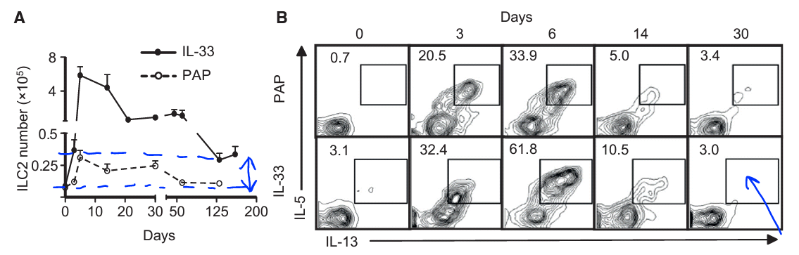
When analyzed ILC2 response to IL-33 or papain in lung tissue, the authors found that ILC2 displayed a typical adaptive-like behavior (expansion, contraction, quiescence).


Importantly, when IL-33 primed mice were challenged with allergen one month later ILC2 showed heighten type II response to allergen (papain) but not to control (saline). This response was "allergen"[protease]-specific but antigen-independent. It is possible that primed ILC2 were responding to IL-33 [or IL-25] released during papain challenge.


Similar data were obtained from mice primed with fungal Aspergillus protease (ASP) allergen and challenged 3.5 months later with papain (but not to saline). Here too, primed ILC2 could be responding [indirectly] to IL-33 or IL-25 released by papain.


In summary, this study showed that at least in mice "primed" innate lymphoid cells retain "heightened" non-specific responsiveness to allergen "long-term" (up to 6 months). This could explain why adaptive TH2 cell targeting immunotherapies may not be fully successful because it ignores contributions from innate cells such as ILC2.

David Usharauli


Wednesday, April 20, 2016

Nanoparticle-caged antigens could treat allergen-sensitized host

The challenge with immune system-[driven] disorders is the fact that ordinarily such conditions are detected at the later stages when immune system had already developed self-perpetuating "memory circuit". Even now we know very little how to break such "memory circuit".

New study in PNAS suggested an experimental model that were able to tolerize already-sensitized host against allergen. The authors showed that biodegradable nanoparticles incorporating caged antigen, but not simple conjugates, could deliver both prophylactic and therapeutic treatment to allergen-prone host.

For this study the authors have used widely-accepted experimental sensitization protocol in mice to induce TH2-driven allergy to ovalbumin  antigen (OVA). Next, they proceeded to test three different combination of  nanoparticles + ovalbumin: (1) OVA conjugated to polystyrene nanoparticles (Ag-PS), (2) OVA conjugated to biodegradable poly(lactide-co-glycolide) nanoparticles (Ag-PLG) and (3) OVA caged within biodegradable poly(lactide-co-glycolide) nanoparticles [PLG(Ag)].

First, the authors found that all three nanoparticle combinations displayed potent prophylactic action against allergen when delivered before allergen sensitization (Ag-PLG is shown here).


However, when applied to already allergen-sensitized host, polystyrene nanoparticles induced severe allergic reaction. In contrast, application of Ag-PLG nanoparticles did not induce allergic response in sensitized hosts and even reduced TH2 cytokines. However, Ag-PLG nanoparticles could not inhibit lung eosinophilia.


Finally, the authors showed that application of PLG nanoparticles with caged OVA antigen could both prophylactically and therapeutically inhibit TH2-driven allergic response.


In summary, this study showed that nanoparticles incorporating antigens (Ag caging) avoids detection by allergen-specific immune effector molecules (IgE) and instead drives tolerance to allergen.      

David Usharauli

Wednesday, April 6, 2016

Type II immunity is fueled by ILC2-specific arginase-1

In recent years much attention has been focused on group of innate cells called innate lymphoid cells (ILCs). There are group 1 (TH1 behavior), group 2 (TH2 behavior) and group 3 (TH17 behavior) ILCs, so far.

This week, new paper in Nature Immunology from David Artis lab (extremely prolific research lab) showed that ILC2-specific arginase-1 (Arg1) contributes to lung allergic inflammation.

Arg1 is an enzyme that metabolizes the amino acid L-arginine. Most L-arginine metabolism occurs primarily in the liver, though immune cells can express it too during immune response (i.e. Arg1 activity is a key signature of alternatively activated macrophages). Here, fate-mapping study of Arg1-YFP-expressing cells in lungs of naive mice showed that Arg1 was mostly expressed by ILC2.


Papain challenge (TH2 trigger) induced expansion of Arg1+ lung ILC2.


Using genetic mouse model where Arg1 is selectively absent in ILC2, Arg1ΔILC, the authors showed that Arg1 deficiency impaired ILC2 expansion and allergic lung inflammation upon papain challenge.


Finally, the authors showed that lung allergic inflammation was not impaired if Arg1 was missing from macrophage lineage (Arg1ΔLyz2), implying dominant role of ILC2-derived Arg1 in papain induced lung allergic inflammation.


In summary, this study revealed ILC2-specific role of Arg1 in promoting type II immunity in response to TH2 triggers (papain and helminth parasite N. brasiliensis).

David Usharauli


Saturday, March 26, 2016

Papain protease activity and mast cells, but not IL-33, are necessary for papain allergic sensitization

Papain is a proteolytic enzyme from papaya. Like other protease allergens such house dust mites (HDM) group 1 allergen, Der p 1, papain is allergen that belongs to family of cysteine proteases. Papain can induce skin contact sensitization or airway hyper-reactivity. 


When injected in mouse ear lobes, intact papain, but not protease-inactive papain (E64-papain), induced skin inflammation and IgE production.


Papain induced IgE production after skin challenge depended on presence of functional mast cells as (a) such response was diminished in mast cell–deficient WBB6F1-W/Wv (W/Wv) mice, and (b) such response could be recovered after transfer of WT mast cells. 


However, unlike mast cells, IL-33 was dispensable for IgE production after papain skin [primary] challenge (though IL-33 played the role in lung eosinophil infiltration later on, upon airway re-challenge with intact papain).


In summary, this study indicate that initial skin sensitization with papain required mast cells but not proto-TH2 innate "primer" cytokine IL-33. This knowledge should be incorporated in therapeutic strategies targeting IL-33 pathway (IL-33 and its receptor ST2).

David Usharauli


Tuesday, November 3, 2015

Helminths are in cahoots with commensal microbes against allergy

Conventional wisdom suggests that Th2 response (aka, type II immunity) developed in response to worms (helminths). At the same time, notoriety of Th2 response comes from its involvement in allergies [that is spreading far and wide in western-style living environments]. Strangely enough, the same people enjoying western-style living conditions suffer minimally or not at all from helminths. This is by definition a paradox. If Th2 is directed towards helminths, then what activates Th2 response in total absence of such parasites? crazy, right? Here another paradox for you. It appears that presence of helminths are actually beneficial against allergy. What?!


Initially, the authors showed that helminth[Hpb]-infected mice showed reduced inflammation in response to house dust mite (HDM) allergen [eosinophils↓, IgG1↓, IL-5↓, H&E↓].


Interestingly, antibiotic treated mice infected with helminth failed to show attenuated allergic response to HDM.
Parallel experiment showed that helminth infection modified mouse gut flora composition and increased gut concentration of short chain fatty acids (propionate, butyrate, acetate).


Finally, mice deficient for GPR-41 (Ffar3-/-), a receptor for short chain fatty acids, also failed to down-regulate allergic response to HDM allergen after helminth infection.


In summary, this study proposed that helminth infection could modify host's allergic phenotype by its influence on gut flora-derived short chain fatty acids. If application of short chain fatty acids alone could reproduce the key observation of this study, it could potentially provide easily available dietary supplements for allergy treatment.

David Usharauli  

Monday, October 19, 2015

Food allergies caused by mast cells with a serial number 9

Allergies are complex immune responses. Whether allergic reaction have any "protective" function or represent a purely pathological reaction is still debated. Classical form of allergy is IgE mediated and require IL-4 and Th2 cells. However, the list of effector molecules and cells responsible for various forms of allergic reactions are continuously expanding.

For example, in a recent paper in Immunity the authors showed that certain mouse models of food allergy were driven by mucosal mast cells secreting high level of IL-9.

This is quite messy paper with lots of figures. In fact, it was under review for one year [and it shows by its lack of harmonized relationship between figures]. 

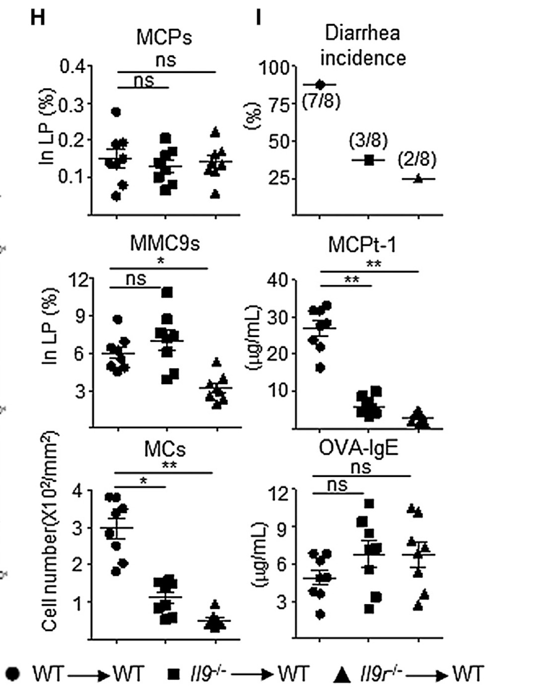
Initially, using mouse model of food sensitization [intra-gastric antigen gavage], the authors showed that allergic reactions (e.g. diarrhea) to antigen in susceptible mouse strains correlated with the number of GI mast cells, not serum IgE. Moreover, such correlation was highly significant for IL-9 producing lamina propria Lin-/- population in susceptible mouse strains [since not every mouse strain develop allergic response in this setting].


Further investigation with IL-4eGFP reporter mice revealed that this IL-9 producing Lin-/- population belonged to mast cell lineage, rather than innate lymphoid cell type 2 (that express IL-25 cytokine receptor IL-17RB).


Next, the authors showed that induction of IL-9+ mast cells (MMC9) required IL-4, STAT6 and T cells.

Finally, using BM chimera experiments the authors showed that while active signaling through IL-9 was dispensable for MMC9 induction, it was required to produce allergic phenotype.


In summary, this study suggests that GI tissue associated mast cell secreting high levels of IL-9 play a role in allergic response to sensitized antigens. The development of this MMC9 requires signaling via IL-4, STAT6 and T cells, implying typical Th2-driven immune response. It is possible [but not formally tested in this study] that therapeutic targeting of IL-9 pathway may benefit patients suffering from GI tract allergies to food.

David Usharauli

Wednesday, September 2, 2015

Adaptive Th2 cells can double as innate cells during allergy and helminth infection

Antigen specificity is a dominant feature of adaptive immune system. However, on occasions, fully differentiated effector T cells do show TCR-independent responsiveness to cytokines. Whether this is a good or bad thing is not easy to tell. 


For this study the authors had used a dual-reporter mouse model, called 4C13R, where both cytokine IL-4 and IL-13 are genetically labeled. Initially, the authors showed that adoptively transferred in vitro differentiated Th2 OT-II transgenic T cells, on 4C13R background, could secrete IL-13 (but not IL-4) in response to cytokine IL-33 (in combination with STAT5 activator IL-7 or TSLP).


Similarly, the authors observed TCR-independent IL-13 secretion by in vivo differentiated Th2 OT-II 4C13R cells to a Papain challenge (Papain is a protease and has been shown to induce type II immune response).


This TCR-independent secretion of IL-13 by differentiated Th2 cells was driven by IL-33.


Since type 2 innate lymphoid cells (ILC2) are known to respond to IL-33, the authors analyzed secretion of IL-13 by both Th2 and ILC cells after Papain challenge (here, mice were initially exposed to N. brasiliensis, a type II activator helminth). Both population could respond to Papain by secreting IL-13 (but not IL-4).


Similar results were obtained with another allergen, house dust mite extract (HDM).


Further experiments with RAG2-IL-2Rγ DKO hosts, which lack endogenous T and ILC2 cells, revealed that adoptive transfer of in vitro differentiated Th2 cells was sufficient to induce eosinophilia in lung tissue upon HDM exposure (allergen exposure).


The authors reasoned that in physiological situations initial antigenic priming would increase the number of Th2 cells to the level comparable to that of ILC2 and this would play a significant role in host protection or response to type II irritants (worm or allergen). Indeed, the authors showed that Th2 cells developed after first helminth infection could provide partial host protection to a second helminth infection, even in absence of ILC2 cells (in RORα bone marrow chimera).


In summary, these results suggest that adaptive immune cells can take the function of innate cells when necessary. Here, IL-13 appears to be a dominant cytokine that drives type II immune effector function (in 1990s ad early 2000s, the prevalent hypothesis positioned IL-4 in the center of type II immune effector response).

Such TCR-independent responsiveness has important implications for treatments of type II immune disorders such as allergy or dermatitis. For example, antigen (TCR ligand) desensitization protocol for allergy treatment may not provide full protection against allergic reaction due to IL-13 secretion (since it can be secreted independent of TCR signaling). On the other hand, blockade of IL-13 and IL-33 signaling may represent clinically relevant therapy (alone or in combination with antigen desensitization).  

David Usharauli