Showing posts with label microbiota. Show all posts

Wednesday, January 11, 2023

If T cell clones are so diverse, what prevents anti-tumor immune response?

Identifying cancer and pathogen-specific epitopes or TCRs may sound intuitive, but it is a futile approach. Diversity of T cell or B cell clones guarantees that the adaptive immune system will always have relevant clones to detect cancer or pathogen.  

Epitopes have no meaning attached to them with one exception.  It is when the said epitope is self. Each body will have different sets of self-epitopes. Every self-epitopes relevant for host's survival are encoded in the thymus, and thymic Tregs are trained to prevent any T cell activity against those epitopes in the periphery. This is called tolerance, and it is antigen[epitope]-specific. 

Then what prevents effective responses to cancers or pathogens? It is commonly but mistakenly believed that Tregs prevent effective T or B cell responses to cancers or pathogens. But Tregs only prevent anti-self response, and it is epitope-specific action.  So, by definition, if Tregs do their job as required, we cannot blame them. But it has nothing to do with cancer or pathogens, which obviously have other epitopes different from self, we call nonself. So, if cancer cells or pathogens express nonself epitopes that are always detected by adaptive immune system, why not everyone can fight it off effectively?

This is because T cells themselves prevent it. Yes, T cells, not Tregs, prevent effective response to cancers or pathogens in certain conditions. What are those conditions? These are condition when polarized T helper cells prevent other T cells functions. Polarization is a pathological state. A Polarized T cell's effect on other T cells is epitope non-specific, meaning, a polarized T helper cell specific to cancer or pathogen nonself epitope A will prevent T cells specific to epitope B, C, D, E, F, etc., to function properly. It is exactly Treg's job to shut down those polarized T helper cells to allow other T cells to manifest their functions and get rid of either cancer or pathogen. And Treg do it, as we already said, epitope-specific manner.

For this reason, it is not so important to identify any cancer or pathogen-specific nonself epitope, but rather to identify an epitope that could activate polarized T helper cells.

To make things even more complicated, one may ask if Tregs are self-specific and act epitope-specific manner, how can Tregs shut down polarized T helper cells that are nonself-specific? It is possible because Tregs are cross-reactive and can inhibit only those polarized T helper cells which share TCR specificity with Tregs.

In other words, control of self-tolerance and control of effective anti-nonself response are one and the same.  

These are 3 papers that together provide a full discussion related to the SPIRAL model we have developed to explain how Tregs work within an adaptive immune system:
 
 
Concurrent cross-reactivity of microbiota-derived epitopes to both self and pathogens may underlie the "Hygiene hypothesis"  


Could cross-reactivity rescue Foxp3+ regulatory T cell precursors from thymic deletion? 


Microbiota-Specific Foxp3+ Regulatory T Cells Could Control Pathological T Helper Responses


Friday, September 18, 2020

A specific bacteria-infecting virus, bacteriophage, found in gut microflora, augments anti-tumor T cell immunity

Molecular mimicry between microbial and host's antigens could contribute to autoimmunity but also to the protection against tumors through epitope cross-reactivity. A new study in journal Science indicates that those cross-reactive epitopes could come from viruses that infect endogenous microbial species. 

In this study the authors made a surprising observation that only certain Enterococcus hirae microbial strains (E. hirae 13144 or IGR11) augmented anti-cancer effect in experimental cancer model.


Next, the authors showed that this biological activity was linked to one dominant epitope, TSLARFANI, derived from TMP protein that originated in 39.2-kb prophage only in those specific E. hirae strains. Mice immunized with heat-inactivated E. hirae 13144 strain, or peptide TSLARFANI, or irrelevant E.coli engineered to express TMP, all augmented anti-cancer effect. 


Mechanistically, the authors showed that epitope, GSLARFRNI, derived from cancer cells used in these experiments, was recognized by the same CD8 T cells which labeled with TSLARFANI epitope tetramers confirming cross-reactivity between these 2 epitopes.


In summary, this study suggests that microbiota and bacteriophages they carry represent new modality in fight against cancer. In this study overall anti-tumor effect is modest but we need to take into account that this is an effect of just one cross-reactive epitope in one type of MHC inbred mice, and it is likely that many other epitopes will be involved in outbred species such as humans. However, it is still extremely hard to do such analysis in humans in real world scenario due to lack of exact knowledge about human microbiota strains and poor reliability and performances of available bioinformatics approaches. However, once the mechanistic principles underlying anti-cancer effects are uncovered and accepted, then it is much easier to move the field forward.

posted by David Usharauli

Tuesday, January 28, 2020

Microbiota-derived peptide and autoimmune heart inflammation: a tale of missing data

Papers published in journal Science supposed to undergo thorough high-level vetting process. However, to err is human. Both reviewers and editors are humans and hence they frequently err, for the annoyance of scientists and for the joy of postdocs doing journal clubs.  

Here is an example of a paper that squeezed through the cracks of the Science vetting process. It claims that peptides derived from certain commensal microbiota species cross-react with heart muscle protein, MYH6, causing autoimmune heart inflammation. It has a great Figure 1 showing that MYH6-specific TCR transgenic mice on a germ-free background, lacking microbiota, is protected from heart autoimmunity.


Furthermore, they showed that the re-introduction of microbiota into germ-free makes these mice susceptible to heart inflammation similar to microbiota+ mice.


The authors then tried to identify the microbiota species that contribute to this inflammatory condition. An in silico search identified cross-reactive β-galactosidase (β-gal) mimic peptides in Bacteroides thetaiotaomicron (B. theta) and B. faecis with high similarity to MYH6.


The authors even introduced into germ-free TCR transgenic mice Bacteroides thetaiotaomicron (B. theta) lacking the β-gal. Up to now, it feels that the authors have checked all the boxes necessary for high-quality research. But then for some reason, they do not show survival data comparing Bacteroides thetaiotaomicron (B. theta) with and without the β-gal gene as in figure 1. They just showed how a lack of β-gal Bacteroides thetaiotaomicron modifies MYH6-T cells accumulation in the heart tissue.


So, why the authors don't show survival data of germ-free MYH6 TCR transgenic mice colonized with Bacteroides thetaiotaomicron -/+ β-gal gene? Isn't it the most important result for their hypothesis? Where were reviewers and editors looking?

posted by David Usharauli


Saturday, July 13, 2019

Pathological changes in the gut could initiate autoimmune diabetes microbiota-specific manner

The mouse strain, NOD, has been used to study the mechanism of type 1 diabetes (T1D). These mice spontaneously develop autoimmune diabetes though it is not clear how islet-specific T cells get activated. To track autoimmune T cell fate, TCR transgenic mice, BDC2.5XNOD, have been used. These mice harbor islet-specific TCR expressing T cells in high numbers that are easily monitored.

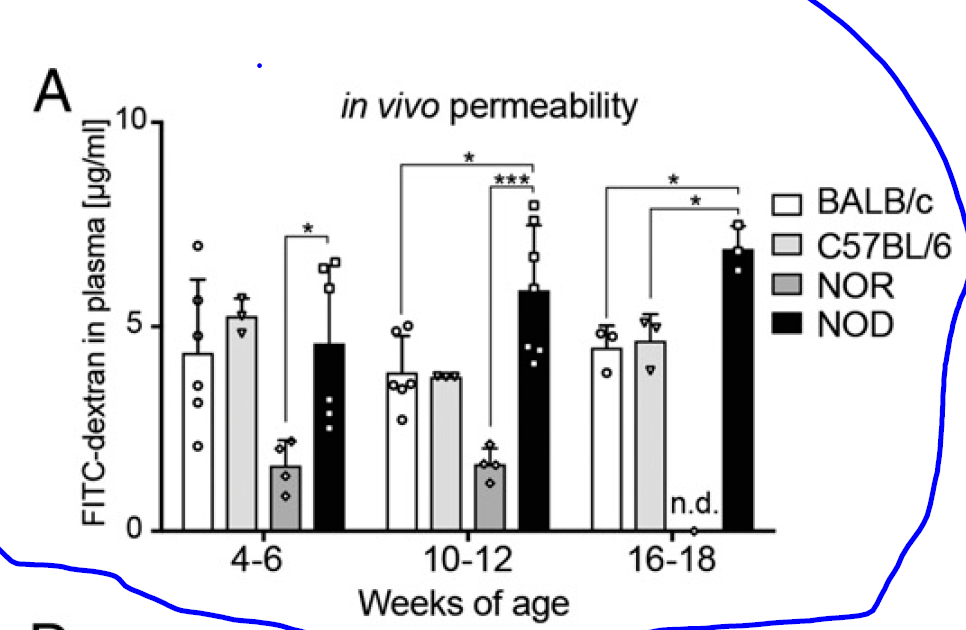
A new study in PNAS did some experiments on BDC2.5XNOD mice to understand the initiation of autoimmunity. First, they showed that the NOD background showed increased gut wall barrier permeability by measuring the FITC-dextran level in the blood after oral application (though they did not show the same permeability test for BDC2.5XNOD mice).


Interestingly, BDC2.5XNOD mice do not develop spontaneous autoimmune diabetes. However, oral application of low-dose dextran-sulfate sodium (DSS) activates T cells and initiates diabetes.


Diabetes, however, does not develop if DSS-treated BDC2.5XNOD mice are depleted of endogenous microbiota or if naive BDC2.5XNOD mice received DSS-modified microbiota.


In summary, this study showed that both gut inflammation and microbiota are necessary for the initiation of autoimmune diabetes in BDC2.5XNOD mice. The most likely scenario is that changes introduced by DSS allow endogenous microbiota to activate islet-specific T cells via cross-reactive antigens. DSS modifies both gut wall permeability and microbiota. Both of these phenomena have been observed by the authors. They conclude that "restoration of a healthy gut barrier through microbiota and diet modulation in diabetes-prone individuals could ultimately reduce intestinal activation of islet-reactive T cells and prevent T1D occurrence".

Others, in news/views for this article, even suggested using antibiotics to deplete endogenous microbiota, but in my opinion, this is a premature suggestion because the authors did not show that microbiota depletion after diabetes has already developed could stop it.

posted by David Usharauli


Saturday, May 25, 2019

Allergy: A newborn's microbiota prevents hyper IgE antibody response to certain food antigens

Prevalence of allergy to food antigens is increasing in the world (alongside autoimmune diseases). Many believe it has to do with changes in microbiota composition due to environmental and processed food effects. But we still don't know how exactly microbiota prevents immune dysregulation characteristic of allergy or autoimmunity.     

For the past couple of years the team from South Korea has published several important papers addressing the role of microbiota and food antigens in modifying the gut immune system. This week they published yet another relevant paper in Science Advance. Below I present the highlights of the study.   

For this study the authors compared the level of IgE in sera from conventional mice fed regular diet, germ-free mice also fed regular sterile diet and germ-free mice fed with sterile antigen-free diet. As seen in the figure below GF mice develop, over time, hyper IgE condition. However, this effect was abolished in GF mice fed antigen-free diet. It indicated that antigens found in food interact with the immune system differently in the absence of microbiota.        


This was confirmed in reciprocal experiments where GF mice were introduced to the antigen-free diet or when antigen-free diet fed GF mice were introduced to a regular diet. In both conditions, a regular diet that contains antigens enhanced IgE level in the absence of microbiota.


Interestingly, the authors found that only certain food antigens, such as wheat gluten, could initiate hyper IgE response in absence of microbiota. Of note, the wheat gluten was shown to be digestion resistant.  


Another noteworthy observation was related to the age at which point mice were introduced to food antigens. Only young, but not older antigen-free fed mice, showed hyper IgE response when introduced to food antigens in the absence of microbiota. It indicated that there were some differences between young and older mice that made older mice resistant to hyper IgE production when responding to food antigens. 


So far we discussed how antigen-free fed mice respond to food antigens in the absence of microbiota. As expected, the introduction of microbiota to GF mice blocked hyper IgE response to food antigens.   


In summary, this study showed that in the absence of normal microbiota mice fed a regular diet that contains antigens will show hyper IgE response to certain food antigens. This response is abolished in GF mice fed antigen-free diet. The main question of how microbiota prevents IgE response or why certain food antigens are more immunogenic has not been addressed here. Also, the authors did not discuss it but it is important to mention here that hyper IgE response by itself does not mean pathological allergic response. The authors did not say that antigen-free mice fed regular diet became allergic to it (or to wheat gluten). It means that allergic sensitization requires additional mechanisms beyond hyper IgE response.

posted by David Usharauli     


Saturday, April 20, 2019

Crohn's disease-like phenotype could be initiated by one gut microbe in genetically susceptible mice


Wednesday, May 23, 2018

Flagellin-specific T cells induce colitis by recognizing antigen other than flagellin

This is very interesting study from Journal of Immunology (JI). Here, researcher led by Timothy Hand at the University of Pittsburgh Medical School, showed that CBir1 transgenic T cells, thought to cause gut inflammation by recognizing flagellin expressing microbiota, were in fact specific for non-flagellin antigens

CBir1 transgenic mouse have been used for past 10 years to model human Crohn's disease in mice. CBir1 tetramer+ T cells recognize epitope from flagellin, antigen thought to be a target in Crohn's disease. Interestingly, all of those experiments were done using CBir1 T cells on WT background. This could be an issue because it has been known for some time now that transgenic T cells on WT background could use alternative Vα chain to form TCR with new specificity.

To avoid such limitation, here the authors generated CBir1 mouse on RAG KO background that only expressed transgenic Vα:Vβ chains. Surprisingly, unlike CBir1:WT T cells, CBir1:RagKO T cells when transferred in immunodeficient hosts did not induce colitis, and it was not because CBir1:RagKO T cells differentiate into Foxp3+ T cells. It appears that CBir1:RagKO T cells just did not see flagellin epitope in adoptive host.


In vitro tests showed that both CBir1:WT T cells and CBir1:RagKO T cells could respond to DCs pulsed with flagellin epitope [it would have been better and more relevant here to use DCs pulsed with gut flora component].


Other tests showed that CBir1:WT T cells in gut tissue could express alternative Vα chain to form a completely new TCR specificity together with transgenic Vβ chain such as against Ags derived from Helicobacter (HH1713 tetramer). 


In summary, it appears that CBir1 T cells initiate colitis by recognizing non-flagellin antigen from the gut flora through non-CBir1 TCR and only following gut inflammation and gut leakage do flagellin-specific CBir1 transgenic T cells get activated and participate in overall colitis.

So, what is missing from this study? One, it would have been relevant to transfer in vitro activated CBir1:RagKO T cells or activate them in vivo directly to see if then they could initiate colitis. Second, the authors could have tried monocolonization of germ-free mice to see the source of non-flagellin microbiota. Third, there is inconsistency between Fig. 3B and Fig. 6A with regard of proliferation of  CBir1:WT T cells in response to Vanc-treated samples (in vitro it did not proliferate but in vivo it did).

posted by David Usharauli


Saturday, May 5, 2018

IgA protects resident commensal microbiota against competitors

This week journal Science published new study from Sarkis Mazmanian lab at Caltech describing role of IgA in providing strain-specific competitive advantage to certain resident commensal microbiota. 

His lab has been studying immunobiology of Bacteroides fragilis (B. fragilis), a gut commensal. In initial series of experiments they have compared germ-free mice mono-colonized with either wild-type B. fragilis or its mutant variants such as, Δccf, shown to modify biosynthesis of its capsular polysaccharides. They noticed that in co-housing experiments wild-type B. fragilis from one mouse could out compete mutant variant in another mouse in a horizontal transfer assay.   


Since B. fragilis polysaccharides are known to interact with host's immune system, the authors wanted to find out whether host's immune system influenced co-housing experiments. Not surprisingly, the authors found that mutant B. fragilis did not efficiently bind IgA (induced by wild-type B. fragilis) and that it in turn induced IgA repertoire that bound wild-type B. fragilis even less effectively, suggesting some kind of association between IgA and missing antigens on mutant B. fragilis.  


To verify these observations, the authors compared co-housing experiments between germ-free IgA+ and IgA-KO mice (or treated with B cell depleting antibody) mono-colonized with wild-type B. fragilis. Indeed and surprisingly this time, wild-type B. fragilis resident in IgA KO mice were easily overtaken by wild-type B. fragilis from mono-colonized wild-type mice. These results suggested that in absence of IgA wild-type B. fragilis has lost competitive advantage against wild-type B. fragilis resident in IgA+ mice.


What could these results mean in biological context: it appears that certain resident commensal microbiota benefit from interacting with IgA. The authors proposed that "during health, IgA fosters mucosal colonization of microbiota with beneficial properties....while disease states may induce (or be caused by) IgA responses to pathogens or pathobionts that disrupt healthy microbiome equilibria." This is an interpretation that does not provide clear mechanistic explanation as to how IgA response could make such discrimination at the level of antigens between which microbes to keep and which ones to eject from the host. 


posted by David Usharauli

Thursday, March 15, 2018

Reduced CTLA-4 signaling predisposes to Th2 driven gastric tumorigenesis

Anti-CTLA4 antibodies such as Yervoy, has been used in clinical practice to treat solid tumors. It supposed to work either by augmenting and revitalizing effector T cells function directly or indirectly through temporal silencing of inhibitory Foxp3+ Treg population or both. However, new study from Journal of Experimental Medicine showed that at least in [genetically predisposed] mice reduced CTLA-4 signaling by itself could cause Th2 driven tumorigenic transformation of stomach epithelial tissue.

For this study the authors created transgenic CTLA4 shRNA knockdown (CTLA4KD) mice on the BALB/c × C57BL/6 (B6) mixed genetic background. This they did because it appears that BALB/c but not B6 mice were susceptible developing gastric tumors in this model. CTLA4KD mice showed gastric epithelial transformation by 20w of age. Similarly, month long treatment of newborn BALB/c mice with anti-CTLA4 antibody also led to gastric epithelial transformation.


This tumorigenic transformation was CD4 T cell dependent and effector T cells from CTLA4KD but not from WT mice could mediate it. It indicated that changes in effector T cell composition and functionality were driving de novo inflammatory tumorigenesis.


Interestingly, gastric epithelial transformation were happening even in germ-free CTLA4KD mice lacking microbiota. However, since these mice also harbor increased numbers of inflammatory T cells, in all subsets analyzed such as Th1, Th2, Th17, and independent of microbiota it could indicate that T cells could be responding to antigens from food or environment.


Finally, elimination of canonical T helper cytokines showed that surprisingly neither IFN-γ nor IL-17 but IL-4 deficiency could abolish gastric epithelial transformation under conditions of reduced CTLA-4 activity.


In summary, this study suggests that inherited or clinically-induced reduction of CTLA-4 signaling in predisposed individuals could paradoxically lead to inflammatory tumorigenesis driven by type II immunity.

posted by David Usharauli


Saturday, March 10, 2018

Translocation of a specific gut pathobiont, Enterococcus gallinarum, exacerbate autoimmune phenotype

A new study in journal Science suggests that a specific gut pathobiont, Enterococcus gallinarum, could exacerbate autoimmune phenotype in predisposed mouse strain. This autoimmune phenotype in mice are thought to represent mouse version of human systemic lupus erythematosus (SLE).

SLE is associated with genetic polymorphism linked to excessive signaling of RNA sensing Toll-like receptor 7 (TLR7) and type I interferons (IFNs). In the specific pathogen-free (NZW × BXSB)F1 hybrid mouse, responses to endogenous retrovirus glycoprotein 70 (ERV gp70) via TLR7 signaling leads to progressive autoimmune response by pathogenic anti-phospholipid [β2-glycoprotein I (β2GPI)] and anti–double-stranded DNA (dsDNA) antibodies.

The authors observed that certain antibiotic treatment significantly improved survival of (NZW × BXSB)F1 hybrid mice.


Further experiments showed that there was bacterial translocation from gut tissue into portal veins and livers in these mice that could be reduced by antibiotic treatment.


16S rRNA sequencing and species-specific PCR consistently revealed Enterococcus gallinarum (E. gallinarum) in the feces, small intestine and liver of (NZW× BXSB)F1 mice. Monocolonzation of germ-free mice with E. gallinarum (EG, here) revealed that it could specifically drive Th17 response, unlike E. faecalis or B. thetaiotaomicron.


Moreover, E. gallinarum could specifically drive  ERV gp70 expression in the liver cells,


and augment anti-nucleic acid antibody response.


Finally, the authors showed that liver tissues from human SLE patients harbored E. gallinarum.


In summary, this study proposes the following patho-mechanism of SLE: when residing in predisposed individuals E. gallinarum causes degradation of gut barrier function, then translocates internally, activates Th17 pathway and initiates "innate" autoimmune phenotype by activating expression of retroviral genes and amplifying endogenous nucleic acid detection system that breaks tolerance checkpoints and leads to auto-antibody formation, Ab-Ag complex deposition in tissues and inflammatory disease exaggeration. The authors proposed antibiotic treatment could provide relieves in certain SLE patients.

However, there are few unanswered questions in this study: first of all, it is clear that E. gallinarum does not induce autoimmunity by itself. Second, If Th17 activity is relevant for E. gallinarum action, then it would have been more valuable for the authors to compare E. gallinarum to segmented filamentous bacteria (SFB) a known inducer of Th17 response in the gut tissue.

posted by David Usharauli


Saturday, March 3, 2018

Specific microbiota species induce serum IgA that protects against sepsis

Some 10 years ago the scientists made observation that microbiota difference between different mouse colonies is responsible for selective TH17 expansion in the gut. Since then, field of immunology was flooded with numerous observations linking gut [and other tissue] microbiota to functional status of immune system.

One such study was recently published in journal Cell Host and Microbe. The researchers observed that serum IgA secreted by bone marrow residing plasma cells (BM PCs) were selectively enriched in mouse colony harboring members of Proteobacteria phylum. More importantly, these serum IgA protected mice during sepsis following gut damage.

Initially, the authors observed that their institute's B6 mouse colony (PENN-SPF) differed from commercial vendor B6 mice in their serum [but not small intestine] IgA status. Co-housing experiment indicated potential involvement of microbiota.


Indeed, 16S ribosomal gene sequencing showed enrichment of Proteobacteria phylum in local mouse colony (and also Deferribacteres).


Serum IgA bound microbiota and sequencing of serum IgA+/IgA- microbiota species confirmed selective enrichment of species within Proteobacteria phylum.


Development of microbiota-specific serum IgA were T cell-dependent.


Finally, serum IgA protected mice against sepsis following gut tissue damage and microbiota invasion (translocation).


In summary, serum IgA, but not intestinal IgA, is produced by bone marrow plasma cells in response to selective microbiota species, mostly from Proteobacteria phylum in mice. These serum IgA could bind microbiota, it developed in a T cell-dependent manner and protected host during gut flora invasion (translocation) in condition such as sepsis. However, it is not clear why serum IgG [in IgA KO mice] could not protect against sepsis in this study since one previous study already showed that serum IgG protected against gram negative bacteria such as E. coli. It is possible that serum IgA and IgG play non-redundant functions by protected against different microbial species.

posted by David Usharauli


Thursday, January 25, 2018

Recognition of microbiota-derived N-formyl methionine peptides by non-classical MHC class I, H2-M3-restricted CD8 T cells

A new study in journal Cell revealed that a specialized subset of CD8 T cells recognize conserved microbial derived N-formyl methionine peptides in context of non-classical MHC class I molecule H2-M3.

Using skin commensal Staphylococcus epidermidis (S. epidermidis) challenge model, the authors showed that recognition of this microbe by CD8+ T cells specifically relied on H2-M3 rather than other MHC molecules.


H2-M3 has been known to bind peptides that contain an N-formyl methionine (fMet), which is required to initiate protein translation in bacteria and mitochondria. Indeed, fMet peptide:H2-M3 tetramer (f-MIIINA:H2-M3) could stain a population of CD8 T cells from skin or lymph nodes. Interestingly, half of tetramer positive CD8 T cells were CD44hi even in germ-free mice indicating cross-reactivity with other antigens or "ready-made" origin similar to thymus-derived Tregs.


To show biological significance of these CD8 T cell population recognizing commensals, the authors used H2-M3 KO mice and wound healing experiment. The authors claim that in the absence of H2-M3 wound healing was delayed, though data do not strongly support such assertion.


In summary, the authors described yet another T cell population recognizing products from bacteria. Because such recognition does not produce inflammation the authors suggest it could be involved in wound repair. But wound repair model provided very minimal support for such hypothesis. Basically, they missed the central point to make this paper relevant. So how it ended up in Cell? Ask editors. I could come up with couple of suggestions. When the paper's authors list includes Giorgio Trinchieri, John O’Shea and senior author, Yasmine Belkaid, all heads of big labs at NIH, it is quite difficult to say no.

posted by David Usharauli


Saturday, January 6, 2018

Microbiota-wide association studies and PD-1 immunotherapy

This week Science published 3 back-to-back studies with the findings that responses to PD-1 immunotherapy in cancer patients could be stratified based on presence of certain microbiota species (at least two of these studies were published couple of weeks back as first release papers). 

The trouble is that all three papers found different set of microbiota who they thought mediated responsiveness to PD-1 immunotherapy (dominated by Bifdobacterium, Akkermansiaor Faecalibacterium). 


In one study, it was actually a difference between set of beneficial microbiota versus nonbeneficial ones (not simply a single species), above certain ratio (>1.5), that determined responsiveness to PD-1 therapy.


Moreover, study of germ-free mice transplanted with opposite sets of microbiota (derived from 3 responders/nonresponders) were inconclusive because 1 set of microbiota from each group showed reverse effect with PD-1 immunotherapy. 


In summary, we have no clear understanding of these results. There is no way to predict if the same microbiota set would provide any benefit to a given patient. In general, presently microbiota research lacks direction and rules necessary to untangle its complexity

As far as I know, the model we have developed, SPIRAL, is the only one that provides a rational how to identify specific microbiota species. Right now it is just a guideline but when fully developed it will look similar to period table-like map that will make it easy to accurately pinpoint microbiota species relevant for antigen-specific immune response in any given individual.

posted by David Usharauli


Sunday, November 5, 2017

Hidden arm against tumors: microbiota-enabled checkpoint immunotherapy

This week Science published two studies showing how diverse microbiota directly contributes to efficacy of PD-1 checkpoint immunotherapy in several tumors.

First, we need to mention that senior authors from both papers disclosed associations with for-profit pharma/biotech companies (as cofounders, stockholders, paid consultants or advisory board members). Such associations could, in general, be seen as problematic if one promotes therapy lacking particularities.     

Second, data presented do not advance our understanding how microbiota contributes to the effectiveness of checkpoint immunotherapy. The sole conclusion from both papers is that the more diverse cancer patient's microbiota the more benefit it provides during PD-1 immunotherapy. However, when it comes to narrow down beneficial correlation to particular species we find that one paper reported enrichment of Akkermansia muciniphila while other paper reported enrichment of Faecalibacterium and Clostridiales in Responders (as opposed to Non-Responders).

We still don't know much about the role of microbiota in cancer immunotherapy. I think real advance will come when we define how antigens derived from specific microbiota contribute to anti-cancer immunotherapy either by amplifying existing cross-reactive effector T cells or Foxp3+ Tregs. 

posted by David Usharauli


Thursday, October 12, 2017

Microbiota-generated butyrate works on Aire to amplify Treg numbers

A short but very interesting paper in Journal of Immunology caught my attention this week. In this study the scientists showed that butyrate, a short chain fatty acid derived from fiber fermentation by microbiota acts on GPR41 receptor in the thymus to increase Aire expression and amplify Foxp3+ Treg frequency (of note, compared to WT, GPR41-KO thymus already contains almost 5-fold less Tregs).  


Generally it is believed that microbiota works locally in the gut or other peripheral tissues to either convert naive T cells into Foxp3+ Tregs or expand existing Treg numbers. However no one yet managed to definitely show which pathway is functionally operational in vivo in physiological conditions. This study now could help to further narrow down biological mechanisms responsible for Treg biology. If microbiota-generated butyrate could work on thymus to increase Aire expression that in turn increases level of Thymus-derived Treg generation, then peripheral conversion pathway may play even less relevant role in physiological mechanisms of tolerance.  

The most obvious question after reading this article is why the authors did not test Aire KO mice to verify their conclusions.

posted by David Usharauli


Friday, September 8, 2017

Loss of microbiota depletes cross-reactive Foxp3+ Tregs leading to selective immunopathologies

Check out our follow-up manuscript in PeerJ Preprints that provides a brief guide to SPIRAL, a novel interpretive framework that demonstrates the central role of microbiota-Treg axis in the initiation of immune disorders.

Kamala T, Usharauli D. (2017)
 
Loss of microbiota depletes cross-reactive Foxp3+ Tregs leading to selective immunopathologies.
PeerJ Preprints 5:e3237v1
 
The 'Hygiene hypothesis', a cornerstone model to account for the role of exogenous pathogens and later of endogenous microbiota in immune disorders, is currently presumed to operate at the innate immunity and metabolite levels to properly 'educate' the immune system. Doing so however fails to satisfactorily account for the antigen-specific nature of such disorders. SPIRAL is a novel interpretive framework that resolves this dilemma. It represents the periodic table of cross-reactive Foxp3+ regulatory T cell (Treg) epitopes selected from commensal microbiota over evolutionary time to mediate self-nonself discrimination and effector class regulation. Here, we utilize the SPIRAL's predictive power to provide a mechanistic antigen-specific basis for the initiation of allergies and autoimmune diseases as well as for the failure to mount effective anti-tumor and vaccine responses through selective loss of microbiota and corresponding cross-reactive Foxp3+ Tregs.


Wednesday, August 23, 2017

MHC class II epitope presentation modulates microbiota and protects against autoimmunity

It is not clear how exactly microbiota modulates host's immune system. Evidence are largely based on empirical observations and nonspecific factors secreted by microbiota. Presently very little is known if microbiota-immune system interface is also controlled at the level of antigen-specific adaptive immune system.    
 
New study from Diane Mathis lab published in PNAS suggests it may be the case.
 
Her lab studies human type I diabetes (T1D) mouse model known as NOD. NOD mice lack MHC II allele, Eα. In this study they used NOD mice expressing Eα, referred as Eα16/NOD. In mating experiments, they noticed when Eα was expressed by female but not by male parent, baby mice with NOD genotype showed significant protection from developing T1D, suggesting protection was transmitted vertically from Eα16/NOD mother to NOD pups. Interestingly, this protection was lost when pregnant mothers (dams) were treated with antibiotics pointing towards role of microbiota.


Experiments with germ-free sterile mice confirmed this observation.


In summary, this study showed that MHC class II [epitope] presentation modulates composition of microbiota in such a way to harbor species protective against T1D. Again, the authors were unable to specifically pinpoint any specific mechanism of protection, though they reported increase in Foxp3+ Treg numbers in Eα16/NOD mice compared to NOD (but found no difference in microbiota bound to IgA between mouse strains). It is likely that epitope presentation at the level of adaptive CD4 T cells contributed to development of protective environment.

posted by David


Monday, July 10, 2017

How Foxp3+ Tregs and microbiota work together to control immune system

Check out our new paper in PeerJ Preprints that unlocks the mystery of how Foxp3+ regulatory T cells work that enables proper immune functioning.

Usharauli D, Kamala T. (2017) An identical mechanism governs self-nonself discrimination and effector class regulation. PeerJ  Preprints 5:e3081v1  https://doi.org/10.7287/peerj.preprints.3081v1

Prevailing immunological dogma dictates self-nonself discrimination, meaning to respond or not, and effector class regulation, meaning choosing the most effective response, are two separate decisions the immune system makes when faced with a new antigen. Representing a cardinal departure from the past, our model instead predicts both self-nonself discrimination and effector class regulation are in fact one and the same process controlled by Foxp3+ regulatory T cells (Tregs) whose antigen-specific repertoire is entirely maintained by commensal microbiota-derived cross-reactive antigens.
 
 
posted by David Usharauli


Thursday, February 9, 2017

Newborns' resistance to pneumonia is driven by acquired microbiota

This week Science Translational Medicine published new study that showed that in newborn mice resistance to pneumonia is driven by neonatally acquired microbiota. It revealed how antibiotic therapy given to mothers near time of delivery could alter and weaken baby's defenses against airway pathogens.

Newborn mice derived from germ-free or from antibiotic-exposed pregnant mice display increased susceptibility to Streptococcus pneumoniae serotype 19A-induced pneumonia that could be reversed by microflora.


Application of epithelial-focused cytokine IL-22 had similar effect on reversing newborn mice susceptibility to pneumonia.


In the lungs of newborn mice, majority of IL-22 is made by RORgt+ group 3 innate lymphoid cells (ILC3).


Antibiotic therapy of pregnant mice reduced IL-22+ ILC3 population in the newborn lungs that could be reversed by microflora.


Moreover, depletion of endogenous ILC3 increased host's susceptibility to pneumonia that could be reversed by adoptive transfer of WT ILC3.


In summary, this study showed that antibiotic therapy of pregnant females at the time of delivery could profoundly affect newborns' ability to mount proper defense against airway pathogens by depleting microflora and disrupting microflora → ILC3 → IL-22 axis.


David Usharauli