Showing posts with label inflammation. Show all posts

Thursday, January 10, 2019

Inflammation modifies pattern of differentiation of commensal-specific T cells


Tuesday, March 20, 2018

IL-33 is a natural target of allergen proteases

IL-33 is a member of IL-1 family and requires proteolytic cleavage to form active form. It has already been linked to allergy manifestation. Now, new study in nature immunology expands on earlier observations to show that full-length non-active IL-33 (IL-33FL) is a natural target of group of allergens with proteases activity.

In general, many known allergens display protease activity such as from fungi, mites, pollens, insects. Co-incubation of IL-33FL with such allergen proteases in presence of innate lymphoid cells type II (ILC2) generated biologically-active smaller fragments and release of type II cytokines.


IL-33FL is an intra-nuclear proto-chemokine found in epithelial and endothelial cells. So, how allergens get access to it? It requires cell damage to release IL-33FL extra-cellularly. However, it is not clear if any allergen proteases can damage cells. At least one such allergen from fungus, Alternaria alternata (A. alternata) can damage cell and then cleave IL-33FL.


Similar effects were seen in vivo using IL-33KO mice. In these mice, recruitment of eosinophils, a readout for IL-33-driven allergic response, were only observed when injected with pre-incubated IL-33FL and A. alternata mixture.


In summary, these results suggest that allergen from A. alternata with protease activity can damage cells to release IL-33FL and cleave it into biologically-active shorter peptides. However, this study did not show that other allergens can deliver similar double punch. Also, it is not clear how this innate mechanism translates to adaptive immune system to generate allergen-specific T cell and antibody responses.

posted by David Usharauli


Saturday, February 10, 2018

Antigen-specific Foxp3+ Tregs control tolerance to gut pathobiont H. hepaticus

Helicobacter hepaticusH. hepaticus, is an opportunist commensal, a pathobiont, that causes gut inflammation in IL-10 deficient but not in WT mice. A New study in journal Nature from Dan Littman's lab tried to understand basis for such dual nature of host response to H. hepaticus.

The authors have generated transgenic T cells specific for H. hepaticus-unique protein, HH_1713. Next, the authors co-transferred H. hepaticus-specific T cells (HH7-2tg cells) alongside with segmented filamentous bacteriaSFB-specific T cells (7B8tg cells) into WT mice exposed to H. hepaticus. Interestingly, HH7-2tg cells mostly differentiated into RORγt+ Foxp3+ Tregs while 7B8tg cells mostly differentiated into Th17 cells, as observed in earlier studies.


However, similar transfer of H. hepaticus-specific T cells into IL-10KO mice exposed to H. hepaticus mostly yielded inflammatory T cells (Th17 and Th1 cells).


It appears that transcription factor c-MAF control differentiation of H. hepaticus-specific T cells into RORγt+ Foxp3+ Tregs and its deficiency recapitulates H. hepaticus-specific T cells differentiation into inflammatory T helper cells observed in IL-10KO hosts when exposed to H. hepaticus.


In summary, this study indicates that in WT mice harbor H. hepaticus specific Foxp3+ T cells that keep tolerance to H. hepaticus. These induced Foxp3+ T cells are probably generated (or expand) by chain reaction orchestrated by existed antigen-specific natural Tregs since induced Tregs don't develop efficiently when transferred into RAG KO hosts lacking natural Tregs. IL-10 or c-MAF deficiency could alter overall microbiota composition (unrelated to H. hepaticus) and drive shift from Treg program into inflammatory pathway.

posted by David Usharauli


Tuesday, December 20, 2016

Lactobacillus reuteri extends lifespan of FOXP3-deficient scurfy mouse via microbiota–inosine–A2A receptor axis

Scurfy mice harbor natural mutation in FOXP3 gene that clinically resembles FOXP3 deficiency. Both scurfy mice and genetically modified FOXP3-KO mice die prematurely within first month of life. In humans, clinically observed FOXP3 deficiencies do not seem to be as lethal as in mice though one could argue that scurfy mice lifespan could be extended if these mice were given human-like medical attention (for example, i.v. feeding, anti-inflammatory medication, so on). 

In this regard, a new paper in Journal of Experimental Medicine is of great interest. It showed that a member of gut microflora, Lactobacillus reuteri, a probiotic microbe, when given orally could drastically extend lifespan of scurfy mice (30d vs. >125d) via microbiota–inosine–A2A receptor axis.


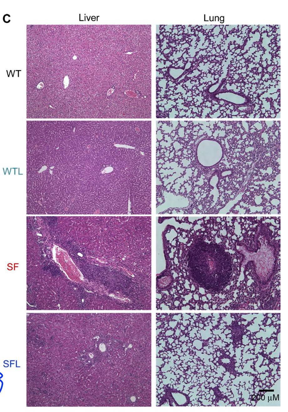
Morphologically, oral Lactobacillus reuteri significantly reduced tissue inflammation in scurfy mice.


Blood test showed that one molecule Lactobacillus reuteri could restore to WT level in scurfy mice was a purine metabolite inosine.


Indeed, oral inosine was able to recapitulate Lactobacillus reuteri effect on scurfy mice lifespan (and both Lactobacillus reuteri and inosine effects were specifically mediated via adenosine A2A receptor).


In summary, this study revealed that purine metabolites, inosine or adenosine could protect scurfy mice from tissue immunopathology and drastically prolong their lifespan. It is quite rare to see that one molecule could produce such effect. It would be interesting to see how caffeine consumption affects immunopathologies in humans as it acts as a natural antagonist to adenosine A2A receptor.

David Usharauli


Friday, August 12, 2016

Foxp3+ Tregs undergo memory-loss following inflammation

Adaptive immune system is characterized by its ability to recall prior antigen encounter and to mount stronger and swifter response for the second time. This is what typically happens to conventional T cells. But what about regulatory T cells (Tregs)? Do Tregs also display enhanced recall response when encountering [antigen] for the second time?


To test this hypothesis, the authors subjected Tregs to inflammatory environment and tracked their behavior. First, they found that phenotypically Tregs returned to "resting" state within 60 days following activation and resolution of inflammation. 


Gene expression analysis confirmed Tregs tested prior to inflammation (resting Tregs) or following inflammation (memory Tregs) resembled each other, while Tregs going through inflammatory process (activated Tregs) had a distinct gene profile. 


Functionally too resting Tregs and memory Tregs showed similar potential to inhibit hyper-proliferation of naive T cells when co-transferred into T cell-deficient host.


However, unlike conventional memory CD4 T cells, memory Tregs did not undergo more robust recall response when subjected to inflammation + undefined antigen for the second time.


Finally, the authors observed that Tregs displayed common gene profile with conventional memory CD4 T cells that differentiate them from naive T cells.


In summary, this study suggests two things: first, Tregs do not acquire secondary enhanced recall response capability [at least when exposed to inflammation and undefined antigen] and second, Tregs and conventional memory CD4 T cells share similar gene expression profile.

David Usharauli 


Wednesday, April 6, 2016

Type II immunity is fueled by ILC2-specific arginase-1

In recent years much attention has been focused on group of innate cells called innate lymphoid cells (ILCs). There are group 1 (TH1 behavior), group 2 (TH2 behavior) and group 3 (TH17 behavior) ILCs, so far.

This week, new paper in Nature Immunology from David Artis lab (extremely prolific research lab) showed that ILC2-specific arginase-1 (Arg1) contributes to lung allergic inflammation.

Arg1 is an enzyme that metabolizes the amino acid L-arginine. Most L-arginine metabolism occurs primarily in the liver, though immune cells can express it too during immune response (i.e. Arg1 activity is a key signature of alternatively activated macrophages). Here, fate-mapping study of Arg1-YFP-expressing cells in lungs of naive mice showed that Arg1 was mostly expressed by ILC2.


Papain challenge (TH2 trigger) induced expansion of Arg1+ lung ILC2.


Using genetic mouse model where Arg1 is selectively absent in ILC2, Arg1ΔILC, the authors showed that Arg1 deficiency impaired ILC2 expansion and allergic lung inflammation upon papain challenge.


Finally, the authors showed that lung allergic inflammation was not impaired if Arg1 was missing from macrophage lineage (Arg1ΔLyz2), implying dominant role of ILC2-derived Arg1 in papain induced lung allergic inflammation.


In summary, this study revealed ILC2-specific role of Arg1 in promoting type II immunity in response to TH2 triggers (papain and helminth parasite N. brasiliensis).

David Usharauli


Monday, March 21, 2016

IL-23p19 has an inside job in endothelial cells

IL-12 cytokine family is full of surprises. Ordinarily, these cytokines (IL-12, IL-23, IL-27, IL-35) are made of two heterodimeric sub-units. But one after another, each of the sub-units were found to have an independent function.  

Latest in these series is a study from Science Signaling that showed that human endothelial cells express intra-cellular IL-23p19 that signals via gp130 molecules mimicking IL-6.

Initially, the authors showed that endothelial cells from Giant-cell arteritis (GCA) patients express IL-23p19 subunit, but not another IL-23p40 subunit (while I support fully-human studies, in controversial situations such as this, I preferred if the authors have included confirmation staining on IL-23p19 KO cell from KO mice or CRISPR/Cas9 edited IL-23p19 KO human cells). 


Next, in vitro experiments with primary human umbilical vein endothelial cells (HUVECs) and human dermal microvascular endothelial cells (HDMECs) confirmed selective expression of IL-23p19 within endothelial cells in response to pro-inflammatory signaling.


IL-23p19 was detected in endothelial cell lysate but not in supernatants, suggesting that it was intra-cellular protein and wasn't secreted in culture medium.


Mechanistically, IL-23p19 transduced endothelial cells up-regulated adhesion molecules (VCAM-1, ICAM-1, PECAM-1) and induced gp130-dependent STAT3 activation.


The authors hypothesized that IL-23p19 could be similar to viral IL-6 (vIL-6), a viral cytokine product of human herpesvirus 8 (HHV-8, also known as Kaposi’s sarcoma-associated herpesvirus).


In summary, this study provided evidence that in humans IL-23p19 could have an independent biological function in endothelial cells by promoting adhesion and recruitment of inflammatory cells thus contributed to vasculitis.

David Usharauli