Showing posts with label tumor. Show all posts

Saturday, October 26, 2019

Tumor elimination requires simultaneous expression of both class I and II neo-epitopes

The most tumors express mutant epitopes that could be detected by T cells. According to current paradigm, CD4+ T cells provides help to CD8+ T cells that in turn attack tumors. As tumor cells ordinarily express class I recognized by CD8+ T cells but not class II molecules recognized by CD4+ T cells, primary focus on CD8+ T cell epitopes made a lot of sense. But what about CD4+ T cell 'help' to CD8 T cells? 

Indeed, a new 'classically-done' immunology study from Robert Schreiber's lab clearly showed that irrespective class II expression, tumor cells must express both CD8+ and CD4+ T cell neo-epitopes to achieve efficient local tumor control following immunotherapy.

As a starting point, they used nonimmunogenic oncogene-driven KP9025 sarcoma cells (KP), which lack mutational neoantigens. Next they re-expressed in KP cells 2 mutant epitopes, one for class I, mLAMA4, and another for class II, mITGB1 (identified using a hidden Markov model (HMM)-based MHC binding predictor the authors claim is better than other available algorithms). 


A mutant but not wild-type version of ITGB1 was detected by CD4+ TILs.

  
Next, the authors showed that only KP tumors expressing both neo-epitopes but not single expressors, could be eliminated by T cells following immunotherapy.


As expected, presence of CD4+ T cell epitope enhanced CD8+ T cell response.


Interestingly, both class I and II  neo-epitopes must be expressed by the same tumor to mediate protection when used as immunized agents (mixing of single expressor tumors was not enough).


And notably, expression of both class I and II neo-epitopes were necessary to mediate efficient local tumor control (single expressor tumors were resistant against CD8+ or CD4+  T cells)


In summary, this is a simple, easy to follow experments that indicate the authors' thought process.  It shows that CD4+ T cells 'help' to CD8+ T cells are required both at priming and as well as at effector stage. It is not clear if it is simply a quantitative or rather qualitative issue. It is not known either whether CD4+ T cells do something directly against tumor beyond simply helping CD8+ T cells here. 

posted by David Usharauli


Thursday, January 24, 2019

11-strain consortium from human faecal microbiota drives IFN-gamma production in CD8 T cells


Wednesday, December 5, 2018

A minor population of tumor infiltrating CD8+ T cells actually express tumor antigen specific TCR

Tumor immunotherapy with checkpoint inhibitors (anti-CTLA-4, anti-PD1/PDL1 antibodies) showed remarkable therapeutic effect in narrow slice of cancer patients (~20% - 25% of cases). It relies on reactivation of tumor infiltrating T cells called TILs (tumor-infiltrating lymphocytes). Obviously, the more we know about nature of these TILs the better medical approaches could be implemented.

A new study in Nature Medicine by Ton Schumacher's group in Netherlands analyzed single cell sorted TIL TCR specificity derived from 4 treatment-naive patients. Their limited and [technically inadequate, in  my view] analysis revealed that tumor specific T cells represent a minor population among TILs from such cancers as ovarian cancer (OVC) and microsatellite stable colorectal cancer (CRC). 

First, the authors validated their approach with T cells derived from melanoma samples. 88 CD8+ TILs were single cell sorted, TCR sequenced and TCR α/β chain pairs established. 15 such TCR pairs were trasduced into primary T cells and exposed to melanoma samples. 9 different TCR transduced T cells showed IFN-γ expression (60%). It is not clear what tissue they used as non-melanoma negative samples. They mentioned B cells in methods section though it is not obvious why would B cells act as a good negative samples for skin tissue. Also, I find it highly inadequate to limit tumor antigen-specificity detection to intracellular IFN-γ expression when using primary T cells as a TCR transduction carrier. Based on TCR affinity it might engage other types of response.


With these caveats in mind lets examine their results. When they tested single cell sorted TIL TCRs derived from ovarian cancer sample (from a single patient, OVC21), out of 20 TCR transduced T cells only 1 TCR showed IFN-γ response to tumor tissue. Another patient's TCR assay showed zero reactivity. Again, it is not clear whether low reactivity is a biological effect or simply technical deficiency as discussed earlier. We cannot even be sure whether that 1 TCR is actually tumor specific either. It also appears that pairing of TCR α/β chains is not a straightforward either because out of 95 sorted cells only 37 (39%) TCRα/β pairs could be identified.


A slightly more encouraging results were obtained with TCRs from one CRC sample (patient CRC11). Here 5 out 16 TCR tested showed IFN-γ response to cancer organoid tissue. however, similar test on another patient's TCRs showed zero response.


In summary, this study tried to show [but utterly failed in my view] that for some tumors tumor-reactive TCRs among TILs are quite rare. What are then such TILs' TCR specificity are unknown. Why are they recruited in tumor tissue is not known either. The authors have not even formally tested what neoantigens, if any, these tumors from those 4 patients actually expressed. Very unsatisfactory study.

posted by David Usharauli


Friday, September 8, 2017

Loss of microbiota depletes cross-reactive Foxp3+ Tregs leading to selective immunopathologies

Check out our follow-up manuscript in PeerJ Preprints that provides a brief guide to SPIRAL, a novel interpretive framework that demonstrates the central role of microbiota-Treg axis in the initiation of immune disorders.

Kamala T, Usharauli D. (2017)
 
Loss of microbiota depletes cross-reactive Foxp3+ Tregs leading to selective immunopathologies.
PeerJ Preprints 5:e3237v1
 
The 'Hygiene hypothesis', a cornerstone model to account for the role of exogenous pathogens and later of endogenous microbiota in immune disorders, is currently presumed to operate at the innate immunity and metabolite levels to properly 'educate' the immune system. Doing so however fails to satisfactorily account for the antigen-specific nature of such disorders. SPIRAL is a novel interpretive framework that resolves this dilemma. It represents the periodic table of cross-reactive Foxp3+ regulatory T cell (Treg) epitopes selected from commensal microbiota over evolutionary time to mediate self-nonself discrimination and effector class regulation. Here, we utilize the SPIRAL's predictive power to provide a mechanistic antigen-specific basis for the initiation of allergies and autoimmune diseases as well as for the failure to mount effective anti-tumor and vaccine responses through selective loss of microbiota and corresponding cross-reactive Foxp3+ Tregs.


Monday, April 17, 2017

HPV+ cancers express non-viral neo-antigens targeted during adoptive immunotherapy

Human papillomavirus can induce epithelial cancers. HPV oncoproteins (E6, E7) are thought to be clinically-relevant antigenic targets for antigen-specific immunotherapy. 

New study in Science showed however that in patients "with HPV+ metastatic cervical carcinoma who experienced complete cancer regression" after adoptive immunotherapy, T cells target cancer-specific mutated neo-antigens, not just viral antigens.


These results indicate that HPV+ cancer patients could benefit from adoptive transfer of T cells specific for non-viral oncoproteins  (though it is not clear from this study whether non-viral tumor neo-antigen specific T cells or HPV oncoprotein specific T cells were responsible tumor regression).

David Usharauli


Wednesday, August 10, 2016

Tumor-induced T cell dysfunctions are antigen-specific and expressed early on


For this paper the authors have used tamoxifen-inducible, autochthonous [indigenous] liver cancer model (ASTxCre-ERT2; AST = Albumin-floxStop-SV40 large T antigen, Tag). In this model tamoxifen application induces Tag expression that inactivates two central tumor suppressor proteins, retinoblastoma protein and the p53. Analysis of Tag-specific CD8 T cells at weeks 1 or 4 showed that already by week 1 tumor antigen-specific CD8 T effector cells display signs of "exhaustion" phenotype, that was further exaggerated by week 4 (up-regulation of PD-1, 2B4, LAG3, TIM-3 and failure to express cytokines).


Furthermore, when such tumor "educated" CD8 T cells were transferred into tumor-free hosts and challenged with live Tag+ infection, only week 1 CD8 T cells could respond. Week 4 T cells were irreversibly dysfunctional. Week 4 tumor antigen-specific CD8 T cells could not be revived even with checkpoint anti-PD-1 antibody.   


Finally, the authors showed that tumor induced T cell dysfunction was antigen-specific because presence of tumor did not affect non-specific CD8 T cell functionality (Tag-specific vs. OVA-specific T cells).


In summary, this study indicates that tumors could imprint irreversible "epigenetic" effector dysfunction on tumor antigen-specific T cells early on. The results in this study have important implications for clinical immunotherapy. We need to recognize that even with recent progress in application of checkpoint inhibitors for tumor management success is not guaranteed.

One drawback of this study is that the authors did not determine relationship between "antigen dosage" and CD8 T cell dysfunction. Albumin could drive huge amount of tumor antigen expression and the speed of tumor formation in these mice is clear example for such rapid "non-physiological" model. Next step would be to design tumor models where tumor specific antigen expression [dosage] could be regulated as well.

David Usharauli


Friday, June 3, 2016

Tumor protein encoding [negatively charged] RNA-nanoparticles induced robust anti-tumor response


In earlier studies for in vivo delivery of RNA/DNA nucleic acids into antigen-presenting cells scientists have used positively charged [cationic] nanoparticles. However, such particles were mostly trapped within lungs rather than lymphoid tissues where most antigen-presenting cells, such as dendrtic cells, reside. Surprisingly, here the authors showed that negatively charged RNA-nanoparticles selectively accumulated in spleen within antigen-presenting cells after in vivo injection.


Strangely, accumulation of RNA-nanoparticles were reduced when mice were depleted of conventional DCs (cDCs) after diphtheria toxin injection (CD11-DRT BM chimera mice), even though these mice retain both plasmacytoid DCs (pDCs) and macrophages which can themselves capture RNA-nanoparticles.


RNA-nanoparticle injection was associated with rapid IFN-α secretion in a manner that depended on TLR7 expression.


In addition, RNA-nanoparticles induced robust priming of antigen-specific adaptive immune response.


More importantly, injection of tumor protein encoding RNA-nanoparticles delivered incredibly robust prophylactic and therapeutic anti-tumor effect (article has some preliminary  results from phase I study showing T cell priming in cancer patient after RNA-nanoparticle delivery).


In summary, the authors of this study believe that they found universal antigen delivery RNA-nanoparticle construct that induces robust adaptive immune response.

There is one thing that is puzzling about this study which the authors did not tried explain: in mice depletion of cDCs eliminated RNA-nanoparticle uptake and T cell priming. However, pDCs are not depleted in CD11c-DTR chimera mice and the data showed that pDCs are themselves could capture RNA-nanoparticles and are the main source of type I IFNs. So, the question then is why cDCs depletion has such disproportional effect?

David Usharauli


Thursday, May 26, 2016

Cish is a NK cell checkpoint inhibitor

This week several science news outlets spotlighted new study from Nature Immunology showing anti-tumor effect of Cish deficient NK cells. This study showed that Cish deficient NK cells are hyper-responsive to its canonical cytokine IL-15 and show improved control of experimental tumors.

I analysed this study to determine if the buzz was deserving. In my view this study is in fact two independent [and not connected] studies put together artificially. The finding that Cish deficient NK cells have superior anti-tumor behavior is based on non-physiological experimental model.

First part of this study deals with cytokine sensitivity of Cish deficient NK cells. This is exclusively in vitro experiments. It does show that Cish deficient NK cells are hyper-responsive to NK cell canonical cytokines such as IL-15, or IL-12/IL-18 combo [and other activatory receptors].


Second part of this study, however, is completely dissociated with IL-15 story and jumps directly to tumor protection experiments with WT or Cish deficient NK cells. These experiments showed that Cish deficient mice are resistant to exogenously injected tumor cells and this protection depended on asiolo-GM1+ cells and IFN-γ.


Finally, the authors showed that adoptive transfer of Cish deficient NK cells into NK-deficient hosts (Ncr1Mcl1Δ/Δ mice) also provided protection against i.v. injected melanoma cell metastasis.


In summary, this study proposed that Cish functions as a checkpoint inhibitor for NK cells.

My view:

(A) It is not clear whether Cish deficient NK cells alone is sufficient for anti-tumor effect [it is independent of CD8 T cells, but CD4 T cells involvement was not tested].

(B) It is not clear what role cytokines such as IL-15 or IL-12/18 play in vivo in Cish deficient mice.  

(C) One of the major differences between NK and T cells is that NK cell effector functions are not regulated in an antigen-specific manner but rather by sensing signaling balance between activatory and inhibitory membrane receptors [functions like a rheostat].

During ontogeny individual NK cell undergoes its own "adjustment" to its environment and can even "tolerate" naturally arising tumor cells. This is why experimental tumor models when tumors are injected exogenously, and appear in the body out of the blue so to speak, do not recapitulate natural interaction with NK cells and easily could produce biased, non-physiological response from first-time encountering NK cells. The more physiological tumor models will be to use spontaneously arising mouse tumor models.

David Usharauli


Saturday, January 9, 2016

Tumor suppressor PTEN promotes anti-viral immunity

In recent years new and powerful immunothereutic strategies such as checkpoint inhibitors (anti-CTLA4/anti-PD1) and CAR-T cells entered into clinics. Moreover, just few months ago, another immunotherapeutic strategy, called oncolytic viral therapy, received FDA approval. This latter approach is very interesting. Basic claim is that this "so called" oncolytic viruses selectively infect and target tumor cells. Of course, this is a nonsense. No virus, including modified oncolytic viruses, could selectively infect tumor cells (and sparing healthy cells). However, analysis of the following paper would explain how oncolytic viruses would "prefer" cancer cells.


To understand significance of this finding in oncolytic immunotherapy, we need to remember that PTEN is a classic tumor suppressor (like p53), which is frequently mutated in cancer cells. Here, the authors initially found that PTEN -/- cells were deficient in type I IFN (but not IL-6) production upon viral infection.

PTEN -/- cells could be rescued to produce type I IFN by transfection with WT PTEN, but not phosphatase[activity]-deficient PTEN (though this PTEN anti-viral phosphatase activity was independent of its phosphatase activity for PI(3)K-Akt pathway).

In vivo experiments with tamoxifen-inducible Cre recombinase PTENLoxP/LoxP mice confirmed essential role of PTEN in anti-viral immunity.

These results could explain why tumors maybe selectively "susceptible" for oncolytic virus immunotherapy. If tumor cell lacks PTEN it becomes sensitive to viral infection since it will fail to produce basic defense anti-viral molecules such as type I IFNs. In a sense, a tumorigenic transformation of healthy cells (as a result of PTEN mutation) comes at cost of reduced anti-viral defense that could be exploited by therapy. However, oncolytic viruses are not regular WT viruses either. They are modified for reduced virulence (WT viruses would not get FDA approval, for sure). So, in the end, positive outcome with oncolytic virus therapy is a sum of (1) reduced anti-viral defense of cancer cells, (2) reduced virulence of oncolytic virus, (3) direct viral cytotoxicity of infected cancer cells, and (4) priming of oncolytic virus-specific T cells (and then cancer epitope spreading).

David Usharauli

Sunday, June 7, 2015

Not all CAR-T cells are created equal

CAR-T cell-based adoptive immunotherapy against B cell-derived malignancies shows remarkable effectiveness in clinical trials. But, for some reason, other CAR-T cells, specific for diverse tumor-associated antigens, are not as effective as α-CD19 CAR-T cells. So why is that?

New paper in Nature Medicine may provide some clues to this mystery. The authors, led by Crystal Mackall, showed that unlike α-CD19 CAR-T cells, several CAR-T constructs displayed antigen-independent basal CD3zeta signaling leading to early exhaustion and lack of anti-tumor in vivo effectiveness.   

When comparing two CAR constructs α-CD19 and α-GD2 (sarcoma antigen), the authors showed that while both CAR-T cells displayed similar in vitro cytotoxicity, their in vivo anti-tumor activity against sarcoma cell line double-positive for CD19 and GD2 were vastly different: only α-CD19 CAR-T cells could control tumor growth.  


These results suggested that CAR construct, rather than tumor targets, was responsible for this difference. Indeed, T cells double-transduced with both α-CD19 and α-GD2 CAR constructs displayed diminished in vivo anti-tumor effectiveness against CD19+sarcoma (compared to CD19-only CAR-T cells).  


Further analysis showed that unlike CD19 CAR-T cells, GD2 CAR-T cells showed spontaneous, basal CD3zeta activity. Such basal CD3zeta activity were absent in GD2 CAR-T cells with mutations in CD28/CD3 signaling domains indicating that GD2 CAR was responsible for this spontaneous signaling.

Furthermore, the authors showed that spontaneous clustering of GD2 CAR construct (oligomerization of scFv fragments) probably was responsible for basal signaling in GD2 CAR-T cells (and not vice versa).


Finally, the authors showed that substitution of CD28 domain with 4-1BB (CD137) domain in CAR-T construct could partially rescue GD2 CAR-T cells exhaustion phenotype and improve its in vivo anti-tumor effectiveness.


In summary, data provided in this paper indicate that biochemical property of CAR constructs impart their anti-tumor effectiveness. This suggests that design of effective CAR-T construct would take more than just specificity. In my view the issue is that scFv are of BCR origin, but precise nature of BCR signaling is not clear even today. One model suggests that BCR signaling requires their oligomerization and other model suggests exactly opposite: BCR signaling requires disruption of BCR oligomers. But since assembly of TCR singaling complexes may differ from that of BCR signaling complex assembly, simply transplanting BCR domain into T cells may not be as successful as initially thought.

David Usharauli        

Tuesday, May 19, 2015

Tumors repel immune system but attract lytic viruses

Clinically relevant tumors have two characteristics: uncontrolled proliferation (expansion) and immune evasion.

Surprisingly, several recent papers indicated that tumor transformations are associated with inhibition of cell-autonomous anti-viral/anti-modified RNA/anti-modified DNA recognition pathways such as RIG-I or STING.


First, the authors showed that cancer-associated fibroblast (CAF) showed [TGF-β dependent] increase in sensitivity to virus replication compared normal fibroblast harvested from the same cancer patient.


Next, the authors showed that cancer cells too become susceptible to viral infection when co-cultured with cancer associated fibroblast.


This viral titre enhancement was mediated by soluble factor.


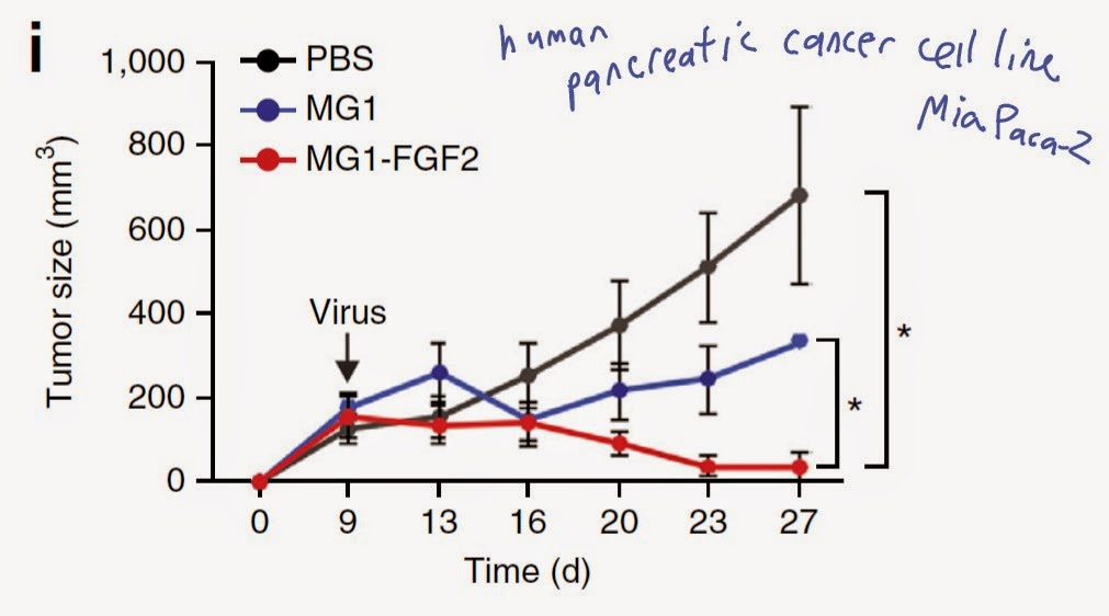
Screen for active factors indicated that enhancing factor was fibroblast growth factor-2 (FGF-2). Indeed, inhibition of FGF-2 by RNAi reduced viral titre in tumor-CAF co-culture.


Finally, the authors showed that virus expressing FGF-2 could induce regression of established tumor in mouse model.


In summary, these results suggest that exploiting cancer vulnerability towards lytic viruses may be an alternative path for biological cancer therapy

Note: It is puzzling that virus targets only cancer cells. The authors explained such selective sensitivity of cancer cells based on reduced baseline anti-viral activity in cancer cells. However, we need to consider the fact that cancer patients maybe immune deficient in general, making them generally susceptible to viral infections.

David Usharauli