Showing posts with label Foxp3 Tregs. Show all posts

Wednesday, April 3, 2019

Innate help to Tregs in the gut


Thursday, March 21, 2019

A physiological T cells' "weaning reaction" to microbiota and solid foods in newborn pups requires FOXP3+ Tregs


Friday, September 8, 2017

Loss of microbiota depletes cross-reactive Foxp3+ Tregs leading to selective immunopathologies

Check out our follow-up manuscript in PeerJ Preprints that provides a brief guide to SPIRAL, a novel interpretive framework that demonstrates the central role of microbiota-Treg axis in the initiation of immune disorders.

Kamala T, Usharauli D. (2017)
 
Loss of microbiota depletes cross-reactive Foxp3+ Tregs leading to selective immunopathologies.
PeerJ Preprints 5:e3237v1
 
The 'Hygiene hypothesis', a cornerstone model to account for the role of exogenous pathogens and later of endogenous microbiota in immune disorders, is currently presumed to operate at the innate immunity and metabolite levels to properly 'educate' the immune system. Doing so however fails to satisfactorily account for the antigen-specific nature of such disorders. SPIRAL is a novel interpretive framework that resolves this dilemma. It represents the periodic table of cross-reactive Foxp3+ regulatory T cell (Treg) epitopes selected from commensal microbiota over evolutionary time to mediate self-nonself discrimination and effector class regulation. Here, we utilize the SPIRAL's predictive power to provide a mechanistic antigen-specific basis for the initiation of allergies and autoimmune diseases as well as for the failure to mount effective anti-tumor and vaccine responses through selective loss of microbiota and corresponding cross-reactive Foxp3+ Tregs.


Thursday, September 7, 2017

IL-27 protects against autoimmunity through its effect on Tregs

IL-27 is a heterodimeric cytokine composed of the p28 and Ebi3 subunits produced by APCs. It binds to IL-27 receptors (IL 27Rα:gp130) expressed on several cell types, including T lymphocytes. IL-27Ra-/- mice are shown to be highly susceptible to experimental autoimmune encephalomyelitis (EAE), a mouse of human MS. Earlier studies suggested that effect of IL-27 is mediated through its suppression of pro-inflammatory Th17 cells and generation of anti-inflammatory IL-10 producing Tr1 cells.

A new study in PNAS, however, showed that mice with Treg-specific IL-27Rα-deficiency displayed similar susceptibility to EAE as total IL-27Rα-deficient mice pointing to the role of Tregs in mediating IL-27 effect on EAE.

Compared to WT mice, mice with Treg-selective IL-27Rα-deficiency were not able to recover from EAE, a similar trend seen in total IL-27Rα-deficient mice. IL-10 levels were not different between WT and KO littermates.


Furthermore, in contrast to WT mice, injection of "therapeutic" dosage of IL-27 had no effect on EAE dynamics in Treg-selective IL-27Rα-deficient mice. Nor did antibody-blockade of IL-10 diminish effect of IL-27 on WT mice.


These results suggest that in addition to other cell types or even in contrast to other studies, the role of IL-27 in controlling severity of EAE could be solely mediated through its effect on Foxp3+ Tregs.

posted by David Usharauli 


Wednesday, February 22, 2017

Antigen-specific Tregs maintain immune privileges of male reproductive tract

This week Journal of Clinical Investigation published very good research article that shed light on how tolerance to sequestered self-antigens expressed by immune privileged  tissues are established

Some tissues such as brain, eye, testis or ovaries are thought to be "immune privileged" organs meaning that ordinarily immune system does not see their antigens. However, in this paper, the authors showed that in fact some testes antigens in male mice, for example lactate dehydrogenase 3 (LDH3), are actually secreted and detected by immune system.   

Series of experiments confirmed that WT male mice did not respond to LDH3 immunization, while female and LDH3-null male mice mount detectable immune response to it. This suggested that male mice were physiologically tolerant to LDH3. Notable, both male and female mice could be immunized against another testis antigen, zonadhesin (ZAN), implying absence of tolerance to ZAN in male mice. 


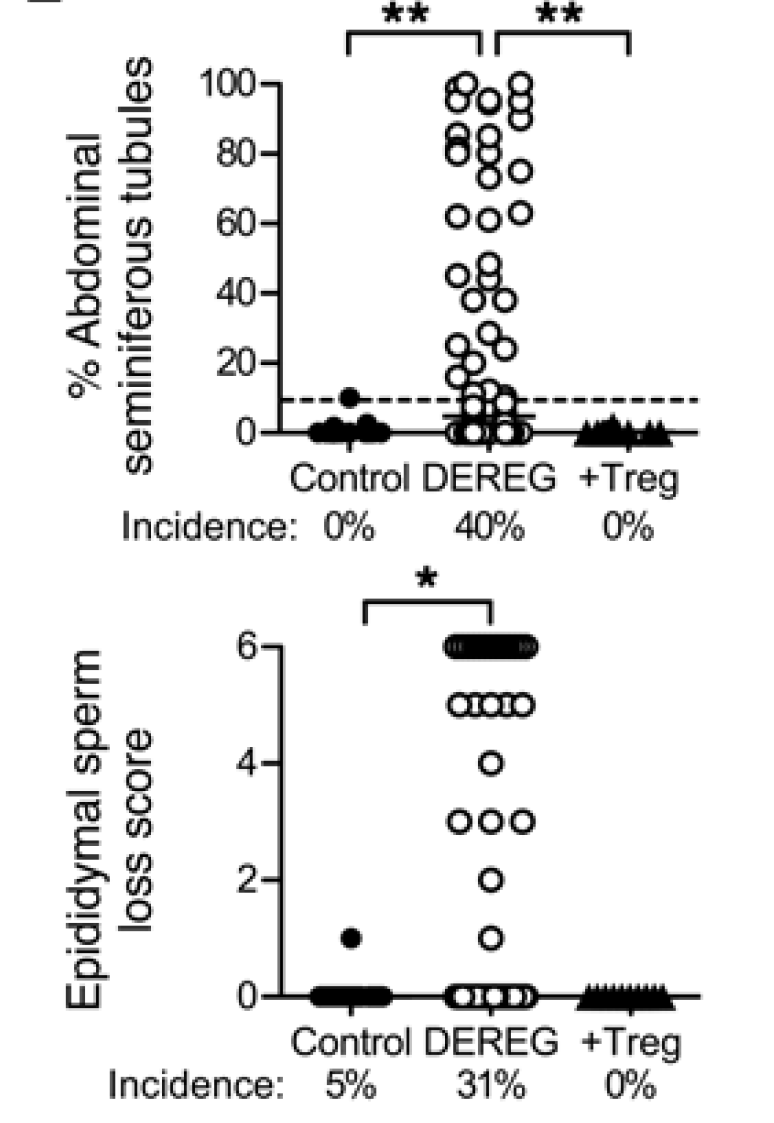
So, how male mice were tolerant to LDH3? To answer it, the authors temporally depleted Tregs and it led to immune response to LDH3 in immunized WT male mice. since no changes were seen in response to ZAN, the authors concluded that tolerance to LDH3 in WT male mice was dependent of presence of FOXP3+ Tregs.


Furthermore, depletion of Tregs even in absence of testes antigen immunization still led to autoimmune pathology in testes in ~40% of male mice ("autoimmune orchitis occurs in autoimmune polyendocrine syndrome 1 (APS1) patients due to mutations of AIRE, possibly associated with impaired thymic deletion of autoreactive T cells and deficient Treg function")


In summary, this study suggests that "immune privilege" is not absolute and self-antigens that naturally leak maintain tolerance by induction of antigen-specific Tregs.

David Usharauli


Saturday, October 22, 2016

FOXP3+ Tregs and allergic TH2 cells recognize mutually exclusive allergic proteins


One reason why it is so difficult to study human FOXP3+ Tregs is that we don't have a good surface marker that selectively identifies human FOXP3+ Tregs from that of conventional T cell memory. As a consequence, we can't easily study antigen-specific human FOXP3+ Tregs.

In this new paper, the authors proposed that expression of CD137 versus CD154 identifies FOXP3+ Tregs versus Tconv in antigen-specific stimulation assay. Using CD137 as a marker, the authors showed that presence of FOXP3+ Tregs specific for variety of nonself antigens, including allergens.


The vast majority of human antigen-specific FOXP3+ Tregs displayed memory CD45RO+ phenotype.


Interestingly, TCR Vbeta analysis revealed mutual exclusivity of FOXP3+ Tregs and Tconv memory.


Next, when analyzing T cells from allergic individuals the authors first confirmed that indeed allergic individuals harbor significantly more allergen-specific TH2 cells.   


However, surprisingly, the authors found that allergic individuals harbor normal numbers of allergen-specific FOXP3+ Tregs.  


To understand this finding, the authors conducted more stringent allergen-specific assay by culturing T cells with individual allergen protein rather than the whole allergen extract as customary (the whole extract contains several related proteins). Indeed, this modified assay showed mutual exclusivity of antigens recognized by human FOXP3+ Tregs and allergic TH2 cells.   


In summary, this paper showed that unlike conventional wisdom, failure of FOXP3+ Tregs to control TH2 cells is antigen-specific. In essence, allergic individuals have specific "holes" in FOXP3+ Tregs repertoire that do not allow them to prevent allergen-specific naive T cells differentiation into allergic TH2 cells. This is first checkpoint. However, simple absence of allergen-specific FOXP3+ Tregs is not sufficient to induce allergy. Second checkpoint for allergy development requires additional factors, not yet fully understood, that promote TH2 response.

David Usharauli 


Tuesday, October 4, 2016

Antigen-specificity of human FOXP3+ Tregs

Foxp3+ regulatory T cells (Tregs) control immune response to prevent immunopathology. However, unlike conventional T cells, it is hard to determine antigen-specificity of Foxp3+ Tregs in a random T cell pool. Tregs do not secrete anything unique and they do not even proliferate when exposed to antigens in vitro, two functional readouts that are still used as a gold standard for determining antigen-specificity of conventional T cells. 

Hence, we have no clue as to antigen[epitope] specificity of vast majority of human Tregs. Specificity of T cells could be also determined by non-functional readout such as tetramer staining. This is what the authors in new PNAS paper have used to determine antigen-specificity of Tregs

They found that adult human peripheral blood contains FOXP3+ T cells that stain with tetramers specific for self as well as nonself peptides (Flu, melanoma protein, HIV epitopes).


Interestingly, frequency of antigen[epitope]-specific FOXP3+ Tregs varied among donors, but they were, on average, equally distributed among self and nonself [epitope]-specific Tregs, except Flu HA epitope.  


Finally, comparison of neonatal [cord blood] and adult blood revealed that actual number of [epitope]-specific Tregs / per ml of blood did not change much between newborn and adult indicating that most of Tregs tested in this study were generated already by the time of birth.


In summary, this study revealed that human peripheral blood contain Tregs specific for both self nonself antigens. Since tested donors were negative for some of the infection such as HSV, CMV or HIV, it begs the question what [cross-reactive?] antigens maintain CMV or HIV-specific Tregs in antigen-naive [antigen-unexposed] hosts?    

David Usharauli

Wednesday, September 28, 2016

Wiskott-Aldrich syndrome protein (WASp) deficient FOXP3+ Tregs fail to control IgE mediated allergies

The inherited immunodeficiencies are frequently characterized with dysregulated Th2 responses, atopy, and elevated IgE levels. Mutations in Foxp3, STAT3, DOCK8, PGM3, LAT, ZAP70, or RAG result in hyper IgE phenotypes. Mechanism is unclear.


For this study the researchers analyzed "the overall burden of clinical food allergy within a cohort of 25 patients with mutations in the WAS gene" and found that individuals with WAS mutations were more likely to demonstrate serum sensitization to peanut, milk, and egg (compared to the general population).


Lab mice deficient for WASp also display elevated IgE (and IgG1) antibody levels to components of their chow food (even on germ-free background).


Since WASp is expressed in different cell types, the authors tested mice selectively deficient for WASp in B cells, DCs or FOXP3+ Tregs. Out of these gene-modified mice, only Wasfl/fl Foxp3-Cre mice showed deviation to Th2 phenotype and development of IgE to chow food.


In vitro T cell stimulation confirmed that total T cells from Wasfl/fl Foxp3-Cre mice showed selective enhancement in Th2 cytokines.


In sum, these results indicate that mice with selective deficiency of WASp in FOXP3+ Tregs display excess in Th2 subsets. It is possible that absence of WASp destabilizes FOXP3+ Tregs and this somehow drives their de-differentiation into Th2-like cells (and not into Th1 or Th17, for instance).

David Usharauli


Tuesday, September 6, 2016

Foxp3+ Tregs control CD8 T cells, but not CD4 T cells, by IL-2 deprivation

This week Nature Immunology published another interesting study from Rudensky's lab done in collaboration with Jason Fontenot (who apparently moved from Biogen to Juno Therapeutics). So, if you are a fan of Tregs, below is my short scientific overview of that paper.

This study tries to answer what role IL-2 signaling plays in already established Foxp3+ Tregs. For such study mice with germline deficiency in IL-2 signaling cascade would have been impractical since IL-2's effect on thymocytes and etc. Instead, the authors went to already well established path of using mice lacking "molecule of interest" specifically in Tregs. In this case several Foxp3-cre mice were used, such as: l2rbfl/flFoxp3Cre, Il2rafl/flFoxp3Cre, Stat5afl/flStat5bfl/flFoxp3Cre, Rosa26Stat5bCAIl2rbfl/flFoxp3Cre and Rosa26Stat5bCAIl2rafl/flFoxp3Cre, Rosa26Stat5bCAFoxp3Cre–ERT2. All these gene-modified mice allows specific targeting of Foxp3+ Tregs.

Unsurprisingly, mice lacking IL-2Rβ or IL-2Rα or STAT5 signaling specifically in Tregs developed autoimmunity.


Surprisingly, however, while constitutive expression of STAT5 in Tregs lacking IL-2 signaling receptors (IL-2Rβ or IL-2Rα) could rescue mice from "CD4 effector phenotype" and early death, these mice still developed immunopathology later due to massive expansion of CD8 effector/memory cells, suggesting that sensing of IL-2 by Tregs, so called "IL-2 sink", was necessary to specifically control CD8 T cells, but not CD4 T cells.


In summary, this study suggests that Tregs are using distinct mechanisms to control CD4 and CD8 T cells. It kind of makes sense because MHC class-II restricted CD4+ Tregs cannot interact the same way with MHC class-I restricted CD8+ T cells as they could with conventional MHC class-II restricted conventional CD4+ T cells.

David Usharauli

Saturday, July 30, 2016

Remote control of tissue tolerance by regulatory T cells

This week journal PNAS published an interesting study on Foxp3+ regulatory T cells (Tregs). It showed that presence of Tregs within secondary lymphoid organs (in lymph nodes) was sufficient to prevent peripheral tissue immunopathology.

As a starting point for this study, the authors showed that mice with Tregs-specific deficiency of kruppel-like factor 2 (KLF2) develop non-fatal peripheral tissue immunopathology, even though in vitro such Tregs [Foxp3-cre; Klf2fl/fl] displayed normal suppressive activity).


Interestingly, in vivo adoptive transfer model, KLF2-KO Tregs also failed to prevent colitis and weight loss when co-transferred with WT naive T cells [but not with KLF2-KO naive T cells].


These results suggested that Tregs re-circulation between tissue and lymphoid tissues could have been involved. Surprisingly, the authors found no difference for Tregs presence [%-wise, no data about #] between KLF2-KO mice or WT (this is probably why this article ended up in PNAS).


Finally, the authors showed that combined deficiency of KLF2 and CCR7 in Tregs accelerated tissue immunopathology [by preventing Tregs access to lymph nodes].


In summary, the authors speculated that Tregs access to lymphoid tissue played a crucial role in preventing tissue immunopathology.

David Usharauli

Thursday, May 19, 2016

Foxp3+ Tregs go against tumor in the "dark"

This week PNAS published follow-up study from Harvey Cantor's lab examining role of Helios (the Sun in Greek mythology) in Foxp3+ TREG stability. In this new study they showed that antibody [anti-GITR]-mediated down-regulation of Helios in Foxp3+ TREG causes them to acquire effector function and participate in anti-tumor response.

I will highlight relevant findings. The first finding was that mice with Helios-deficient Foxp3+ TREG showed additional resistance to tumor (however, please note that tumor growth pattern and mice survival do not correlate in Heliosfl/fl.FoxP3-Cre mice).


Similar tumor growth retardation was observed in adoptive co-transfer experiment with WT T cells and Helios-KO Foxp3+ TREG.


Mechanistically, Heliosfl/fl.FoxP3-cre TREG up-regulated effector cytokines specifically at tumor site.


Finally, treatment of mice with antibody, DTA-1, directed to GITR (glucocorticoid induced TNF receptor), caused Helios down-regulation in FoxP3+ TREG and improved tumor protection.


In summary, the authors proposed that therapeutic targeting of Helios expression in tumor-associated FoxP3+ TREG could convert them from tolerogenic cells into anti-cancer fighting cells (however, please note the authors did not formally test whether anti-cancer effect of anti-GITR antibody was indeed mediated via its effect on Helios in FoxP3+ TREG).

David Usharauli

Thursday, February 11, 2016

Flexing muscles with IL-33 responsive Tregs

Just read another paper about Foxp3+ Tregs published in journal Immunity. Not clear what to make out of it. It came from Christophe Benoist and Diane Mathis lab, both wellknown immunologists, but it feels if it was done by non-immunologist. It does have Amy J. Wagers as a co-authors. She is known for her studies of stem cells.


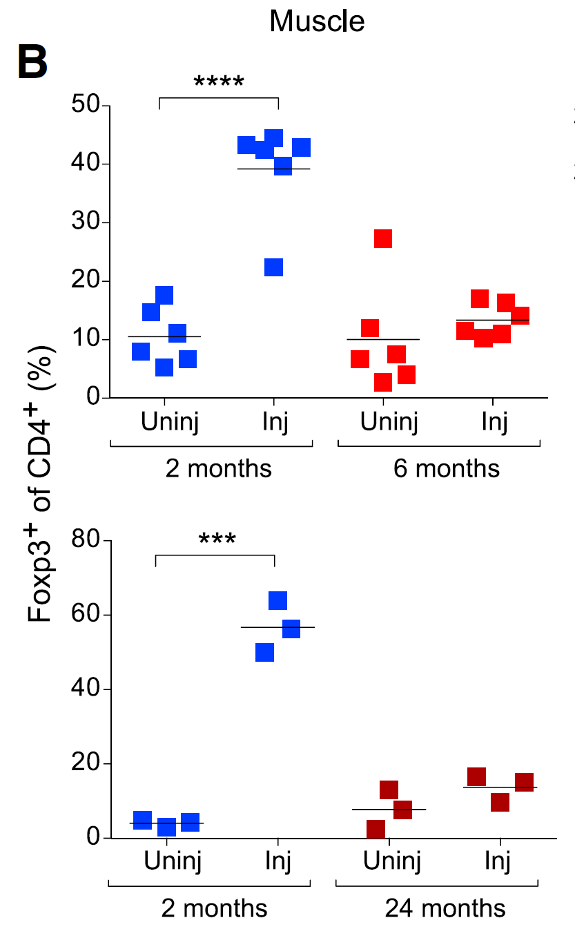
Now, how does one test this hypothesis? I will highlight what is good in this paper and what is missing. Initially, the authors showed that in old mice muscle injury (cryo-injury or by toxin) does not attracts as much Tregs as in young mice. Strangely, Tregs recruitment within injured muscle tissue was examined using anti-Foxp3 antibody rather more widely accepted model of Foxp3 reporter mice [which they indicated in methods section they had].


Next, the authors found that cytokine IL-33 was up-regulated in injured muscle tissue (young mouse).


Correspondingly, Tregs recruited and enriched within injured muscle tissue expressed IL-33 receptor called ST2.


Next, the authors speculated that there was maybe some connection between IL-33 responsive Tregs (ST+ Tregs) and muscle regeneration. Indeed, when they have repeated the same experiment with mice deficient for IL-33 receptor specifically on Tregs, muscle regeneration were impaired [after injury]. Though the authors did not show data whether there was no difference between muscle regeneration between young and old mice when both were deficient for IL-33 receptor specifically on Tregs. Without such data it is not possible to conclude whether difference in IL-33 responsiveness in Tregs accounts for difference between young and old mice with respect to muscle regeneration.  
  

Analysis of IL-33 producing cell within muscle tissue showed that fibro/adipogenic progenitor (FAP) cells were the main producer of muscle IL-33 and they produced less of IL-33 after muscle injury in old mice.

Finally, injection of IL-33 into old mice muscle improved its regenerative potential after injury (however, the authors did not repeat the same experiment with Treg-specific IL-33 receptor deficient mice to formally confirm that beneficial effect of exogenous IL-33 was indeed mediated via Tregs).


In summary, this study suggests that IL-33 improves muscle regeneration, in general. Whether reduced number of IL-33 responsive Tregs in old mice represents only variable between young or old mice with respect of muscle regeneration is not clear from this paper. Basically, In my opinion, the results do not support the paper's conclusions with respect to Tregs.

David Usharauli

Wednesday, February 10, 2016

Foxp3+ Treg-derived IL-35 limits anti-tumor T cell immunity

IL-35 is a cytokine composed of the p35 subunit of IL-12 (encoded by Il12a) and Ebi3 (Ebi3). It was discovered in 2007 at Dario Vignali's lab and since then ~ 200 papers have been published about IL-35, according PubMed database. Most studies indicate that IL-35 has immune inhibitory function.

Now, new study in journal Immunity from the same Dario Vignali's lab provided evidence that Foxp3+ Treg-derived IL-35 inhibits effectiveness of anti-tumor T cell activity, pointing to it as a new immunotherapeutic modality. I am going to highlight the most notable results.

First, the authors showed that tumor (B16 melanoma) growth or its metastasis were inhibited when (a) mice were injected with antibody against IL-35 or when (b) mice lacked IL-35 specifically in Tregs (Foxp3Cre-YFP. Ebi3L/L mouse).


Similar effect of IL-35 blockade on tumor growth was seen in genetically-induced tumor model, in KP mouse, that spontaneously develop lung cancer after adenovirus-Cre delivery (KP mouse contains an activating mutation in K-RAS and a loss of function mutation in p53 controlled by Cre mediated recombination).

Mechanistically, the authors showed that IL-35 blockade improved T cell recruitment into tumor tissue.


Finally, the authors found that presence of Treg-derived IL-35 contributes to T cell exhaustion during tumor growth.


In summary, this study indicate the following scenario: certain tumors recruit Tregs. These Tregs secrete IL-35 and inhibit recruitment of effector T cells and/or contributes to their exhaustion. Blocking of IL-35 could provide immunotherapeutic benefit (interestingly, it appears that IL-35 does not synergize with anti-PD1 treatment in tumor models discussed in this paper).

David Usharauli

Thursday, February 4, 2016

Some anergic T cells are precursors for Foxp3+ Tregs

T cell anergy is defined as T cell non-responsiveness to [secondary] antigen challenge. This concept has originated  in 1980s to explain T cell behavior following primary encounter with antigen (signal 1) in absence of co-stimulation (signal 2). Later, anergy concept was expanded to include T cell behavior following repetitive antigen encounter, similar to what today we would call T cell exhaustion.    

Generally speaking, anergic T cells are different from canonical thymic Foxp3+ Tregs. Anergic T cells do not express Foxp3 marker. While there are several surface and transcription factors that could "identify" anergic T cells, classical test for anergic T cells is to run several functional cytokine secretion assay and if memery/effector T cells do not produce cytokines, especially IL-2, and do not express Foxp3, you can call it anergic T cells. It is primitive but its all we have right now.  

In the past scientists thought that anergic T cells were necessary to maintain peripheral tolerance against self-antigens. It was believed that anergic T cells would compete for antigen access with self-reactive naive T cells (recent thymic emigrants) and would not allow their activation. This model lost its appeal once Foxp3T cells were re-discovered and re-defined. So, presently, functional significance of anergic T cells is in a kind of limbo.

So, it was interesting to see new paper in Nature Immunology that revisited the role of anergic T cells. In this study the authors tried to make sense of anergic T cells by postulating that some portion of anergic T cells represent precursors for classical Foxp3+ T regs.

First, the authors showed that among Foxp3-negative T cells, FR4hiCD73hi markers define anergic T cells (by low IL-2 production) that develop following antigen encounter in absence of co-stimulation (for example, pregnant females harbor pregnancy-associated anergic T cells specific for antigen [2W1S] expressed by male fetus). Interestingly, majority of those Foxp3-negative anergic T cells express neuropilin-1 (Nrp-1), a marker for thymus Tregs.


Next, the authors showed that in functional assays, FR4hiCD73hi anergic T cells behave very similar to classical Foxp3T cells.

Furthermore, when polyclonal Foxp3-FR4hiCD73hi anergic T cells were transferred into lymphopenic host, anergic T cells gave rise to classical Foxp3T cells (compared to naive or effector T cells).

Importantly, if those newly formed Foxp3T cells [derived from anergic T cells] were selectively deleted it led to systemic autoimmune disease, implying functional role for such daughter Foxp3T cells.

Finally, the authors showed that new Foxp3T cells formed from anergic T cells could prevent arthritis development when co-transferred with cartilage antigen-specific KRN T cells (that themselves do not become anergic or develop into Foxp3T cells).  


In summary, this study showed that some anergic T cells (especially Nrp1+ subpopulation) could give rise to peripheral Foxp3T cells. However, not all T cells can produce anergic T cell that could serve as a precursors for Foxp3T cells (for example KRN T cells or 5C.C7 CD4 T cells). It remains seen what role, if any, anergic T cells play in un-manipulated host. So far we lack tools to selectively deplete anergic T cells in un-manipulated host to test their real-world biological significance. Also, since anergic T cells resemble thymic Foxp3T cells in Npr1 expression, it will be interesting to found out whether anergy induction is a part of thymic Foxp3T cell development program, in general.

David Usharauli

Saturday, January 30, 2016

Food antigens, not microbiota, regulate Foxp3+ regulatory T cells in small intestine

Foxp3+ regulatory T cells, shortly Tregs, are an unique subset of CD4 T cells that regulate magnitude and possibly class of immune response. 

In general, Tregs are divided into two subsets referred as thymus-derived Tregs (tTregs) and peripheral tissue-induced Tregs (pTregs). Historically speaking, this division of Tregs is arbitrary [it is assumed that thymus alone could not generate Tregs specific for peripheral antigens, such as tissue-specific antigens or food derived antigens] but till today we have no definite proof whether there is indeed such a thing as a peripheral Tregs.


I would like to point out that in this study the authors have used Neuropilin-1 (Nrp-1) to differentiate between tTregs and pTregs. However, more recent studies questioned the validity of this marker. So, for me, the results in this paper are simply "observations", not necessary "mechanisms".

The main contribution of this study is the use of antigen-free diet [chemically defined food devoid of macro-molecules] to analyse Tregs physiology in mice. It showed that mice on antigen-free diet had reduced number of Tregs in small intestinal tissue (but not in spleen or large intestine).


Next, the authors reported that small intestine of mice fed with antigen-free diet lacked Tregs expressing low level of Neuropilin-1 (that according to the authors represent peripheral Tregs). At least, these results indicate that food derived antigens regulate the fate of Neuropilin-1low Tregs in small intestine.


Finally, by transferring ovalbumin-specific OT-II CD4 T cells in mice fed with antigen-free diet, the authors showed that lack of host Neuropilin-1low Tregs in small intestine could lead to excessive proliferation of donor antigen-specific T cells in these mice (that could lead to exaggerated, allergic-type immune response).

In summary, this study showed that solid food derived antigens are important in regulating physiology of small intestinal Tregs. This knowledge could help to understand how food allergies develop.

David Usharauli  

Tuesday, January 12, 2016

Type I IFNs produced during viral infection promotes bystander regulatory T cells

Foxp3+ regulatory T cells are one of my favorite topics. Foxp3+ T cells are the most powerful regulatory subset within immune system. It is unbelievable but true that as of today only sure thing we know about Foxp3+ T cells is the fact that their absence or ablation leads to severe and total autoimmune organ disorders and death. No other T or B cells or innate subsets produce such a dramatic effect on the body.

At the same time, we are still have no clear answer whether Foxp3+ regulatory T cells operate in an antigen-specific manner or whether they non-specifically suppress "overly" excessive immune response (both innate or adaptive driven).

There are several hypothetical questions regarding Foxp3+ T cells that would require experimental proof before we can make any significant breakthrough. 

For example, 
"if Foxp3+ T cells are so potent, how immune response is initiated in the first place?" 

"If initial inflammatory stimuli temporary inactivates Foxp3+ T cells to allow initiation of immune response, how long such inactivation lasts?" 

"if Foxp3+ T cells regulate excessive immune response (excessive inflammation), how they can sense what is excessive?"

In this regard new paper in Nature Immunology from Steven Ziegler's lab is interesting to read. Here, the authors showed that pre-exposure of naive antigen-inexperienced CD4 T cell to type I IFNs initiates their differentiation towards regulatory pathway rather than effector. Such scenario possibly prevents excessive bystander activation of naive T cells and reduces overall tissue damage.

In vitro studies showed that CD4 T cells initially pre-exposed in vivo to polyI:C (as a source of type I IFN) and then co-cultured with antigen-pulsed DCs and TGF-β tended to preferentially develop into Foxp3+ T cells.

Such preference for Foxp3+ T cell development were abolished with CD4 T cells from IFNαR1KO mice.

RIP-mOva × Rag2KO hosts receiving naive OVA-specific T cells pre-exposed to polyI:C do not develop diabetes (c), while RIP-mOva × Rag2KO hosts receiving control naive OVA-specific T cells together with polyI:C rapidly developed diabetes. Importantly, secondary exposure to polyI:C of RIP-mOva × Rag2KO hosts transferred with naive OVA-specific T cells pre-exposed to polyI:C still did not break tolerance (d).

These experiments indicated that simultaneous exposure of naive T cells to antigen and type I IFNs drove effector differentiation, but if they were first pre-exposed to type I IFNs and then to antigens it drove their Foxp3+ T cell differentiation. Indeed, this observations were confirmed in subsequent experiments that revealed that exposure to type I IFNs 2-3 days before antigenic exposure provided the most optimal condition for Foxp3+ T cell development.


In summary, this study suggests the following scenario: type I IFNs released during viral infection conditioned bystander, nonspecific naive T cells for Foxp3+ T cell development. This mechanism potentially prevents excessive activation and effector differentiation of naive T cells encountering antigens later during immune response (including tissue-derived self-antigens).

David Usharauli