Showing posts with label nanoparticles. Show all posts

Friday, June 3, 2016

Tumor protein encoding [negatively charged] RNA-nanoparticles induced robust anti-tumor response


In earlier studies for in vivo delivery of RNA/DNA nucleic acids into antigen-presenting cells scientists have used positively charged [cationic] nanoparticles. However, such particles were mostly trapped within lungs rather than lymphoid tissues where most antigen-presenting cells, such as dendrtic cells, reside. Surprisingly, here the authors showed that negatively charged RNA-nanoparticles selectively accumulated in spleen within antigen-presenting cells after in vivo injection.


Strangely, accumulation of RNA-nanoparticles were reduced when mice were depleted of conventional DCs (cDCs) after diphtheria toxin injection (CD11-DRT BM chimera mice), even though these mice retain both plasmacytoid DCs (pDCs) and macrophages which can themselves capture RNA-nanoparticles.


RNA-nanoparticle injection was associated with rapid IFN-α secretion in a manner that depended on TLR7 expression.


In addition, RNA-nanoparticles induced robust priming of antigen-specific adaptive immune response.


More importantly, injection of tumor protein encoding RNA-nanoparticles delivered incredibly robust prophylactic and therapeutic anti-tumor effect (article has some preliminary  results from phase I study showing T cell priming in cancer patient after RNA-nanoparticle delivery).


In summary, the authors of this study believe that they found universal antigen delivery RNA-nanoparticle construct that induces robust adaptive immune response.

There is one thing that is puzzling about this study which the authors did not tried explain: in mice depletion of cDCs eliminated RNA-nanoparticle uptake and T cell priming. However, pDCs are not depleted in CD11c-DTR chimera mice and the data showed that pDCs are themselves could capture RNA-nanoparticles and are the main source of type I IFNs. So, the question then is why cDCs depletion has such disproportional effect?

David Usharauli


Wednesday, April 20, 2016

Nanoparticle-caged antigens could treat allergen-sensitized host

The challenge with immune system-[driven] disorders is the fact that ordinarily such conditions are detected at the later stages when immune system had already developed self-perpetuating "memory circuit". Even now we know very little how to break such "memory circuit".

New study in PNAS suggested an experimental model that were able to tolerize already-sensitized host against allergen. The authors showed that biodegradable nanoparticles incorporating caged antigen, but not simple conjugates, could deliver both prophylactic and therapeutic treatment to allergen-prone host.

For this study the authors have used widely-accepted experimental sensitization protocol in mice to induce TH2-driven allergy to ovalbumin  antigen (OVA). Next, they proceeded to test three different combination of  nanoparticles + ovalbumin: (1) OVA conjugated to polystyrene nanoparticles (Ag-PS), (2) OVA conjugated to biodegradable poly(lactide-co-glycolide) nanoparticles (Ag-PLG) and (3) OVA caged within biodegradable poly(lactide-co-glycolide) nanoparticles [PLG(Ag)].

First, the authors found that all three nanoparticle combinations displayed potent prophylactic action against allergen when delivered before allergen sensitization (Ag-PLG is shown here).


However, when applied to already allergen-sensitized host, polystyrene nanoparticles induced severe allergic reaction. In contrast, application of Ag-PLG nanoparticles did not induce allergic response in sensitized hosts and even reduced TH2 cytokines. However, Ag-PLG nanoparticles could not inhibit lung eosinophilia.


Finally, the authors showed that application of PLG nanoparticles with caged OVA antigen could both prophylactically and therapeutically inhibit TH2-driven allergic response.


In summary, this study showed that nanoparticles incorporating antigens (Ag caging) avoids detection by allergen-specific immune effector molecules (IgE) and instead drives tolerance to allergen.      

David Usharauli

Thursday, February 18, 2016

pMHC tetramer-coated nanoparticles treat autoimmune conditions in mice

This week Nature published "figure-dense" paper showing that nanoparticles coated with auto-antigen+MHC complexes (pMHC) could treat spontaneous or experimentally-induced autoimmune diseases in mice. In fact, this study contains so many experimental results it should have been published in two part, in my view. I will try to condense its basic findings in few paragraphs and provide its interpretation below.

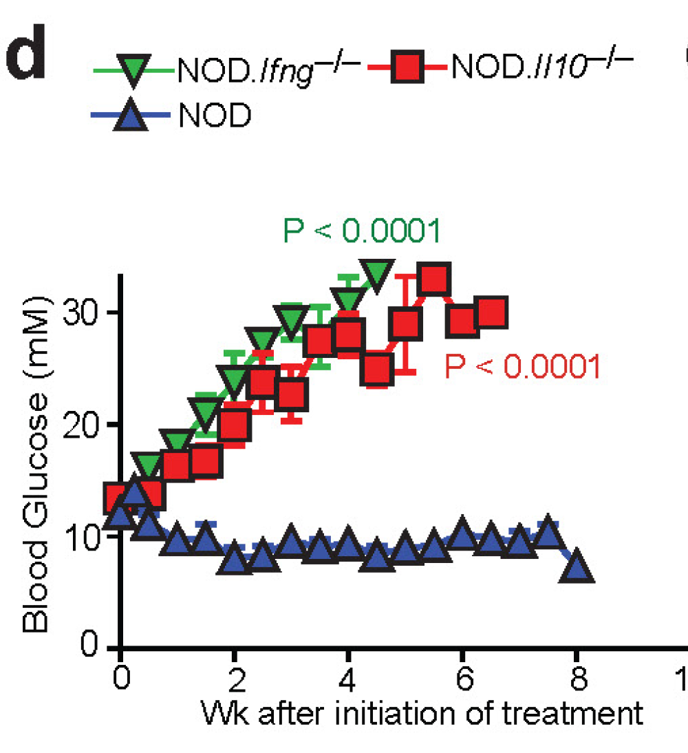
Non-obese diabetic (NOD) mice spontaneously develop [autoimmune] diabetes. Part of T cells that infiltrate and damage β cells in NOD mice express T-cell receptor (TCR) resembling diabetogenic BDC2.5-specific TCR that can be recognized by pMHC–2.5mi/IAg7 tetramers. When the authors injected nanoparticles (dextran-coated or pegylated iron oxide NPs) coated with 2.5mi/IAg7 tetramers (pMHC–NP) it induced expansion of cognate CD4+ T cells in blood and spleens of NOD mice. These cells had a memory-like (CD44hiCD62Llow) FOXP3− TR1-like phenotype.


These "activated/experienced" CD4+ TR1 cells from donor mice treated with pMHC–2.5mi/IAg7-NPs suppressed diabetes development in T-cell-reconstituted NOD-scid hosts upon adoptive transfer. This effect of TR1 cells was augmented by treating hosts with pMHC–NPs.


Moreover, 90–100% of the already diabetic mice that received nanoparticles coated with β cell antigens, 2.5mi/IAg7, IGRP4–22/IAg7 or IGRP128–145/IAg7, reverted to stable normoglycaemia.


Of note, treatment withdrawal resulted in loss of the normoglycaemic state in 25–60% of mice, in association with the loss of the tetramer+CD4+ T-cell pools. In other words, these data suggest that almost half of treated diabetic mice maintained normoglycaemic state after 5 weeks of treatment.


Beyond diabetes, the authors showed that nanoparticles coated with myelin oligodendrocyte glycoprotein, pMOG38–49/IAb–NP, dampended progression of experimental autoimmune encephalomyelitis (EAE, a model of multiple sclerosis), when given on day 14 after immunization and even restored motor function in paralytic mice when given on day 21.


Similar therapeutic effects were seen in 3rd autoimmune disease model, collagen-induced arthritis (CIA). HLA-DR4-IE-transgenic mice receiving nanoparticles displaying mouse collagen (mCII)259–273/DR4-IE showed reduced joint inflammation. In all these models, pMHC–NPs effect was antigen-specific.

Indeed, the effects of pMHC–NP therapy were not associated with impaired systemic immunity because pMHC–NP-treated mice showed unimpaired anti-viral immunity and mounted antibodies against an exogenous antigen as efficiently as control mice.

Interestingly, pMHC–NPs could not expand tetramer+ T cells in non-diabetic control mice which harbor mostly naive T cells. Further experiments confirmed that therapeutic effect of pMHC–NP required presence of "experienced" T cells. For example, whereas pMHC–NP therapy afforded 100% diabetes protection to T-cell-reconstituted NOD-scid hosts bearing memory BDC2.5 T cells, such therapy was inconsequential in hosts receiving naive BDC2.5 T cells.


The role of "experienced", but not naive T cells, in therapeutic effect of pMHC–NP was also supported by observation that whereas diabetic NOD G6pc2−/− mice (which lack IGRP antigen and corresponding memory/experienced T cells, but most likely harbor IGRP4–22 specific [aka, nonself-specific] naive T cells) responded to 2.5mi/IAg7–NPs like wild-type NOD mice, they did not respond to IGRP4–22/IAg7–NPs.


At molecular level, the authors showed blockade of IL-10, TGF-β and IL-21R (but not IFNγ) abrogated the anti-diabetogenic properties of 2.5mi/IAg7–NPs or IGRP4–22/IAg7–NPs in diabetic NOD mice.

However, to make things more complicated, the authors also showed that the development of the TR1 precursors and/ or TR1-like cells that expand in response to pMHC–NP therapy required IFNγ in addition to IL-10 in pre-diabetic NOD mice.

Finally, the authors found that at the cellular level, pMHC–NP treated NOD mice harbored increased numbers of so called regulatory B cells, BREG cells (but only in pancreatic lymph nodes, PLNs). These BREG cells were producing IL-10 in response to lipopolysaccharide (LPS). In vivo, these B cells suppressed diabetes development in T-cell-reconstituted NOD-scid hosts as compared to PLN B cells from control mice (and worked together with TR1 cells). The role of BREG cells was supported by observation that treatment of newly diabetic NOD mice with a B-cell depleting anti-CD20 mAb abrogated the anti-diabetogenic activity of 2.5mi/IAg7–NPs (see above).


In summary, this study suggests that when injected into mice these pMHC–nanoparticles expand antigen-specific and antigen-experienced T cells with regulatory properties. These TR1 work with BREG cells to block the chain of events that perpetuates autoimmune cycle. 

David Usharauli


Monday, September 28, 2015

Sialic acid decorated multi-valent nanoparticles prevent sepsis mortality in mice

Sepsis is an uncontrolled tissue-immune [hyper]response that leads to the organ failure and frequently patients' death. At present, there is no specific treatment for sepsis, just supporting care. Obviously, sepsis represents important unmet medical condition.

New study in Science Translational Medicine provided evidence that sialic acid coated nanoparticles could prevent sepsis-induced death in mice model of acute sepsis.  

This study is based on prior evidence that binding of sialic acid (type of sugar) to its receptors called Siglecs on macrophages or neutrophils could inhibit inflammatory pathways. Here, the authors showed that unlike bi-valent anti-Siglec antibodies, administration of multi-valent sialic acid coated nanoparticles (in a form of α2,8 sacharide linkage), at both T= 0h or T= +2h, prevented endotoxin induced animal death (i.p. injection).


Similar protection was observed in anesthetized mice undergoing cecal ligation and puncture (CLP) procedure (here too, α2,8-NANO was administered i.p.).


Additionally, intra-lung administration of α2,8-NANO showed significant (but not as impressive) protection against lung inflammation (a secondary complication following cecal ligation and puncture procedure).


The authors showed that protection provided by α2,8-NANO was IL-10 dependent in endotoxin injection model (though, for some reason, the authors had used 1mg α2,8-NANO here, not 2mg α2,8-NANO, as in other figures). Also, application of exogenous IL-10 in IL-10KO mice would have provided more clear role of IL-10 here. 


Finally, the authors tried to show that α2,8-NANO application could be useful for human condition as well. They showed that α2,8-NANO could reduce inflammatory cytokine secretion from primary monocytes stimulated with endotoxin in vitro (but for some reason the authors decided to show "processed", not raw data, implying that data were either not consistent or showed too much variability).


In summary, this study suggests that sialic acid decorated multi-valent nanoparticles may play beneficial role in sepsis management (I want to note here that I am against conducting such distressing experiments on lab animals as are i.p. injection of endotoxin or CLP. We must find better alternative models).

As mention earlier, this study comes with 2 major drawbacks (that make this study not easily "translational"):

(1) while animal studies, shown here, were quite impressive, primary human cells showed less sensitivity towards α2,8-NANO application.
(2) even in animal models, α2,8-NANO was applied through i.p. injection. This route of injection is completely non-applicable for human use and I am puzzled that labs still continuing using it. Why not just use injection via i.v. route?

David Usharauli