Showing posts with label cancer immunotherapy. Show all posts

Sunday, November 5, 2017

Hidden arm against tumors: microbiota-enabled checkpoint immunotherapy

This week Science published two studies showing how diverse microbiota directly contributes to efficacy of PD-1 checkpoint immunotherapy in several tumors.

First, we need to mention that senior authors from both papers disclosed associations with for-profit pharma/biotech companies (as cofounders, stockholders, paid consultants or advisory board members). Such associations could, in general, be seen as problematic if one promotes therapy lacking particularities.     

Second, data presented do not advance our understanding how microbiota contributes to the effectiveness of checkpoint immunotherapy. The sole conclusion from both papers is that the more diverse cancer patient's microbiota the more benefit it provides during PD-1 immunotherapy. However, when it comes to narrow down beneficial correlation to particular species we find that one paper reported enrichment of Akkermansia muciniphila while other paper reported enrichment of Faecalibacterium and Clostridiales in Responders (as opposed to Non-Responders).

We still don't know much about the role of microbiota in cancer immunotherapy. I think real advance will come when we define how antigens derived from specific microbiota contribute to anti-cancer immunotherapy either by amplifying existing cross-reactive effector T cells or Foxp3+ Tregs. 

posted by David Usharauli


Thursday, June 16, 2016

Pre-clinical animal model of CD47-blocking immunotherapy in small-cell lung cancer

This week Journal of Clinical Investigation (JCI) published a new study examining CD47-blocking immunotherapy in small-cell lung cancer. This is a joint study by people affiliated with Forty Seven Inc. and Alexo Therapeutics Inc., two biotech companies which focus on clinical application of anti-CD47 targeting immunotherapy.

Just to remind the readers, that simply put, CD47 is an inhibitory molecule expressed on multiple cell types, including tumors, that act as a "do not eat me" signaling. It is believed that by removing this negative signaling, macrophages could phagocytose tumor cells efficiently and thus restrict tumor growth.

In this study, the authors analyzed effectiveness of blocking of CD47/SIRPα pathway in NSG mouse small-cell lung cancer (SCLC) model. First, the authors showed that human small cell lung cancer cell lines or patient-derived SCLC cells express CD47 and when co-incubated with macrophage and anti-CD47 antibody, such tumor cells are efficiently phagocytosed (though there is no correlation between level of CD47 expression and phagocytosing activity).


Next, the authors showed that treatment with anti-human CD47 antibody inhibited growth of human SCLC cells implanted in immunodeficient NSG mice.


Anti-human CD47 antibody was active in PDX model as well (patient-derived xenograft tumor model in NSG mice).


Similar trend, though less potent tumor growth inhibition was observed with CD47KO SCLC cell line derived via Cas9 editing [suggesting that anti-CD47 antibody provides additional signaling beyond CD47 blocking]. 


To answer some of the criticism of earlier studies, the authors conducted CD47KO mouse tumor cell transplantation experiments in immunocompetent mice and observed similar outcome.  


In summary, this study showed that anti-CD47 therapy could be effective against CD47+ small cell lung cancer which originates from neuroendocrine cells of the lung (however keep in mind that in many models, CD47 blockade does not completely eliminate tumors). 

David Usharauli


Tuesday, May 24, 2016

Harnessing donor TCR specificity for cancer immunotherapy

Cancerous tissues harbor protein mutations that can be recognized by immune system as neoantigens. However, when tumor progresses it indicates that either (A) patient lacks T cells with adequate affinity to tumor neoantigens or (B) tumor environment actively suppresses immune response [or both]. 

For example, novel drug class of checkpoint inhibitors targeting CTLA4 and PD1/PD-L1 inhibitory circuits operating in T cells (Keytruda, Opdivo, Yervoy, Tecentriq) work on option B by modulating tumor suppressive micro-environment.

Another approach obviously would be an option A by using engineered T cells expressing tumor specific T cell receptors. Ideally, patient's own T cells can be expanded and re-infused back to attack tumor cells. But, more likely, patient will lack T cells with  adequate affinity to tumor neoantigens due to TCR editing. 

To overcome this limitation, new study published in journal Science suggested to use instead tumor-specific TCRs harvested from healthy donors. Here, the authors led by T cell expert Ton Schumacher, showed that HLA-matched healthy donors contain T cells with sufficient affinity and specificity to recipient's tumor neoantigens (of note, Ton Schumacher is also affiliated with biotech company Kite Pharma).

This study focused on HLA-A*02:01-restricted neoantigens from stage IV melanoma patients. Neoantigens were identified with whole-exome and RNA sequencing and selected for further analysis based on high predicted binding affinity to HLA-A*02:01. Autologous monocyte-derived dendritic cells transfected with mRNA encoding the candidate epitopes and cultured with healthy donor T cells. All 4 healthy donor  T cells specifically detected mutated tumor neoantigens with greater sensitivity.


Tumor neoantigen-specific T cell response was confirmed in epitope pulse experiment using WT or mut epitopes.


Furthermore, when donor T cell derived TCRs were re-introduced by gene transfer, resulting T cells were specific to patient's mut neoantigen and did not recognize, for example, 3rd party tumor cells.


In summary, this short but definitive study points to a growing and undeniable evidence in support for T cell based cancer immunotherapy. By incorporating donor derived TCR specificity this strategy vastly expands the reach of T cells immunotherapy. The challenge remains how to streamline this process (exome sequencing, MHC:peptide binding prediction, TCR identification, TCR transduction and re-infusion) to make it affordable for every cancer patient.  

David Usharauli


Wednesday, April 27, 2016

Tumor burden, not cancer per cell antigen density or T cell receptor affinity, determine immunotherapy outcome

The checkpoint inhibitors and adoptive T cell therapies, though quite expensive, are transforming cancer management and outcome. However, even in the best scenario, currently available immunotherapies against few solid tumors work in ~30% of cancer patients. By "work" clinician usually mean treatment when immunotherapy delays patient's death from cancer for  >1 year.

Right now, there is no specific marker that can reliably predict a success or a failure of cancer immunotherapy. So far, best evidence points to a density of cancer-associated mutations as a positive predictor of success in immunotherapy. However, it is too early to generalize this correlation.

For example, following paper published in Journal of Immunology highlights our incomplete understanding of cancer-immune system relationship. Here,  the authors showed that it was overall tumor burden [tumor mass + antigen] rather than cancer [per cell] antigen density or affinity of adoptively transferred T cells that determined outcome of immunotherapy.

For this study the authors used non-Hodgkin mouse lymphoma model, Eµ-myc, engineered to express different amount of OVA antigen. For adoptive T cells, OT-I (high affinity) and OT-3 (low affinity) CD8 T cells were used that differed in their TCR affinity to OVA.

First, the authors injected lymphoma cells and 2 days later transferred high and low affinity T cells. Surprisingly both T cells were able to eliminate lymphoma cells.


Next, the authors repeated the same experiment but this time they transferred T cells 5 days later. This time, both T cells failed to eliminate lymphoma cells.


Moreover, both T cell types exposed to day 5 lymphoma underwent functional "exhaustion" and failed to eliminate antigen-pulsed target cells.


In a separate experiments, the authors also used lymphoma cells expressing different amount of OVA. However, while OT-I could eliminate both types of lymphoma cells when transferred on day 2 post tumor inoculation, the same OT-I were impotent against lymphoma cells when transferred on day 3 post tumor inoculation.


Interestingly, whereas only OT-I CD8 T cells exposed to OVAhigh tumor cells down-regulated their TCR receptor, both type of tumors (OVAhigh and OVAlow) induced up-regulation of PD1 on OT-I cells.


In summary, this simple study suggests following: neither TCR affinity nor per cell density of mutated antigens determine immunotherapy outcome. Rather it is overall tumor burden [meaning, total level of cancer antigen + total tumor mass] could influence immunotherapy outcome. Another conclusion from this study, though not formally tested, is that co-administration of PD1 checkpoint inhibitors may benefit adoptive T cell therapy.

David Usharauli

Wednesday, April 20, 2016

Monotherapy with anti-CD47, a "do not eat me" signal", may not be sufficient for cancer immunotherapy


For this study the authors generated novel high affinity anti-mouse CD47 nanobody that could enhance in vitro cancer cell [melanoma] phagocytosis by macrophages when combined with cancer-specific antibody.

However, in vivo, CD47 nanobody, A4, failed as a (1) monotherapy against melanoma challenge, (b) it couldn't enhance anti-cancer effect when combined with cancer-specific antibody (TA99), (c) it couldn't enhance anti-cancer effect of GM-CSF–producing B16F10 cells vaccine (GVAX) and (d) it slightly improved tumor protection when combined with checkpoint inhibitor PD-L1 antibody.


But, when CD47 nanobody, A4, was combined in triple combination with cancer-specific antibody (TA99) and checkpoint inhibitor PD-L1 antibody, it delivered long-lasting tumor protection in 60% of recipients against primary as well as to secondary tumor challenge indicating tumor-specific memory generation.


In summary, this study revealed that anti-CD47 antibody alone showed minimal activity when used against tumor in hosts with intact immune system (senior author of this study is involved in biotech company focusing on CD47 application). The authors suggested that earlier studies reached different results because they used immunodeficient mouse models [NOD-scid, IL2rgKO (NSG) mice] that lack intact adaptive immune system.

David Usharauli


Saturday, November 28, 2015

Immunotoxins show anti-cancer synergy with immune checkpoint inhibitors

This week Science Translational Medicine published 2 papers directly or indirectly related to Foxp3T cells [this is in in addition of two Foxp3+ T cell papers in Nature]. Since I am very curious about the role of Foxp3+ T cells in immune regulation I decided to review them.

First paper I review here is not a typical Foxp3+ T cell paper. It is actually a study of tumor immunotherapy. The authors showed that when combined with checkpoint inhibitors CTLA-4/PD-1, direct tumor immunotoxins, such as T-DM1 [trastuzumab emtansine], significantly augment anti-cancer immune response and immune memory.

Trastuzumab [herceptin] is a humanized antibody that targets HER2 antigen over-expressed in breast cancer cells. Initially, the authors showed that anti-cancer effect of ado-trastuzumab conjugated to cancer toxin, T-DM1 (Kadcyla®), was depended on T cells [as revealed by T cell depletion]. This suggested potential synergy with checkpoint inhibitors.


Indeed, a combined application of T-DM1 and CTLA-4/PD-1 antibodies showed close to  100% protection against breast cancer in mice.


Surprisingly, the authors noticed that combo therapy, that fully protected mice against tumors, actually increased infiltration of tumor tissue by functioning Foxp3+ T cells [in addition to conventional T cells]. This was counter-intuitive.


However, T cell depletion confirmed that when CD4 T cells [that includes Foxp3+ T cells] were removed, more than 50% of mice on combo therapy [100% tumor-free] developed severe autoimmune inflammation [results with Treg-specific depletion would have been more valuable here, of course].


In summary, this study points to 2 distinct results: first, direct cancer cytotoxocity by immunotoxins could synergize with checkpoint inhibitors, probably via efficient antigen uptake and DC maturation [antigen processing stage]. Second, tumor infiltrating Foxp3+ T cell could play not yet understood tissue protective role during such combo immunotherapy [in effector stage].

David Usharauli


Tuesday, November 10, 2015

Gut commensal Bifidobacterium promotes anti-cancer efficacy of anti-PD-L1 immunotherapy. Part II


Now this paper starts with experimental results that became classical experiments in gut immunology [since the discovery of TH17 subset]. Namely, the authors showed that B6 black mouse colonies derived from JAX lab or Taconic showed different susceptibility to cancer growth.


This difference between JAX and Taconic mice disappeared when mice were co-housed, implying the role of gut microflora


This hypothesis was confirmed in fecal cross-feeding experiments between JAX and Tac mice. It turned out that gut microbiota from JAX mice could enhance anti-tumor immunity when transplanted into Tac mice [but not vise versa].


Furthermore, JAX mouse microbiota could enhance anti-cancer effectiveness of anti-PD-L1 immunotherapy.

Finally, the authors found that a single commensal species, called Bifidobacterium, was largely responsible for microbiota's effect on cancer immunity.


In summary, the authors proposed that live Bifidobacterium could enhance anti-cancer immunity by supporting "generic" dendritic cell maturation and improving T cells antigen-sensitivity


This model, however, lacks data that could meaningfully explain why and how only Bifidobacterium has such influence on anti-cancer immunity. Also, it would have been more relevant to test FDA approved anti-PD1 antibody here rather than not-yet-approved anti-PD-L1 antibody therapy. 

David Usharauli  

Tuesday, October 27, 2015

Epigenetic modification silence cancer responsiveness to IFN-γ and immunotherapy

Epigenetics is a study of modulation of gene function through non-heritable mechanisms such as histone modification and DNA methylation. These mechanisms are so potent that observed phenotypes could be mistaken for genomic mutations. In clinics, epigenetics plays especially important role during therapeutic treatment where drug effectiveness varies widely among pool of patients, for example, during immunotherapy in cancer patients.      


The authors have used EZH2 inhibitor GSK126 or DNMT1 inhibitor 5-AZA-dC. Using humanized mouse model of ovarian cancer, the authors showed that combination of GSK126 and 5-AZA-dC synergized with tumor-specific T cells in tumor protection (inhibitors alone or in absence of T cells had no/minimal effect).


Interestingly, combination of GSK126 and 5-AZA-dC specifically enhanced tumor expression of Th1 chemokines CXCL9 and CXCL10 in response to IFN-γ.


Further experiments showed that both inhibitors could independently enhanced cancer CXCL10 expression in response to IFN-γ (here, primary ovarian cancer cells were pre-treated with shEZH2 or shDNMT and exposed to 5-AZA-dC and GSK126 respectively).


As an additional control, the authors showed that GSK126 had no effect on DNMT1 and 5-AZA-dC had no effect on H3K27me3


Finally, the authors found negative correlation between expression level of EZH2 and DNMT1 and cancer patients survival.


In summary, this study expands our understanding of cancer tissue sensitivity to immunotherapy (including to anti-PD1 therapy). It appears that in some patients EZH2 and DNMT1 selectively silence TH1 chemokine locus and inhibit cancer cell responsiveness to IFN-γ derived from tumor infiltrated T cells thus blocking positive loop for attracting and recruiting additional tumor-specific T cells. Accordingly, application of selective inhibitors of EZH2 and DNMT1 could improve cancer patients survival.

Of note: it is not clear why would cancer cells silence only Th1 chemokine locus. While it is true that Th1 play important role in cancer protection, several other cancer models have shown protective role for Th17 and even Th2 cells. Since we don't know whether cancer protection in this paper is dependent of IFN-γ, we have no way to make any conclusion in this matter.      

David Usharauli
   

Tuesday, October 13, 2015

Costimulation design for next-gen CAR T cells. A case study

Engineered (C)himeric (A)ntigen (R)eceptor (CAR) T cells show unprecedented level of protection against fluid tumors such as myelomas and lymphomas in clinical trials. The vast majority of these trials are based on CAR T cells with α-CD19 specificity. CD19 is a B cell specific marker and it is involved in B cell receptor signaling.

It is not immediately obvious why α-CD19 CAR T cells show such superior activity compared to CAR T cells with other specificity. One study suggested that α-CD19 CAR construct itself is uniquely effective. Another possibility is that since α-CD19 CAR T cells targets are a fluid populations such as B cells and B cell-derived tumors, they are highly sensitive for T cell mediated cytotoxicity.

In this regard, it is of interest to review a new paper published in Cancer Cell (part of Cell publication). Here, the authors have assessed efficacy of next-gen α-CD19 CAR constructs that incorporated several co-stimulatory molecules.

Initially, the authors compared 1st and 2nd generation α-CD19 CAR T cells designated here as 19z (α-CD19 CAR + CD3zeta), 1928z (α-CD19 CAR + CD28 + CD3zeta) and 19BBz (α-CD19 CAR + 4-1BB + CD3zeta). Here, 1928z CAR construct showed superior activity against myeloma, as measured in overall survival assay.


However, when the authors have analyzed CAR T cells expansion and tumor cell reduction in bone marrow at day 21, no difference was found between 1928z and 19BBz. Thus mice survival data and bone marrow data did not match. This complicates interpretation of this study.


Next, the authors showed that next-gen α-CD19 CAR construct incorporating two set of co-stimulatory molecules showed enhanced anti-tumor activity compared to 2nd generation α-CD19 CAR T cells (but not all combinations were effective and some were even detrimental).


Yet again, when the authors have analyzed CAR T cells expansion and tumor cell reduction in bone marrow at day 21, minimal difference was found between "active" CAR constructs. Thus, mice survival data and bone marrow data did not match. This complicates interpretation of this study.


In summary, we can conclude that incorporation of several co-stimulatory molecules in next-gen CAR constructs may enhance their anti-tumor activity, but we need better models to study relationship between CAR T cells anti-tumor activity and their cellular response.

David Usharauli

Sunday, September 13, 2015

Tumor cell-intrinsic effects of α-PD1 therapy in absence of adaptive immune system

α-PD1 antibody immunotherapy shows significant benefits in cancer patients. In some settings, α-PD1 immunotherapy is even superior to α-CTLA4 immunotherapy. Since both α-PD1 and α-CTLA4 immunotherapy work as the immune checkpoint inhibitors, question then is what determines such selective advantage of α-PD1 immunotherapy?  

New paper in journal Cell may provide the clue. Here, the authors reported that α-PD1 antibody could delay cancer cell growth independent of its role on adaptive immune system.

First, using several complementary approaches (Flow Cytometry, RT-PCR, In Situ staining), the authors confirmed that melanoma cells (both primary and cell lines in both human and mouse) expressed PD1.

Next, using mouse melanoma transplantation model, the authors showed that melanoma growth correlated with the level of PD1 expression (wherein PD1high tumors grow faster [PD1 over-expression (OE)], and PD1low tumors grow slower [PD1 shRNA]) and this effect was independent of adaptive immune system (NSG mice, see below).


Using anti-PDL1 antibody blockade, the authors showed that tumor growth promoting effect of PD1 over-expression (OE) was PD-1 specific (and not off-target effect).


Similar, cancer cell-intrinsic effect of PD1 signaling were seen with human melanoma cells transplanted in NSG mice (NSG mouse lack adaptive immune system and has minimal NK cell activity).


To verify cancer cell-intrinsic effect of PD1 in unmodified cells, the authors transplanted mouse parental melanoma cells (B16-F10) into wild-type, PD1 KO or NSG mice. Here, α-PD1 antibody therapy could delay tumor growth in PD1 KO and NSG mice, but strangely not in wild-type mice (this goes against human clinical trials).


However, more importantly, α-PD1 antibody therapy could delay patient-derived melanoma cell growth in NSG mice (it appears that human melanoma cells are more sensitive to α-PD1 therapy compared to mouse melanoma cells).


In summary, these results suggest that some portion of anti-tumor effect seen with α-PD1 antibody therapy could be attributed to the cancer cell-intrinsic effects of PD1 signaling, independent of its role on adaptive or innate immune system (i.e. independent of its role as a checkpoint inhibitor).

David Usharauli 


Friday, September 4, 2015

NSAIDs show synergy with anti-PD1 antibody in cancer immunotherapy

Tumor progression from a single cell to a multicellular state can take years and several mutations that allow cancer cells to (1) expand uncontrollably, and (2) adopt natural defense mechanisms against immune surveillance (commonly referred as tolerance). 

Ordinarily, healthy (or healing) tissues defend themselves against excessive immune probing by secreting or expressing inhibitory molecules. Each healthy tissue will have its own suitable natural defense(s) against excessive immune probing.

There are several known tissue specific inhibitory signals. Many are still unknown. By understanding how healthy tissues communicate tolerogenic signals to immune system we can gain better control over cancer tissue (because cancer tissues are imitating the same tolerogenic state). 

For example, new pepar in prestigious journal Cell showed that blockade of COX1/COX2 enzymes leads to tumor control by adaptive immune system. COX1/COX2 enzymes are the same enzymes the drugs such as aspirin targets to relieve the pain. 

This is very straightforward article. The reason it is in Cell has to due with CRISPR/Cas9 technology the authors has used to develop COX1 or COX1/COX2 double deficient tumor cell lines. The senior author on this paper is Caetano Reis e Sousa, a wellknown name among immunologists. This study was done at the Francis Crick Institute in London which  will be the largest biomedical institution in Europe when completed.

Initially, the authors developed melanoma-prone cell lines (Brafv600E) deficient for COX enzymes using CRISPR/Cas9 technology.


Next, the authors showed that Brafv600E melanoma tumor cells deficient for COX1/2 enzymes lack the ability to secrete PGE2 (among other bioactive lipids), a prostaglandin molecule known for its immunomodulating property.


The authors observed that when transplanted COX1/2 DKO melanoma Brafv600E tumor cells (but not parental cells) were rejected by wild-type, but not by RAG KO hosts, implying the role of adaptive immune system in recognizing tumor cells in absence of PGE2.


Similar results were obtained with melanoma Brafv600E tumor cells selectively deficient for PGE2 synthase.


Growth of other tumor cell lines (colorectal and breast cancer cells) deficient for COX1/2 enzymes were also controlled by adaptive immune system in wild-type hosts, though less efficiently compared to melanoma cells (the authors do not indicate whether these tumors were ultimately rejected or stayed dormant).


Finally, the authors showed that combination of aspirin (that blocks COX1/2 enzymes) and anti-PD1 antibody could synergize in inhibiting tumor growth (here also it is not clear what term "rejection" means, since tumor growth graph and rejection graph are not identical).


In summary, the data presented in this paper expands our understanding of "natural" defenses of tumor cells against immune probing. Here, tumor growth was controlled by T cells and it could generate memory response to secondary tumor transplantation.

One weakness of this study is the tumor transplantation model itself. The authors did not study already established tumors but rather than they followed the tumor growth or implemented therapy immediately after transplantation. This approach seems to lack a clinical relevance for human cancers where patients are frequently first seen with already established tumors (sometimes quite in advance stages).

David Usharauli
    

Sunday, March 15, 2015

Synergy of checkpoint inhibitors for tumor immunotherapy

Introduction of checkpoint inhibitors, α-CTLA4 and α-PD1/PD-L1 humanized antibodies, injected new hope in cancer immunotherapy. Still, these clinical advances are at early stage and the significant benefits are achieved in small proportion of patients (~ 20%, for solid tumor patients). 


The concept is similar to treatment of antibiotic resistant pathogenic microorganisms. Single mechanism of action usually is not sufficient for successful treatment since microorganisms are quick to adapt and develop resistance to it.

Tumors, too, frequently behave as the separate macro-organisms within the body. When coming under immune pressure exerted by α-CTLA4 antibody, tumors will try to escape immune surveillance by up-regulating parallel inhibitory pathways such as PD1/PD-L1 system. 


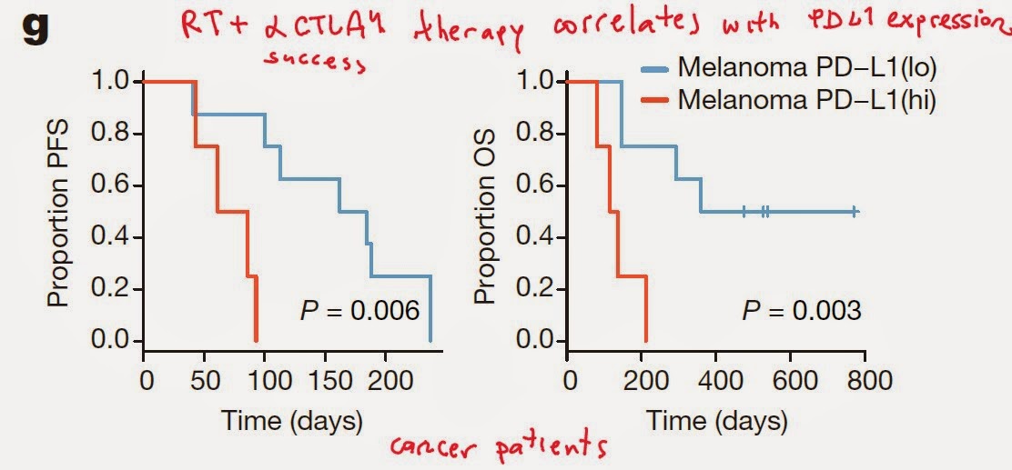
Identification and simultaneous targeting of such inhibitory and/or escape strategies will finally provide clearly measurable clinical benefits in majority of tumor patients.

David Usharauli