Showing posts with label T1D. Show all posts

Saturday, July 13, 2019

Pathological changes in the gut could initiate autoimmune diabetes microbiota-specific manner

The mouse strain, NOD, has been used to study the mechanism of type 1 diabetes (T1D). These mice spontaneously develop autoimmune diabetes though it is not clear how islet-specific T cells get activated. To track autoimmune T cell fate, TCR transgenic mice, BDC2.5XNOD, have been used. These mice harbor islet-specific TCR expressing T cells in high numbers that are easily monitored.

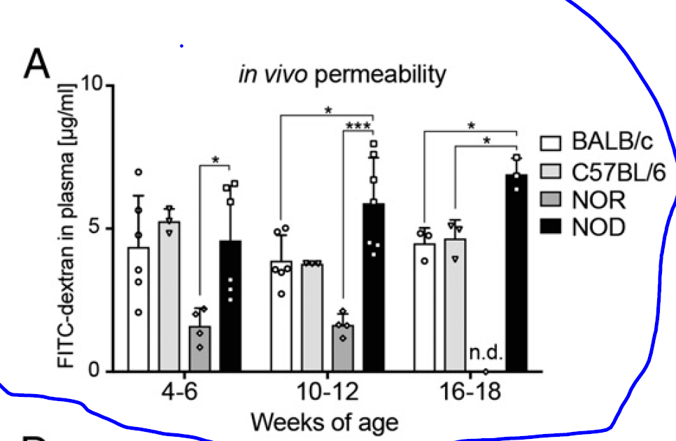
A new study in PNAS did some experiments on BDC2.5XNOD mice to understand the initiation of autoimmunity. First, they showed that the NOD background showed increased gut wall barrier permeability by measuring the FITC-dextran level in the blood after oral application (though they did not show the same permeability test for BDC2.5XNOD mice).


Interestingly, BDC2.5XNOD mice do not develop spontaneous autoimmune diabetes. However, oral application of low-dose dextran-sulfate sodium (DSS) activates T cells and initiates diabetes.


Diabetes, however, does not develop if DSS-treated BDC2.5XNOD mice are depleted of endogenous microbiota or if naive BDC2.5XNOD mice received DSS-modified microbiota.


In summary, this study showed that both gut inflammation and microbiota are necessary for the initiation of autoimmune diabetes in BDC2.5XNOD mice. The most likely scenario is that changes introduced by DSS allow endogenous microbiota to activate islet-specific T cells via cross-reactive antigens. DSS modifies both gut wall permeability and microbiota. Both of these phenomena have been observed by the authors. They conclude that "restoration of a healthy gut barrier through microbiota and diet modulation in diabetes-prone individuals could ultimately reduce intestinal activation of islet-reactive T cells and prevent T1D occurrence".

Others, in news/views for this article, even suggested using antibiotics to deplete endogenous microbiota, but in my opinion, this is a premature suggestion because the authors did not show that microbiota depletion after diabetes has already developed could stop it.

posted by David Usharauli


Tuesday, February 6, 2018

T1D target epitope from zinc transporter 8 (ZnT8) cross-reacts with commensal bacteria

Type 1 diabetes (T1D) is considered autoimmune disease. Of course, in humans, we don't have a direct evidence that islet-specific auto-reactive T cells and auto-antibodies found in peripheral blood from T1D patients are indeed responsible for tissue damage. Such evidence would require T/B cell and Ab depletion experiment that is not feasible. Autoimmune nature of T1D is basically extrapolated from mouse studies or in vitro antigen binding assays.   

Several islet antigens are known to represent targets in T1D, such as preproinsulin (PPI), glutamic acid decarboxylase (GAD), insulinoma-associated protein-2 (IA-2), islet-specific glucose-6-phosphatase catalytic subunit-related protein (IGRP) and more recently described zinc transporter 8 (its peptide ZnT8186–194).

New study in Science Immunology found that ZnT8186–194-specific CD8 T cells are largely similarly present in both T1D patients and healthy controls and that ZnT8186–194-specific CD8 T cells could recognize (cross-react) peptide derived from gut commensal microbe Bacteroides stercoris.

Staining with ZnT8186–194-specific HLA class I multimers (MMr) and other functional antigen-specific assays found that T1D and healthy controls harbored largely similar number and functional ZnT8186–194-specific CD8 T cells.


Interestingly, ZnT8186–194-specific CD8 T cells could be double stained with HLA class I multimer + peptide derived from B. stercoris, a commensal bacterial species found in gut flora. It is not the first time such cross-reactivity has been observed between islet-specific CD8 T cells and commensal bacteria. Previously, at least two bacterial species have been identified to cross-react with IGRP-specific CD8 T cells.


In summary, this study indicates that deletion of auto-reactive CD8 T cells in the thymus is not sufficient to prevent autoimmunity and that regulatory mechanisms operating in the periphery is necessary to prevent initiation of auto-reactive attack by circulating ZnT8186–194-specific CD8 T cells (probably Tregs the authors had in mind). Cross-reactive peptide derived from B. stercoris could play role in priming of ZnT8186–194-specific CD8 T cells in absence of regulatory circuit. But how and why such antigen-specific tolerance breakdown happens in one and not in another is not clear at this stage.

posted by David Usharauli


Tuesday, September 20, 2016

Fusobacteria, a gut commensal, contributes to autoimmune type I diabetes in mice

Initiation of autoimmune disease is still an immunological mystery. Some forms of autoimmune diseases are results of genuine genetic defects in signaling molecules within immune system. Other forms show strong linkage to certain HLA haplotypes that present antigenic epitopes. More recently scientists focused on the role of gut commensals in autoimmune diseases.

A new study in Jounral of Experimental Medicine showed that cross-reactivity at the epitope level between gut commensal Fusobacteria-derived magnesium transporter and β islet-specific glucose-6-phosphatase catalytic subunit–related protein (IGRP) contributed in autoimmune diabetes development in IGRP-specific CD8 T cell transgenic, CD8+ TCR NY8.3 NOD mice.

Initially, the authors observed that unlike MyD88KO NOD mice, MyD88KO CD8+ TCR NY8.3 transgenic NOD mice showed accelerated diabetes development (though unlike the authors, I don't find this surprising).


Interestingly, when co-housed with WT NOD mice, MyD88KO TCR NY8.3 transgenic NOD mice showed enhanced protection against diabetes, suggesting dominant role of fecal bacteria present in WT NOD mice in providing this protection.


Since it is known that NOD mice susceptibility to diabetes is commensal-dependent, the authors sequenced fecal microbiome in MyD88KO NY8.3 NOD mice to determine its composition. Not surprising, certain families of commensals underwent changes on MyD88KO background.


When the authors compared the IGRP206–214 peptide sequence against bacterial protein sequences in the nonredundant protein sequence database, they found several hits shared strong homology with IGRP206–214 peptid, the native autoantigen detected by NY8.3 CD8+ T cells. One such peptide, W15944, was derived from L. goodfellowii, a member of the phylum Fusobacteria (gram-negative anaerobe), a human and NOD mouse oral commensal.


Indeed, W15944 stimulated NY8.3 CD8+ T cells could transfer diabetes in NOD mice.


Finally, introduction of L. goodfellowii into WT NY8.3 NOD mice accelerated diabetes development, further suggesting role of L. goodfellowii in this process (while in general, this paper is of high quality, it lacks some of the crucial experiments such as, for example, (1) introduction of L. goodfellowii into NY8.3 NOD mice on germ-free background and (2) experiments with L. goodfellowii lacking cross-reactive W15944 peptide).


In summary, the author showed that in this artificially constructed NOD mice, IGRP-specific CD8 T cells could be activated by commensal-derived cross-reactive peptide and initiate autoimmune destruction of  β cells (it is remains to be seen why Foxp3+ regulatory T cells are incapable of preventing such T cell attack).

David Usharauli


Sunday, February 14, 2016

Hybrid peptides represent novel target for auto-reactive T cells in autoimmune diabetes


Initially, the authors showed that chemically cross-linked or synthetic peptide that is made one half of proinsulin C-peptide and another half from chromogranin A (ChgA) peptide (both β cell proteins), could activate diabetogenic mouse T cell clones.


Next, the authors showed that T cells specific for such hybrid peptides can be detected in un-manipulated diabetes susceptible NOD female mice (it would have been more informative if the authors have included data from non-susceptible WT mice as a control).


Finally, the authors showed in T cells from T1D individuals could also recognize hybrid peptide made of human proinsulin C-peptide and neuropeptide Y.

In summary, this study suggests that in T1D susceptible individuals, diabetogenic auto-antigens could be generated by fusion of peptide derived from two unrelated proteins (a side reaction of the proteolytic hydrolysis of peptide in secretory granules). Whether generation of such immunogenic hybrid peptides happens only T1D susceptible individuals remains to be determined.  

David Usharauli

Saturday, November 28, 2015

Autologous Foxp3+ polyTreg adoptive transfer immunotherapy: phase I study


Unlike CAR-T therapy with its bulk PBMCs as its main ex-vivo target population, working with flow sorted polyTregs cells is more demanding and few institutions are equipped to conduct such GMP-grade clinical trials. As shown in Table 3, there is variability in ex-vivo expansion potential between polyTregs cells derived from various donors [since no "healthy" control subjects were included in this trial, it is no clear whether this level of expansion of polyTregs cells is specific for T1D patients].


The authors conducted several functional/marker analysis on 14-day expanded polyTregs cells. These results revealed that expanded polyTregs cells retain the same degree of (a) demethylation at Foxp3 locus, (b) they showed improved phosphorylation of STAT5 in response to IL-2, (c) displayed improved suppressive potential in an in vitro proliferation assay.


After adoptive transfer of expanded polyTregs cells patients were monitored for any [metabolic] abnormalities. There were few adverse events related to T1D metabolic events that probably were not caused by polyTregs cell transfer per se. However, due to small sample size, adoptive transfer of polyTregs cells did not reveal any substantial improvement of T1D in recipients either [though transferred polyTregs cells labeled with [6,6-2H2]glucose were detected up to 1 year in cohort 3 and 4].

In summary, this phase I study of polyTregs cells confirms feasibility of adoptive transfer immunotherapy with Foxp3+ polyTregs cells. In general, it is my opinion that only by understanding Foxp3+ Tregs cells can we truly unlock the full benefits of immunotherapy for cancer, allergy and autoimmune diseases.

David Usharauli