Showing posts with label autoimmune disease. Show all posts

Tuesday, January 28, 2020

Microbiota-derived peptide and autoimmune heart inflammation: a tale of missing data

Papers published in journal Science supposed to undergo thorough high-level vetting process. However, to err is human. Both reviewers and editors are humans and hence they frequently err, for the annoyance of scientists and for the joy of postdocs doing journal clubs.  

Here is an example of a paper that squeezed through the cracks of the Science vetting process. It claims that peptides derived from certain commensal microbiota species cross-react with heart muscle protein, MYH6, causing autoimmune heart inflammation. It has a great Figure 1 showing that MYH6-specific TCR transgenic mice on a germ-free background, lacking microbiota, is protected from heart autoimmunity.


Furthermore, they showed that the re-introduction of microbiota into germ-free makes these mice susceptible to heart inflammation similar to microbiota+ mice.


The authors then tried to identify the microbiota species that contribute to this inflammatory condition. An in silico search identified cross-reactive β-galactosidase (β-gal) mimic peptides in Bacteroides thetaiotaomicron (B. theta) and B. faecis with high similarity to MYH6.


The authors even introduced into germ-free TCR transgenic mice Bacteroides thetaiotaomicron (B. theta) lacking the β-gal. Up to now, it feels that the authors have checked all the boxes necessary for high-quality research. But then for some reason, they do not show survival data comparing Bacteroides thetaiotaomicron (B. theta) with and without the β-gal gene as in figure 1. They just showed how a lack of β-gal Bacteroides thetaiotaomicron modifies MYH6-T cells accumulation in the heart tissue.


So, why the authors don't show survival data of germ-free MYH6 TCR transgenic mice colonized with Bacteroides thetaiotaomicron -/+ β-gal gene? Isn't it the most important result for their hypothesis? Where were reviewers and editors looking?

posted by David Usharauli


Thursday, September 20, 2018

Hypocretin-specific T cells mediate autoimmune sleeping disorder narcolepsy

Narcolepsy is a sleeping disorder characterized by "excessive daytime sleepiness, cataplexy, hypnagogic hallucinations and sleep paralysis". In recent years several studies provided evidence suggested that etiology of narcolepsy could be autoimmune in nature. Such conclusion has become more mainstream especially following observed relationship between 2009 pandemic flu vaccine, Pandemrix (from GSK), vaccination and development of sleeping disorder in a subset of vaccine recipients. In one study, it was found that Pandemrix but not Focetria flu vaccine (they differ in only one amino acid from each other) caused anti-flu antibody production that cross-reacted with hypocretin receptor.


Antigen-specific T cells are rare so to reveal their presence the authors first non-specifically amplify T cells from PBMCs and after considerable expansion tested on hypocretin peptide pool pulsed autologous B cells. Majority of individuals with narcolepsy showed reactivity in this assay.


Single cell analysis showed that hypocretin-specific T cells consist of several clones (up to 30 different clones).       


Interestingly, most of T cells specific for hypocretin were HLA-DR restricted, rather than HLA-DQ as earlier association studies would have predicted.


Also, notably, hypocretin-specific T cells did not reacted with autologous B cells (or monocytes) pulsed with hypocretin protein suggested that this in vitro pulsing assay could not recapitulate in vivo hypocretin protein processing pathway


Of note, the authors showed that hypocretin-specific T cells did not cross-react with pandemic flu peptide pool or vaccine (influvac). However, they did not use Pandemrix vaccine here so these results are not conclusive


In summary, this study showed that narcolepsy could indeed be a bona fide autoimmune disease. As in many other autoimmune diseases exact molecular events that initiate them are yet to be discovered.

posted by David Usharauli 


Tuesday, September 12, 2017

It is really hard to replicate human autoimmune diseases in mice

A new study in PNAS highlights the challenges scientists face when trying to reproduce human diseases in mice.  

Here, the authors tried to reproduce human type I diabetes by creating humanized mice by transplantation of HLA-DQ8+ human fetal thymus and CD34+ stem cells into immunodeficient mice (to recreate human immune system in mouse) followed by transfer of autologous [hu-mice]-derived HLA-DQ8/insulin-B:9–23 specific TCR transduced human CD4+ T cells and followed by two successive low doses of streptozotocin (a chemical to damage islet β cells and release auto-antigen).

However, even these steps were not enough to induce diabetes in hu-mice. Only immunization with insulin B:9–23 peptide + adjuvant (HLA class II-restricted T-cell response to InsB:9–23 peptide is highly associated with T1D in humans) in addition to above mentioned "conditioning" were able to induce diabetes in hu-mice.


No one really knows how autoimmune diseases are initiated in humans and these study shows that it is really hard to "reproduce" it in mice. Of course, it is not known what factors could play the role of "streptozotocin" and "B:9–23 peptide immunization" in natural context in humans. 

posted by David Usharauli

Saturday, June 24, 2017

α-synuclein in Parkinson’s disease could be a target of autoimmune attack

Parkinson's disease is a neurodegenerative movement disorder characterized by the death of dopaminergic neurons and accumulation of  intraneuronal aggregates known as Lewy bodies that are composed of α -synuclein (α -syn). 

New study in Nature suggests that T cells recognizing epitopes from α -synuclein could contribute to Parkinson's diseases pathogenesis, especially on DRB1* 15:01 and DRB5* 01:01 backgrounds.


Of course, in humans it is quite hard to prove any causation directly.

posted by David Usharauli


Tuesday, July 26, 2016

Infection-induced cell apoptosis activates self-epitope reactive T cells but barely

This week journal Nature Immunology published a study that had great title but poor data and conclusions. In fact, I rarely read such weak study for a long time, especially from top subject-matter journal. 

Basically, the authors tried to show that infection-induced cell apoptosis leads to self-peptide presentation and self-reactivity or even autoimmunity. Data are however misleading.

For this study the authors used the rodent pathogen Citrobacter rodentium that infects intestinal epithelial cells and induces their apoptosis. As a control, they have used infection with ΔEspF Citrobacter rodentium, a variant that lacks the secreted protein EPEC that mediates apoptosis. Initially, they showed that infection with WT Citrobacter rodentium, but not ΔEspF Citrobacter rodentium, induces Th17 response from large intestinal lamina propria (LI LP).


Next, the authors tried to examine whether infection-induced apoptotic cells will also provide self peptides for T cell activation (alongside of Citrobacter rodentium peptides). To do it, they have used so called double transgenic (DTg) mice derived from crossing OT-II mice with Act-mOVA mice. Now, these DTg mice delete absolute majority of OVA-specific OT-II cells in the thymus (from 1.5x10^6 to ~1,000 cells, i.e >1000X fold reduction of auto-reactive cells). The authors noted that DTg mice did not spontaneously develop autoimmunity and were healthy.


Next, when DTg mice were infected with Citrobacter rodentium, some portion of those OT-II cells left in DTg mice responded to it by up-regulating IL-17. The authors did not quantify the number of responding self-reactive OT-II cells and dot plot analysis reveals that their numbers seemed extremely low (on contour plot analysis). Moreover, it is not even clear whether self-reactive OT-II were responding to self-antigen or simply to inflammatory cytokine milieu [homeostatically] since even un-infected DTg mice showed proliferation and IL-17 expression in LI LP self-reactive OT-II cells.


The authors also showed that when infected with Citrobacter rodentium DTg mice showed little spike in anti-OVA IgA response driven by OT-II cells. However, it is not clear whether this anti-OVA IgA response has any pathogenic role.


Still, the authors believed that Th17 OT-II cells generated in DTg mice upon Citrobacter rodentium infection played pathogenic role in gut inflammation. As a "proof" they provided H&E staining of sections of large intestine from wild-type and DTg mice on day 40 after infection. Now, if scale bar on this H&E staining is 250 μm on both sections, then it is obvious DTg mice intestine is almost 2x more swollen or inflamed. But the authors noted that "DTg mice did not exhibit altered susceptibility to C. rodentium relative to that of wild-type or OT-II mice" and OT-II depletion did not significantly modify gut inflammation. So it is not clear from these data whether anti-OVA IgA or Th17 response after Citrobacter rodentium infection were in fact driving those observed pathogenic changes in the DTg mice guts (use of IL-17KO OT-II cells would have provided some guidance on this matter).


In summary, in my view this study only showed that WT Citrobacter rodentium infection induces little Th17 response from self-reactive T cells, however it failed to show that such Th17 response had any consequential effect.

David Usharauli  

Wednesday, June 15, 2016

Mutation in DNAse1L3 triggers systemic lupus erythematosus (SLE)-like condition in mice

Autoimmune disease, as word implies, is an immune response directed to self. Traditionally we used to think that autoimmune diseases arise as a result of failure of adaptive immune system (T and B cells) to distinguish between self and nonself antigens. However, as with many inherited immunodeficiencies, frequently we see that inherited innate genetic mutations play pivotal part in autoimmune phenotypes as well.  


DNAse1L3, a homologue to DNAse1, contains a short, positively charged C-terminal peptide that allows it to uniquely digest DNA chromatin in microparticles released from apoptotic cells.


Mice deficient for DNAse1L3 develop anti-dsDNA Ab response and Ab deposition in the kidney glomeruli [clinical feature of SLE in humans].


Unlike other genetic DNA/RNA housekeeping mutations (Trex1-/-, DNase-II-/-, RNase H2B-/- ) phenotype of DNAse1L3-KO mice was independent of STING activity (but was dependent on MyD88, though was not clear how or why).


In summary, This study revealed that DCs/Mac derived secreted DNAse1L3 is intimately involved in digestion of apoptotic microparticle associated DNA and in prevention of anti-DNA antibody formation (of note, one of the authors is a co-founder and consultant of Resolve Therapeutics, which develops soluble nucleases for therapeutic purposes).

David Usharauli


Thursday, February 18, 2016

pMHC tetramer-coated nanoparticles treat autoimmune conditions in mice

This week Nature published "figure-dense" paper showing that nanoparticles coated with auto-antigen+MHC complexes (pMHC) could treat spontaneous or experimentally-induced autoimmune diseases in mice. In fact, this study contains so many experimental results it should have been published in two part, in my view. I will try to condense its basic findings in few paragraphs and provide its interpretation below.

Non-obese diabetic (NOD) mice spontaneously develop [autoimmune] diabetes. Part of T cells that infiltrate and damage β cells in NOD mice express T-cell receptor (TCR) resembling diabetogenic BDC2.5-specific TCR that can be recognized by pMHC–2.5mi/IAg7 tetramers. When the authors injected nanoparticles (dextran-coated or pegylated iron oxide NPs) coated with 2.5mi/IAg7 tetramers (pMHC–NP) it induced expansion of cognate CD4+ T cells in blood and spleens of NOD mice. These cells had a memory-like (CD44hiCD62Llow) FOXP3− TR1-like phenotype.


These "activated/experienced" CD4+ TR1 cells from donor mice treated with pMHC–2.5mi/IAg7-NPs suppressed diabetes development in T-cell-reconstituted NOD-scid hosts upon adoptive transfer. This effect of TR1 cells was augmented by treating hosts with pMHC–NPs.


Moreover, 90–100% of the already diabetic mice that received nanoparticles coated with β cell antigens, 2.5mi/IAg7, IGRP4–22/IAg7 or IGRP128–145/IAg7, reverted to stable normoglycaemia.


Of note, treatment withdrawal resulted in loss of the normoglycaemic state in 25–60% of mice, in association with the loss of the tetramer+CD4+ T-cell pools. In other words, these data suggest that almost half of treated diabetic mice maintained normoglycaemic state after 5 weeks of treatment.


Beyond diabetes, the authors showed that nanoparticles coated with myelin oligodendrocyte glycoprotein, pMOG38–49/IAb–NP, dampended progression of experimental autoimmune encephalomyelitis (EAE, a model of multiple sclerosis), when given on day 14 after immunization and even restored motor function in paralytic mice when given on day 21.


Similar therapeutic effects were seen in 3rd autoimmune disease model, collagen-induced arthritis (CIA). HLA-DR4-IE-transgenic mice receiving nanoparticles displaying mouse collagen (mCII)259–273/DR4-IE showed reduced joint inflammation. In all these models, pMHC–NPs effect was antigen-specific.

Indeed, the effects of pMHC–NP therapy were not associated with impaired systemic immunity because pMHC–NP-treated mice showed unimpaired anti-viral immunity and mounted antibodies against an exogenous antigen as efficiently as control mice.

Interestingly, pMHC–NPs could not expand tetramer+ T cells in non-diabetic control mice which harbor mostly naive T cells. Further experiments confirmed that therapeutic effect of pMHC–NP required presence of "experienced" T cells. For example, whereas pMHC–NP therapy afforded 100% diabetes protection to T-cell-reconstituted NOD-scid hosts bearing memory BDC2.5 T cells, such therapy was inconsequential in hosts receiving naive BDC2.5 T cells.


The role of "experienced", but not naive T cells, in therapeutic effect of pMHC–NP was also supported by observation that whereas diabetic NOD G6pc2−/− mice (which lack IGRP antigen and corresponding memory/experienced T cells, but most likely harbor IGRP4–22 specific [aka, nonself-specific] naive T cells) responded to 2.5mi/IAg7–NPs like wild-type NOD mice, they did not respond to IGRP4–22/IAg7–NPs.


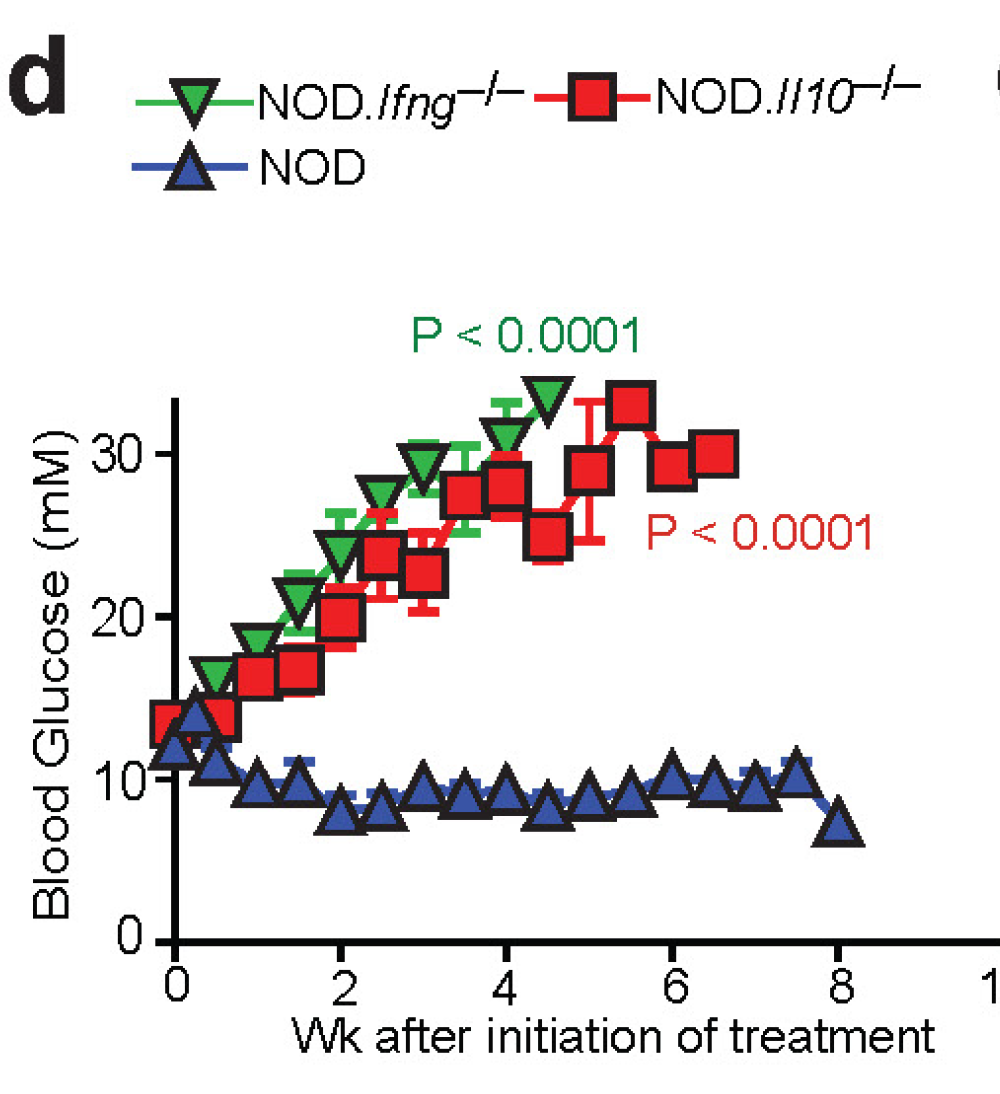
At molecular level, the authors showed blockade of IL-10, TGF-β and IL-21R (but not IFNγ) abrogated the anti-diabetogenic properties of 2.5mi/IAg7–NPs or IGRP4–22/IAg7–NPs in diabetic NOD mice.

However, to make things more complicated, the authors also showed that the development of the TR1 precursors and/ or TR1-like cells that expand in response to pMHC–NP therapy required IFNγ in addition to IL-10 in pre-diabetic NOD mice.

Finally, the authors found that at the cellular level, pMHC–NP treated NOD mice harbored increased numbers of so called regulatory B cells, BREG cells (but only in pancreatic lymph nodes, PLNs). These BREG cells were producing IL-10 in response to lipopolysaccharide (LPS). In vivo, these B cells suppressed diabetes development in T-cell-reconstituted NOD-scid hosts as compared to PLN B cells from control mice (and worked together with TR1 cells). The role of BREG cells was supported by observation that treatment of newly diabetic NOD mice with a B-cell depleting anti-CD20 mAb abrogated the anti-diabetogenic activity of 2.5mi/IAg7–NPs (see above).


In summary, this study suggests that when injected into mice these pMHC–nanoparticles expand antigen-specific and antigen-experienced T cells with regulatory properties. These TR1 work with BREG cells to block the chain of events that perpetuates autoimmune cycle. 

David Usharauli


Saturday, September 19, 2015

CTLA-4 deletion during adulthood leads to a paradoxical resistance to autoimmune disease

CTLA-4 is a T cell inhibitory molecule that competes with CD28 for binding CD80/CD86 co-stimulatory molecules on dendritic cells. It is believed that CTLA-4 functions as a "brake" for T cell activation, a function also referred as a "checkpoint" inhibition. Total genetic deficiency of CTLA-4 or just on Foxp3+ regulatory T cells leads to early onset wasting syndrome and fatal autoimmune diseases. This what we knew up to now. 

However, a new study in Journal of Experimental Medicine, has put upside down the whole concept of "checkpoint" inhibition for CTLA-4. Using genetically-modified mouse models, the authors reported that total or Foxp3+ regulatory T cell-specific conditional deficiency of CTLA-4 in adult mice leads to protection rather than acceleration of autoimmune disease.   

First, the authors have generated mouse model that allowed conditional deletion of total or Foxp3+ Treg-specific CTLA4 (UBCCre/ERT2+ Ctla4fl/fl and Foxp3eGFP/Cre/ERT2+ Ctla4fl/fl).


Next, the authors treated mice with tamoxifen to activate Cre recombinase and delete CTLA4 and challenged mice with myelin self-peptide to induce experimental autoimmune encephalitis (EAE). Surprisingly and contrary to widely held belief, adult mice conditionally deficient for CTLA-4 were highly resistance for EAE induction.


Similar resistance for EAE induction was observed with mice with adult-onset CTLA4 deletion specifically on Foxp3+ regulatory T cells.


To rule out any off-target effect of CTLA-4 deletion, the authors transferred T cells from UBCCre/ERT2+ Ctla4fl/fl into T-deficient hosts and then challenged with tamoxifen and myelin self-peptide. Here too, deletion of CTLA4 on mature T cells protected against EAE. Moreover, adoptive transfer of purified, naive Foxp3- T cells from UBCCre/ERT2+ Ctla4fl/fl mice or transgenic 2D2 T cells specific for myelin self-peptide on UBCCre/ERT2+ Ctla4fl/fl background were capable of inducing EAE, implying that T cells were not inherently incapable of inducing EAE but were actively suppressed by absence of CTLA-4 on Foxp3+ regulatory T cells.


Additional experiments revealed that CTLA-4 deletion in adulthood skewed T cells response towards IL-10 production in UBCCre/ERT2+ Ctla4fl/fl mice.


Finally, the authors showed that CTLA-4 deletion during adulthood did not modify anti-tumor response against MC38 colon adenocarcinoma cells in UBCCre/ERT2+ Ctla4fl/fl mice.


In summary, these results question the assumption that inhibition of CTLA-4 function during adulthood would lead to autoimmune disease. Based on these results, it is unclear how anti-CTLA4 antibody therapy provides benefits during tumor immunotherapy. It maybe the difference between mouse and human immune system or it could be that mechanism of action of anti-CTLA4 antibody is not a "checkpoint inhibition" after all.

David Usharauli
Wednesday, January 21, 2015

T cell-specific Smad4 controls the lethal autoimmunity

I was very excited when I saw this title in journal Immunity and then utterly disappointed after reading the actual paper.

The only noteworthy result from this paper is depicted in Figure 1A. The authors showed that compared to mice single deficient for T cell-specific TGF-beta RII signaling (RII-KO), mice double deficient for T cell-specific TGF-beta RII and Smad4 signaling (RII-S4 DKO) were protected from lethal autoimmunity.


Bone marrow (BM) chimera experiments revealed that unlike RII-KO BM cells, RII-S4 DKO BM cells could generate Foxp3+ Tregs in vivo (though 2-fold less compared to WT BM cells).  


Strangely, however, in vitro experiments showed that RII-S4 DKO T cells were dramatically deficient in generating Foxp3+ Tregs (though they efficiently differentiated into TH1 or TH2 phenotypes).


Finally, the authors showed that mice single deficient for Smad4 (S4-KO) were not able to efficiently reject OVA-expressing tumor (no similar data for RII-S4 DKO mice were produced).


In summary, in my opinion, these results were not ready for publication in Immunity. The authors failed to follow their best Figure 1A result and provide any convincing argument that could explain their observation. It is clear that T cells deficient in Smad4 failed to accumulate in normal numbers in vivo upon antigen encounter (memory failure?), but the fact that Smad4-KO T cells could efficiently differentiate into effector T cells makes all these results confusing. Also, it is not clear whether absence of lethal autoimmunity in RII-S4 DKO mice has to do with the presence of normal number of fully functioning Foxp3 T cells.

David Usharauli


Thursday, December 25, 2014

Copying natural mutations to study autoimmunity

Autoimmune disease is a condition when body's immune system attacks its own tissue. Very little is known how autoimmunity is initiated or maintained. Frequently, we do not even know what are the (self)-antigens that are targeted by self-reactive T cells or auto-antibodies.  

The new paper in journal Science describes an one way how to identify the antigens targeted in autoimmunity. This study, led by Shimon Sakagushi at the Institute for Frontier Medical Sciences, Kyoto University, used SKG mice that has natural mutation in a T cell receptor signaling molecule, called ZAP-70. This mutation alters T cell selection in the thymus and allows peripheral release of self-specific T cells.

Original studies on SKG mice showed that they spontaneously develop arthritis mimicking several hallmarks of human rheumatoid arthritis (RA).  

Here, initial experiments showed that SKG T cells expressing different Vbeta chains could induce arthritis equally well when transferred into RAG-KO hosts, implying the poly-clonal nature of self-reactive T cells in SKG mice.   

The authors focused on one particular TCR family, Valpha2/Vbeta6 that constitute ~ 1% of joint infiltrating T cells. They cloned several Valpha2/Vbeta6 TCR pairs obtained from SKG arthritic joints, transfected them into RAG2-KO/SKG BM cells and transferred these transfected BM cells into RAG2-KO to generate monoclonal, retrogenic mice.

Out of these retrogenic SKG mice, R7-39, but not R1-23, spontaneously developed arthritis and skin inflammation. As expected, 7-39 TCR on wild-type background, W7-39, (without ZAP70 mutation) did not carry arthritogenic potential, probably due to inactivation in the thymus (and neither 7-39 nor 1-23 TCR develop into Foxp3+ T cells).


To examine the specificity of antigen(s) recognized by R7-39 T cells, the authors co-transferred 7-39 TCR transfected RAG2KO-SKG BM cells together with TCRbeta KO BM cells into RAG-KO hosts. In this setting, B cells developed from TCRbeta KO BM cells will secrete antibody specific to antigens recognized by 7-39 TCR, since only this type of help is available to B cells. Indeed, sera obtained from this mice, "R7-39+B", reacted to protein, RPL23A, identified by mass spectrometric analysis as a component of 60S subunit of ribosomes.

As expected, T cells from R7-39 mice, but not from R1-23, produced inflammatory cytokines in response to recombinant RPL23A.


Finally, the authors showed that sera from RA patients too showed reactivity to RPL23A.


In summary, these results shows one way to study mechanisms of autoimmunity. 

Two questions come to mind: it is very strange that SKG mice develop only limited set of autoimmune diseases (joint and skin inflammations), since ZAP70 signaling should affect every T cells. Why is that? Also, RPL23A is a widely expressed protein. So why are target tissues limited to joints and skin in SKG mice? Does it mean that level of expression determines clinical signs? or maybe microbiota play a role?

David Usharauli