Showing posts with label gut microbiota. Show all posts

Thursday, March 21, 2019

A physiological T cells' "weaning reaction" to microbiota and solid foods in newborn pups requires FOXP3+ Tregs


Sunday, October 14, 2018

New auto[cross-reactive]-antigen, GDP-l-fucose synthase, identified in MS patients

Multiple sclerosis (MS) is a debilitating autoimmune disease affecting nervous system. Several auto-antigens have been traditionally suspected for MS pathology, mostly derived from neural tissues such myelin. However, more recently the list of relevant auto-antigens expanded to include such member as RAS guanyl releasing protein (RASGRP1–4) family. Similarly, a new paper in Science Translational Medicine presented evidence that yet another auto-antigen, GDP-l-fucose synthase, is a target antigen in a subset of MS patients.

The authors has used modified version of positional scanning epitope library to identify epitope derived from auto-antigen, GDP-L-fucose synthase, as a target epitope for CD4+ T cell clone, TCC21.1, derived from MS patients with DR15 haplotype. Peptides sequences matching GDP-l-fucose synthase was detected in brain tissue.


MS patients whose T cells displayed high response to peptides from GDP-L-fucose synthase in stimulation assay showed high responses to myelin proteins as well.


Curiously, almost all high responders carried DRB3*02:02 allele. 


Finally, the authors speculated that sequence similarity between human and microbiota-derived evolutionary conserved GDP-L-fucose synthase could be a factor that initiates MS pathology in these patients.


In summary, this study has a lot of nice data that support idea that GDP-L-fucose synthase is a  new auto-antigen that could be relevant in molecular diagnosis of MS pathology.

There are several questions about this study. GDP-L-fucose synthase expression is not restricted to brain tissue. So, it should be relevant to understand if other tissues were affected in those MS patients. Second, cross-reactivity per se is not sufficient to explain how and why autoimmune responses are being initiated. Microbiota expressing GDP-L-fucose synthase most likely reside in patients long before MS, maybe even since birth, and T cells are tolerant to them. So, what has to be changed, in a antigen-specific manner, to make T cells less tolerant to initiate specific autoimmune attack on nervous system and not a total autoimmunity targeting all available auto-antigens? 

posted by David Usharauli

Monday, February 29, 2016

Failure to tolerate gut microbiota transforms immunodeficiency into immunopathology

Here is a paradox for you that many don't even know that it exists: frequently, genetic immunodeficiency (weak immune response) syndromes are associated with immunopathologies (excessive immune response). So how could it be explained?   

New study published in Journal of Experimental Medicine may have some new answers. This study described mouse model of human immunodeficiency syndrome called, Omenn syndrome (after its discoverer) and showed that immunopathology was driven by gut microbiota.

Omenn syndrome is caused by hypomorphic (low active) RAG mutations. Analysis of intestinal tissue from Rag2R229Q [Omenn] mice revealed pathological infiltration with inflammatory T cell subsets, TH17 and TH1.


Adoptive transfer showed that intestinal immunopathology was mediated by Rag2R229Q mutant T cells.


Interestingly, Rag2R229Q mouse harbored comparable numbers of Foxp3+ CD4+ T cells, implying that simple presence of regulatory T cells [generated in Rag2R229Q mice] was not enough to control tissue inflammation.

Defect of Tregs derived from Rag2R229Q mice was confirmed in adoptive transfer experiment with WT Tregs.


Besides T cell-driven immunopathology, Rag2R229Q mice displayed IgA deficiency (failure to properly coat gut microflora). These data pointed to the possibility of microbial translocation causing excessive inflammatory response.


Indeed, antibiotic treatment of Rag2R229Q mice could reduce intestinal immunopathology.


The role of antibiotic-sensitive gut flora in driving immunopathology in Rag2R229Q mice was confirmed in adoptive fecal transfer experiments.


In summary, this study suggests the following scenario: hypomorphic RAG defect in Rag2R229Q mice leads to "narrowing" of TCR and BCR repertoire. This in turn leads to outgrowth of oligoclonal T and B cells in Rag2R229Q mice (wherein Rag2R229Q mice contain T and B cells with limited, restricted, deficient TCR and BCR repertoires). Without proper TCR and BCR repertoire diversity, however, Rag2R229Q mice fails to develop tolerance (IgA and Tregs) to gut flora or commensal microbial antigens present at mucosal surfaces (such as lung, intestine). Repertoire restriction also leads to failure to mount adequate and proper immune response.


David Usharauli

Tuesday, November 17, 2015

Microbiota connects type II immune system to lean body metabolism

There is a renewed interest in gut microbiota research. Initially, this revival of microbiota studies came from observations that obese and lean people have different gut microbiota. It is not clear how exactly microbiota modulates energy metabolism and the research is ongoing in this direction.

For example, journal Nature Medicine has just published another paper where the authors showed that microbiota modulates energy metabolism through its action on a specialized adipose tissue called brown [beige] adipose tissue.

A peculiar aspect of this study [and main reason why it was published in this journal in the first place] has to do with sophisticated methodologies the authors used to study energy metabolism in mice (such as glucose uptake assays with 2-[14C]-deoxyglucose, [18F]fluorodeoxyglucose, 2-[1-3H]deoxyglucose, micro-PET-CT, etc).

Basically, the authors showed that when mice are treated with broad spectrum antibiotics, inguinal subcutaneous and perigonadal visceral adipocytes undergo modification (browning and cell size reduction) resembling adipose tissue in Germ-free mice.

Antibiotic treatment led to the increase in Ucp-1cells in inguinal adipose tissue confirming "browning" of adipose tissue.


In addition, the authors observed "browning" of inguinal and perigonadal adipose tissues in antibiotic treated mice even at thermoneutral conditions (at 30℃ for mice).


Next, the authors showed that microbiota depletion improved "lean body" metabolism both in obese-prone [ob/ob] and high-fat diet fed [HFD] mice.


Finally, the authors showed that antibiotic treatment was associated with type II cytokine profile in "browning" inguinal adipose tissue, including presence of tyrosine hydroxylase (TH) expressing M2 MΦ.


In summary, these results [re]confirmed that microbiota played an important role in "lean body" metabolism associated with development of "brown" adipose tissue. Interestingly, two pathways known for brown adipose tissue development [exposure to low temperature and microbiota depletion] required type II immune system. Type II immune system is mostly known for its involvement in allergy and more recently tyrosine hydroxylase (TH) expressing M2 MΦ has been implicated in neuro-inflammation. It remains to be discovered whether "lean body" metabolism and allergy are interconnected or whether they represent two independent outcome of type II immunity 😕

David Usharauli

Tuesday, November 10, 2015

Gut commensal Bifidobacterium promotes anti-cancer efficacy of anti-PD-L1 immunotherapy. Part II


Now this paper starts with experimental results that became classical experiments in gut immunology [since the discovery of TH17 subset]. Namely, the authors showed that B6 black mouse colonies derived from JAX lab or Taconic showed different susceptibility to cancer growth.


This difference between JAX and Taconic mice disappeared when mice were co-housed, implying the role of gut microflora


This hypothesis was confirmed in fecal cross-feeding experiments between JAX and Tac mice. It turned out that gut microbiota from JAX mice could enhance anti-tumor immunity when transplanted into Tac mice [but not vise versa].


Furthermore, JAX mouse microbiota could enhance anti-cancer effectiveness of anti-PD-L1 immunotherapy.

Finally, the authors found that a single commensal species, called Bifidobacterium, was largely responsible for microbiota's effect on cancer immunity.


In summary, the authors proposed that live Bifidobacterium could enhance anti-cancer immunity by supporting "generic" dendritic cell maturation and improving T cells antigen-sensitivity


This model, however, lacks data that could meaningfully explain why and how only Bifidobacterium has such influence on anti-cancer immunity. Also, it would have been more relevant to test FDA approved anti-PD1 antibody here rather than not-yet-approved anti-PD-L1 antibody therapy. 

David Usharauli  

Monday, November 9, 2015

Gut microbes fuel effectiveness of anti-CTLA-4 cancer immunotherapy. Part I

Recently two back-to-back papers in Science Express received much media attention. These studies claim that gut microbial flora influence clinical effectiveness of checkpoint inhibitors approved for cancer immunotherapy. I am going to review and provide my analysis of these studies.

First paper I will review came from European team led by Laurence Zitvogel. Of note, her team had another tumor immunology paper just 1 week ago in Science. First notable thing about her publications is the number of names included as authors. Her lab had done this since I first heard about her studies in 2007. Either she is a good collaborator or everyone in her lab is get credit for everyone else's work as a matter of right [definitely good for career].


For example, they showed that inhibition of cancer growth by anti-CTLA-4 antibody injected in germ-free or antibiotic-treated mice was not as good as in control WT mice


Analysis of gut flora revealed that presence of specific set of microbes [B. fragilis, for example] were required to show anti-cancer effectiveness of anti-CTLA-4 antibody.


Now these result are quite preliminary and it is not clear how microbes are enhancing anti-CTLA-4 effectiveness. For example, some microbes can live within growing tumor tissue and anti-CTLA-4 therapy simply unleashes immune response against them destroying tumor tissue in the process. There is a possibility that microbes support presence of cross-reactive T cells required to target tumor antigens when they are unleashed by anti-CTLA-4 immunotherapy. In all, based on this paper we can't say how microbes help anti-CTLA-4 and how much this "help" is meaningful in clinical settings. Maybe second paper will provide some ideas in this regard. Stay tuned for next review. 

David Usharauli   

Friday, September 18, 2015

Gut microbiota controls clinical severity of sickle-cell disease via neutrophil "ageing"

Our knowledge of the role of gut microbiome in human health and disease is expanding on a daily basis.

For example, just few days ago journal Nature published a study where the authors showed that in mouse model of sickle-cell disease (Hba-/- Hbb -/-) the presence gut microbiome influenced clinical severity via neutrophil "ageing".      

In sickle-cell disease, neutrophils expressing Mac-1 capture sickle red blood cells that leads to vaso-occlusion and tissue damage. It appears that "ageing", or mature neutrophils, defined as CD62Llow CXCR4high population, express more Mac-1 and those neutrophils can capture more RBCs per cell basis (that contributes to development of sickle-cell disease symptoms). [the authors does not discuss the difference between "ageing", "senescence" and "mature" neutrophils].


Next, the authors showed that germ-free mice or antibiotic treated mice have reduced number of "ageing" neutrophils.


Using BM chimera, the authors found that absence of MyD88 adaptor molecule or TLR4 receptor also mimicked the effect of antibiotic treatment on neutrophil "ageing".


Finally, the authors found that antibiotic treatment of sickle-cell disease model mice reduced number of circulating "ageing" Mac-1 expressing neutrophils and showed reduction in tissue damage.


In summary, this study proposed a novel therapeutic path for treatment for sickle-cell disease by modulation of gut microbiota and neutrophil maturation ("ageing").

David Usharauli


Friday, August 14, 2015

Endogenous gut flora drives colonic RORγ+ Treg development

Foxp3 transcription factor drives development of specialized CD4 T cell subset called regulatory T cells (Tregs) which can protect against immune pathology. 20 years has passed since their re-discovery in 1995 and 10 years has passed since identification of Foxp3 transcription factor as a master regulator of Tregs induction. Still, there is not a single drug or cell therapy protocol approved that involves Tregs. Why? In my opinion it has to do with the fact that we don't how Tregs work.    

Here is another example published yesterday in journal Science. In this article, the authors led by Mathis-Benoist team have reported that colonic gut microbiota induced RORγ, a transcription factor commonly associated with Th17 cell development, in colonic Foxp3+ Tregs and its expression played a beneficial role in maintaining colonic tissue health.  

First, the authors showed that colonic but not spleen Foxp3+ T regs expressed RORγ transcription factor.
However, unlike RORγ+ inflammatory Th17 cells, RORγ+ Foxp3+ Tregs do not express IL-17.


Next, the authors showed that induction of colonic RORγ+ Foxp3+ Tregs was linked to multiple gut microbiota species (of note, the authors did not find any correlation between SCFA expression and RORγ+ Foxp3+ Tregs).


Finally, the authors showed that specific deletion of RORγ in colonic Foxp3+ T regs tipped the balance in favor of IL-17 and IFN-γ and worsened gut inflammation.


In summary, these results indicate that antagonistic transcription factors RORγ and Foxp3 co-expressed and are indeed necessary for proper functioning of tissue specific Tregs. This even more complicates already messy field.

David Usharauli


Friday, July 31, 2015

Cross-reactivity to gut microbiota could explain failure of HIV vaccine

More than 30 years since its discovery and there is still no FDA licensed HIV vaccine. It is not even entirely clear if the failure to develop effective HIV vaccine has really anything to do with frequent HIV antigenic shift. Flu virus, for example, also undergo quite frequent antigenic shift and still there is Flu vaccines with 60-90% efficacy.   


The authors have analysed Ab repertoire to HIV-1 DNA prime, recombinant Adenovirus Type 5 (rAd5) boost vaccine. They found that 93% of Env specific antibodies derived from sorted memory B cells were directed against non-neutralizing gp41 antigen


Interestingly, majority of those gp41-specific antibodies utilized polyreactive, innate-like IGHV1-69 variable segment. VH1-69 locus is involved in Ab repertoire directed to Flu stem region. But unlike Flu specific VH1-69 Abs, gp41-specific VH1-69 Abs were made of allele variants with Leucine substitution at position 54 in HCDR2.


Finally, analysis of antigen specificity of individual gp41 mAbs revealed high level of polyreactivity towards commensal and self antigens.

In summary, these results points to the most important aspect of immune system, namely that strength and specificity of immune response is controlled by environmental antigens, including microflora antigens. It is my opinion that future vaccine testing would require incorporation of cross-reactivity tests against wide range of environmental antigens to select the most effective immunogens.  

David Usharauli


Tuesday, February 17, 2015

microbiota influences non-genomic hereditary immune phenotype

The major advances in fundamental immunology for the past 25 years have to do with major advances in artificial manipulations of gene expressions (knock-out, knock-in, conditional knock-out, CRISPR-Cas9, etc).

Until recently very little attention was given to the extra-genomic influence of such gene manipulation. The scientists rarely bothered and still many don't to study gene effects in homozygous offspring derived from heterozygous parents to balance for exrta-genomic influence

For example, it is conceivable that homozygous gene effect are influenced wholly or in part by extra-genomic factors such as microbiota. This is exactly what a new study in journal Nature suggests.

In this study, the authors made initial observation that two colonies of genetically identical B6 mice kept in two different research facilities differed in their gut derived fecal IgA level (IgAhigh and IgAlow). Interestingly two colonies did not differ in serum IgA level. The authors found that fecal IgA phenotype was vertically transmittable from parents to progeny.


Surprisingly, co-housing experiments revealed that IgAlow phenotype was dominant. This dominance of IgAlow phenotype was reversed when fecal materials were passed through 0.45 micron filter before transplantation into IgAhigh hosts.


In addition, the authors found that pre-treatment of IgAlow mice with broad-spectrum antibiotics or ampicillin could reverse IgAlow phenotype as well, indicating that presence of ampicillin sensitive large microbes were responsible for this microbiota effect.


The authors found that IgAlow mice intestine were more sensitive to chemical injury compared to IgAhigh mice.


However, intestinal sensitivity to DSS were independent of microbiota and dependent of intestinal IgA secretion.

To understand the mechanism behind IgAlow phenotype the authors tested components of fecal IgA complexes. These experiments revealed that IgAlow mice had profound deficiency in secretory component of IgA complexes in fecal samples. Secretory component is known to protect IgA from degradation.  
 

Finally, the authors showed that microbial pellets derived from IgAlow mice could degrade secretory component in a protease-dependent manner (use of germ-free host mice would have been beneficial to know exactly what microbe was responsible for this effect).


In summary, these results showed that non-genomic microbiota could influence hereditary immune phenotype. When studying gene-manipulated mice models, it is of critical importance to evaluate the model in proper manner (fecal transplantation, co-housing or heterozygous parents).

David Usharauli


Tuesday, January 27, 2015

Orphan interferon-lambda vetoes commensal microbes backdoor dealings

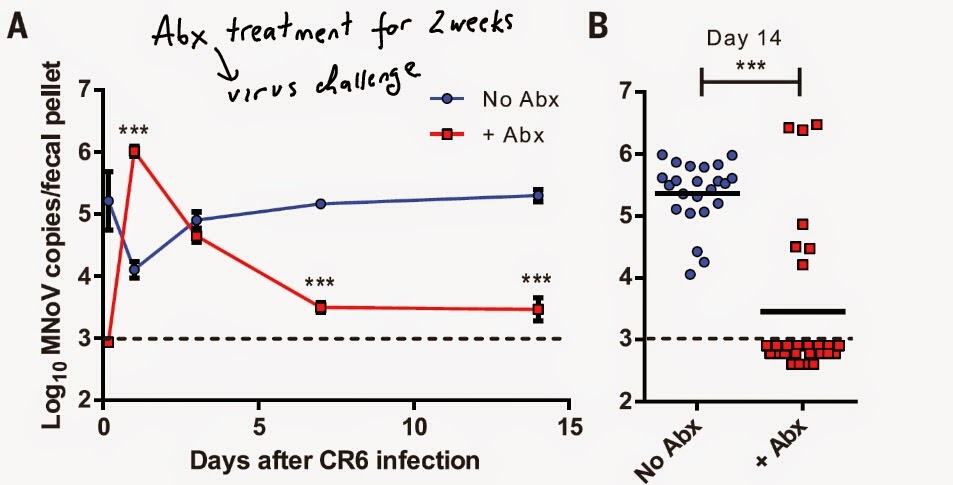
This is a second paper in IFN-lambda series. This new paper published in journal Science provided additional confirmation to the earlier study suggesting the role of commensal microbes in norovirus infection


This resistance to intestine norovirus infection was reversed by normal fecal supplementation implying the role of gut microbes in supporting norovirus infection.


The authors showed that resistance to chronic norovirus infection upon antibiotic treatment were dependent on viral dose and IFN-lambda signaling.


These data suggest that murine norovirus employs endogenous gut microbial flora to establish chronic intestinal infection. It is not clear how microbiota alters IFN-lambda signaling to permit norovirus persistence in the gut.

David Usharauli 

Tuesday, January 13, 2015

Friendly gut microbe defends against Vibrio cholerae infection

This is a very interesting study from journal Nature. It comes from the famous Jeffrey Gordon's lab at the Washington University School of Medicine, St. Louis. 

Usually when we think about the infectious microbes and how to deal with them, we are usually considering antibiotics or vaccination. However, in nature everything is interconnected and mutually dependent, even if any such evolutionary relationships are not immediately obvious. 

In nature, if one considers microbial communities, different species can sense each others presence and can influence each others biology. The most widely described such phenomenon is called quorum sensing wherein microbial community can sense its members density and influence and self-adjust its own or others replication kinetics. There are intra-species and inter-species mechanisms of quorum sensing.


The authors has collected and analysed gut microbiota community from stool (fecal) samples from healthy or Vibrio cholerae-infected patients (collection time ranged from diarrhoeal phase to recovery phase). This analysis showed that abundance of 14 gut microbial species correlated with the recovery from Vibrio cholerae dierrhoeal phase.

Using germ-free mice recolonization strategy the authors found that one of the gut microbial species in particular, Ruminococcus Obeum (R. Obeum), restricted Vibrio cholerae colonization in gnotobiotic mice. 


The authors found that in presence of Vibrio cholerae infection, R. Obeum up-regulated LuxS gene responsible for the inter-species mechanism of quorum sensing through autoinducer-2 (AI-2) synthesis. 


To directly test R. obeum LuxS involvement in restricting expression of virulence factors in V. cholerae, the authors cloned R. obeums LuxS (or V. cholerae LuxS, as a control) into mutant E. coli deficient its own AI-2. Both in vitro and in vivo experiments showed that in presence of R. obeum-derived LuxS gene in mutant E.coli, V. cholerae's replication and expression of virulence factors were reduced during co-culture or co-colonization.     


Finally, the authors found that restriction of V. cholerae infection was mediated through a new mechanism involving quorum sensing transcriptional regulator VqmA in V. cholerae.  


In summary, the results highlight the new mode of treating infectious agents with the help our own gut microbial friends.

David Usharauli