Showing posts with label peripheral tolerance. Show all posts

Wednesday, February 22, 2017

Antigen-specific Tregs maintain immune privileges of male reproductive tract

This week Journal of Clinical Investigation published very good research article that shed light on how tolerance to sequestered self-antigens expressed by immune privileged  tissues are established

Some tissues such as brain, eye, testis or ovaries are thought to be "immune privileged" organs meaning that ordinarily immune system does not see their antigens. However, in this paper, the authors showed that in fact some testes antigens in male mice, for example lactate dehydrogenase 3 (LDH3), are actually secreted and detected by immune system.   

Series of experiments confirmed that WT male mice did not respond to LDH3 immunization, while female and LDH3-null male mice mount detectable immune response to it. This suggested that male mice were physiologically tolerant to LDH3. Notable, both male and female mice could be immunized against another testis antigen, zonadhesin (ZAN), implying absence of tolerance to ZAN in male mice. 


So, how male mice were tolerant to LDH3? To answer it, the authors temporally depleted Tregs and it led to immune response to LDH3 in immunized WT male mice. since no changes were seen in response to ZAN, the authors concluded that tolerance to LDH3 in WT male mice was dependent of presence of FOXP3+ Tregs.


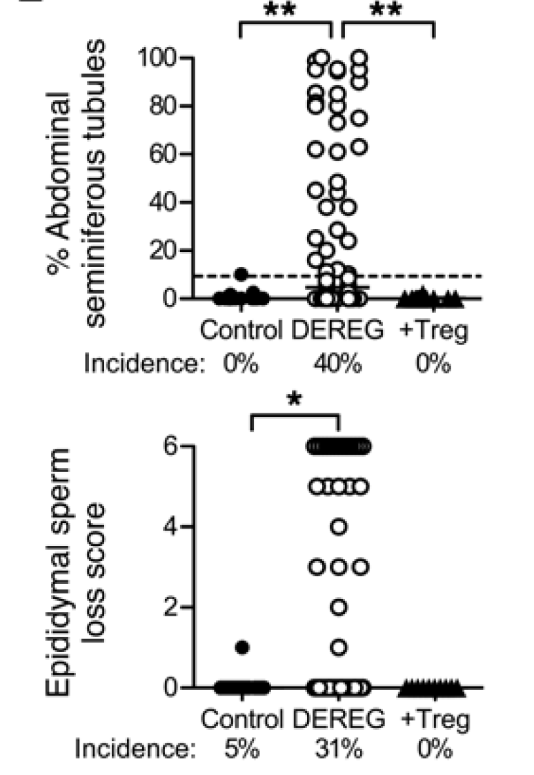
Furthermore, depletion of Tregs even in absence of testes antigen immunization still led to autoimmune pathology in testes in ~40% of male mice ("autoimmune orchitis occurs in autoimmune polyendocrine syndrome 1 (APS1) patients due to mutations of AIRE, possibly associated with impaired thymic deletion of autoreactive T cells and deficient Treg function")


In summary, this study suggests that "immune privilege" is not absolute and self-antigens that naturally leak maintain tolerance by induction of antigen-specific Tregs.

David Usharauli


Monday, January 4, 2016

Thymic epitope expression determines pattern of CD4 T cell peripheral tolerance

A few months ago I posted my analysis of one of the important papers from journal of Immunity discussing mechanisms of CD4 T cell peripheral tolerance. In that paper, the authors led by J.J. Moon, showed that number and functionality of CRE recombinase-specific endogenous CD4 T cells were determined by antigen expression in the thymus

Today, journal Nature Immunology published very similar study from Marc Jenkins' lab (JJ Moon's former supervisor) where the authors showed that number and functionality of eGFP epitope-specific endogenous CD4 T cells were also determined by level of specific epitope expression in the thymus. These studies have important implications predicting the efficacy of vaccines and mechanisms of autoimmune diseases, so lets review it.

As some of you might know Marc Jenkins' lab pioneered technique for analysis of endogenous antigen[epitope]-specific T cells in WT mice. Here, the authors have analysed expansion of endogenous eGFP epitope:MHC II tetramer+ CD4 T cells in dozen of transgenic mice expressing eGFP protein under the guidance of different ubiquitous or tissue-specific promoters

This analysis revealed 3 patterns of T cell tolerance: ignorance, thymic Treg development and thymic deletion. Mechanistically, level of antigen expression in the thymus determined which out of these 3 tolerance patterns were operational.   

Tolerance by ignorance: Ins1eGFP mice express eGFP exclusively in pancreas. Analysis showed that both Ins1eGFP  and WT mice harbor similar number of eGFP-specific naive CD4 T cells, implying ignorance.

Tolerance by Treg induction: Ins2eGFP mice express eGFP in pancreas and in the thymus in a AIRE-dependent manner.
Presence of eGFP in the thymus in Ins2eGFP mice slightly reduced tet+ CD4 T cell numbers and correspondingly increased frequency of tet+ Foxp3+ CD4 T cells. 


Immunization with CFA-eGFP peptide showed that tolerance in Ins2eGFP mice depended on AIRE-driven eGFP epitope expression in the thymus.


Tolerance by thymic deletion: UBCeGFP mice displayed a profound thymic deletion of eGFP-specific tet+ CD4 T cells. 
The authors clearly showed that eGFP expression in the thymus inversely correlated with the number of tet+ CD4 T cells. 

Similar pattern of T cell "tolerance" were found for truly nonself- and self-epitope-specific T cells.

In summary, this study confirmed that level of antigen expression in the thymus (and not in periphery) determined overall T cell tolerance status. By analyzing the number of antigen-specific human T cells [in comparison to reference "self"-specific T cell numbers] the scientist could predict efficacy of vaccines, tumor vaccine for example, or predict the potential for development of autoimmune diseases.

David Usharauli


Wednesday, November 11, 2015

Without thymic assistance peripheral tolerance to self is easily breached

Yesterday journal Immunity published an important paper about mechanisms of immune tolerance. This study showed that peripheral immune tolerance, as we knew it, doesn't really exist. Here is my analysis of main findings from this study.

The authors have used 4 types of mouse strains for this study, each of them expressing nominal neo-self protein, Cre recombinase, under the guidance of ubiquitous or tissue-specific promoters (UBC-Cre (everywhere), RIP-Cre (insulin producing cells), CC10-Cre (lung tissue), Vil-Cre (intestine tissue). To track endogenous Cre:I-Abspecific CD4 T cells the authors designed CD4 T cell Cre [pp61-71]:I-Atetramers. Staining for Cre:I-Atetramer positive CD4 T cells revealed that all strains, except UBC-Cre mice, harbored equal number of Cre:I-Aspecific CD4 T cells.


To test the functionality of Cre:I-Atetramer positive CD4 T cells mice were immunized with Cre peptide/CFA. Both WT and RIP-Cre mice showed similar expansion of Cre:I-Atetramer positive CD4 T cells. UBC-Cre mice showed minimal expansion as expected [due to low frequency]. Interestingly, expansion of Cre:I-Atetramer positive CD4 T cells from CC10-Cre and Vil-Cre were minimal too, even though these mice harbored similar frequency of Cre:I-Atetramer positive CD4 T cells as WT or RIP-Cre mice.


These results suggested that in CC10-Cre and Vil-Cre mice Cre:I-Atetramer positive CD4 T cells were actively "contained". Indeed, CC10-Cre and Vil-Cre mice harbored large proportion of Cre:I-Atetramer positive Foxp3+ CD4 T cells


and depletion of Foxp3+ CD4 T cells in these mice [but not in WT, RIP-Cre or UBC-Cre mice] allowed further expansion of Foxp3-negative Cre:I-Atetramer positive CD4 T cells upon Cre peptide/CFA immunization.


Additional experiments revealed that development of Cre:I-Atetramer positive Foxp3+ CD4 T cells were not affected in Foxp3ΔCNS1 BM chimera mice, implying that Cre:I-Atetramer positive Foxp3+ CD4 T cells were mostly thymic Foxp3+ CD4 T cells.


However, peripheral antigen expression promoted tonic signaling and active cycling of Cre:I-Atetramer positive Foxp3+ CD4 T cells in CC10-Cre and Vil-Cre mice.


Finally, the authors showed that multiple immunization [Lm-Cre infection followed by Cre/CFA immunization] was able to overcome Foxp3+ CD4 T cell-mediated non-deletional tolerance in RIP-Cre, CC10-Cre and Vil-Cre mice [but not in UBC-Cre mice] that resulted in antigen-specific tissue retention of Cre:I-Atetramer positive CD4 T cells and could even induce a transient increase in blood glucose level in RIP-Cre mice [though none of these mice showed any overt sustained autoimmunity].


In summary, these results indicate that (a) peripheral tolerance is primarily maintained by non-deletional action of thymus derived Foxp3+ CD4 T cells, and (b) without ectopic expression of peripheral antigen in the thymus [via Aire or Fezl], such non-deletional tolerance could be easily breached by recurrent infection resulting in sustained autoimmunity.

David Usharauli