Showing posts with label peripheral tissue. Show all posts

Wednesday, February 22, 2017

Antigen-specific Tregs maintain immune privileges of male reproductive tract

This week Journal of Clinical Investigation published very good research article that shed light on how tolerance to sequestered self-antigens expressed by immune privileged  tissues are established

Some tissues such as brain, eye, testis or ovaries are thought to be "immune privileged" organs meaning that ordinarily immune system does not see their antigens. However, in this paper, the authors showed that in fact some testes antigens in male mice, for example lactate dehydrogenase 3 (LDH3), are actually secreted and detected by immune system.   

Series of experiments confirmed that WT male mice did not respond to LDH3 immunization, while female and LDH3-null male mice mount detectable immune response to it. This suggested that male mice were physiologically tolerant to LDH3. Notable, both male and female mice could be immunized against another testis antigen, zonadhesin (ZAN), implying absence of tolerance to ZAN in male mice. 


So, how male mice were tolerant to LDH3? To answer it, the authors temporally depleted Tregs and it led to immune response to LDH3 in immunized WT male mice. since no changes were seen in response to ZAN, the authors concluded that tolerance to LDH3 in WT male mice was dependent of presence of FOXP3+ Tregs.


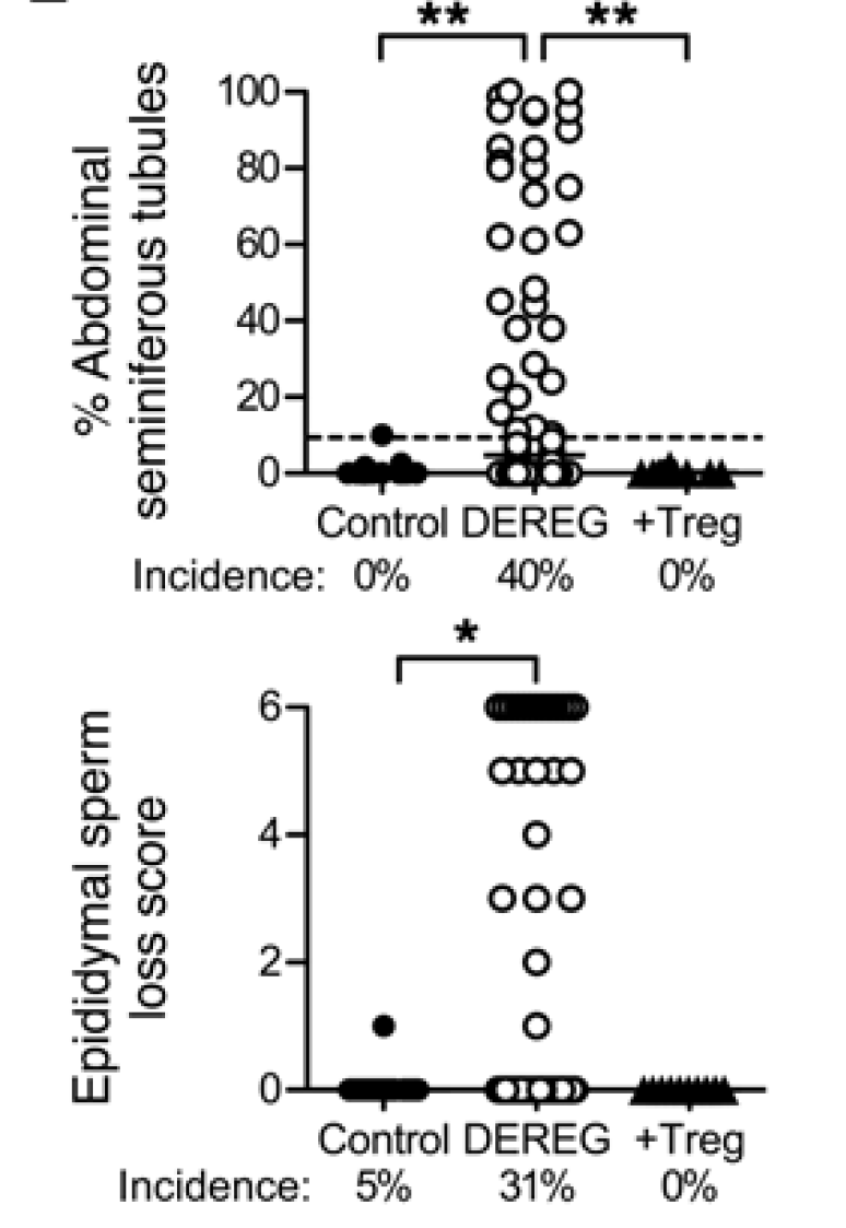
Furthermore, depletion of Tregs even in absence of testes antigen immunization still led to autoimmune pathology in testes in ~40% of male mice ("autoimmune orchitis occurs in autoimmune polyendocrine syndrome 1 (APS1) patients due to mutations of AIRE, possibly associated with impaired thymic deletion of autoreactive T cells and deficient Treg function")


In summary, this study suggests that "immune privilege" is not absolute and self-antigens that naturally leak maintain tolerance by induction of antigen-specific Tregs.

David Usharauli


Monday, April 25, 2016

Hobit controls tissue residency of memory T cells

Tissue resident memory T cells, TRM, represent newly recognized T cell population that play physiologically-relevant role in body's defense against reinfection. TRM cells are local tissue surveyors and act as a first-line defense [alongside of innate immunity] against tissue invading pathogens. Beyond these information nothing much was known about TRM cells.


Using mouse HSV-1 model, the authors found that by day 30 post-infection, Hobit was selectively up-regulated in TRM cells recovered from peripheral tissues, such as skin, but not in central lymphoid tissues such as spleen.


Using Hobit-KO T cells the authors were able to confirm that Hobit was indeed intimately involved in development of tissue-resident memory TRM cells (memory stage, day 40+). Deficiency of another transcription factor, Blimp1, had an additive effect on TRM cells formation when combined with Hobit deficiency. 


Interesting, Hobit, in combination with Blimp1, also controlled tissue residency of innate cells such NK and NKT cells.

In summary, this study revealed that transcription factor Hobit controls tissue residency of memory T cells, TRM cells. These data should be considered in developing T cell-oriented vaccines, including cancer immunotherapy (one limitation of this study is that for some reason the authors failed to show actual virus protection data with the regard of Hobit or Hobit + Blimp1 DKO T cells. ).

David Usharauli 

Thursday, January 28, 2016

Selective PD-L1/2 expression in lymphoid tissues allows tumor escape during allo-HSCT

The goal of allogeneic hematopoietic stem cell transplantation (allo-HSCT) is to achieve graft-versus-leukemia (GVL) effect to eliminate residual tumor cells in host [left after irradiation]. Since most allo-HSCs are derived from HLA-compatible donors, GVL reaction is mediated by donor T cells reacting against host minor histocompatibility antigens (miHAg; self or tumor-specific Ags). However, even reaction to self-miHAg could still induce graft-versus-host disease (GVHD) because self-miHAgs are broadly expressed in different tissues. Interestingly, GVHD preferentially affects peripheral tissues such as liver or gut. But why?


To mimic allo-HSCTs, the authors have used female-to-male BM stem cell transplantation model. In this model, self-miHAg is represented by male antigen, HY. To induce GVHD, female BM cells were transplanted alongside with HY-specific transgenic MataHari CD8 T cells. As expected, only male recipients of female HSCs + MataHari CD8 T cells developed GVHD (in liver, gut and skin tissue).


To address the question why only those peripheral tissues were affected by GVHD, the authors examined the hypothesis that HY specific CD8 T cell cytotoxic activity were differentially affected by different tissues. Indeed, co-transfer of labeled male and female targets revealed that recipients of allo-HSCs + MataHari CD8 T cells showed selective reduction of cytotoxic activity against male targets in lymphoid tissues (but not in liver).

This observation was supported by the fact that in contrast to peripheral tissues, MataHari CD8 T cells obtained from lymphoid tissues expressed low level of granzyme B (molecule involved in cytotoxic activity).

To understand why it is the case, the authors examined expression of inhibitory molecules on CD8 T cells. This revealed that while PD-1 on CD8 T cells were similarly expressed irrespective of tissue origin, its ligands, PD-L1 and PD-L2 were selectively up-regulated in lymphoid tissues.


The role of PD-L1 and PD-L2 in inhibition of CD8 T cell cytotoxicity in lymphoid tissues were confirmed in  experiment with anti-PD1 antibody.


Finally, using anti-PD-1 antibody injection, the authors showed that B cell leukemia cells that were hiding in lymphoid tissues of allo-HSCs male recipients could be now eliminated by MataHari CD8 T cells.


In summary, this study showed that (a) during GVHD donor CD8 T cell activity is differentially regulated by different tissues based on availability of inhibitory PD-1 signaling and that (b) this is exploited by leukemia cells to hide in lymphoid tissues but it could be overcome by anti-PD1 antibody injection.

David Usharauli