Showing posts with label immunopathology. Show all posts

Saturday, July 1, 2017

Role of Th1 transcription factor Tbet in Foxp3+ Tregs' functionality

Sasha Rudensky's lab continues to publish high profile papers in Foxp3+ Treg biology though besides the complicated genetic cell labeling approaches it delivers little of real science. 

This time, the authors followed the fate of Tbet+ Tregs. Tbet is a canonical Th1 transcription factor and its expression in Foxp3+ Tregs is puzzling. In first set of experiments the authors analyzed Tbet expression in mice with "Tbx21tdTomato-T2A-creERT2 knock-in allele combined with the R26Y recombination and Foxp3Thy1.1 reporters". In these mice any cell that express Tbet (Tbx21) will be (1) labelled red (tdTomato), will up-regulate (2) ERT2 (estrogen receptor) and if Treg, it will express Thy1.1 marker in addition. Interestingly, the authors observed that in steady-state "RFP+ Treg cells comprised between 30–70% of CD44hiCD62Llo effector Treg cells in lymphoid organs and non-lymphoid tissues;". 


To understand the stability of any given Tbet+ Treg cell the authors injected these mice with estrogen receptor activator tamoxifen. Tamoxifen will activate cre molecule that will liberate R26Y (yellow color) and cells become double positive (red+yellow). Interestingly, for any given moment only around 1% of  Tbet+ Tregs are labeled with YFP even though as mentioned earlier between 20-70% of Tregs supposed to express estrogen receptor any given moment (see above). It means very low "Tbx21tdTomato-T2A-creERT2 knock-in" gene functionality. In any case, tracing of YFP+ Tregs showed that they were quite stable population over period of 7 months, at least.

When these mice were treated with tamoxifen and then exposed to L. monocytogenes (Lm) infection, YFP+ Treg population did not expand while RFP+ Tregs did, suggesting that conversion rather than expansion plays role in Tbet+ Treg generation (assuming YFP+ and RFP+ Tregs are equal in all regards, especially for TCR specificity). 


In general, Foxp3YFP-cre Tbx21fl/fl mice lacking Tbet in all Tregs are indistinguishable from littermate controls. However, the authors found that Tbx21RFP-creFoxp3fl mice lacking Foxp3 only in Tbet+ [Tregs] did show evidence of clinical pathology and heightened Th1 effector differentiation. However, interpretation of this experiment is difficult because as already mention above large proportion of Tregs in these paper do naturally express Tbet and will undergo loss of Foxp3 following Tbet expression. Pathology could be result of a simple number reduction of functional Tregs independent of Tbet functionality.   


Interestingly, reverse experiment in Foxp3fl-DTRTbx21RFP-creERT2 mice lacking Tbet-negative Tregs only but retaining Tbet+ Tregs showed normal Th1 effector differentiation but heightened Th2 and Th17 differentiation implying that Tbet+ Tregs can only regulate Th1 effector differentiation. Though the authors did not provide any data whether Foxp3fl-DTRTbx21RFP-creERT2 mice have any clinical pathology similar to Tbx21RFP-creFoxp3fl mice.


In summary, this study suggests that Tbet+ Tregs might have unique regulatory capacity and specialization though it is not clear mechanistically how Tbet+ Tregs can specifically regulate Th1 effector cells. The role of TCR specificity is relevant but unknown at this stage.

posted by David Usharauli   


Tuesday, December 20, 2016

Lactobacillus reuteri extends lifespan of FOXP3-deficient scurfy mouse via microbiota–inosine–A2A receptor axis

Scurfy mice harbor natural mutation in FOXP3 gene that clinically resembles FOXP3 deficiency. Both scurfy mice and genetically modified FOXP3-KO mice die prematurely within first month of life. In humans, clinically observed FOXP3 deficiencies do not seem to be as lethal as in mice though one could argue that scurfy mice lifespan could be extended if these mice were given human-like medical attention (for example, i.v. feeding, anti-inflammatory medication, so on). 

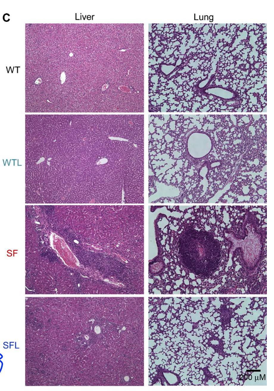
In this regard, a new paper in Journal of Experimental Medicine is of great interest. It showed that a member of gut microflora, Lactobacillus reuteri, a probiotic microbe, when given orally could drastically extend lifespan of scurfy mice (30d vs. >125d) via microbiota–inosine–A2A receptor axis.


Morphologically, oral Lactobacillus reuteri significantly reduced tissue inflammation in scurfy mice.


Blood test showed that one molecule Lactobacillus reuteri could restore to WT level in scurfy mice was a purine metabolite inosine.


Indeed, oral inosine was able to recapitulate Lactobacillus reuteri effect on scurfy mice lifespan (and both Lactobacillus reuteri and inosine effects were specifically mediated via adenosine A2A receptor).


In summary, this study revealed that purine metabolites, inosine or adenosine could protect scurfy mice from tissue immunopathology and drastically prolong their lifespan. It is quite rare to see that one molecule could produce such effect. It would be interesting to see how caffeine consumption affects immunopathologies in humans as it acts as a natural antagonist to adenosine A2A receptor.

David Usharauli


Tuesday, June 21, 2016

Tissue-specific immunopathology initiated by microbiota

Frequently, autoimmune disorders come in combinations. For example, a subset of human patients with type 1 diabetes (T1D) develop autoimmune polyglandular syndrome (APS) that depending on type (APS types 1-4) involves other tissues such as thyroid, adrenal, submandibular, and lacrimal glands. 

New study published in Journal of Immunology and conducted in autoimmune-prone mouse strain, NOD, showed that presence of microbiota modifies immunopathology in a tissue-specific manner.

For instance, female NOD mice devoid of microbiota (GF NOD mice) showed reduced pathology in salivary glands (sialitis) but as expected no change or worsening of pathology in pancreas (insulitis).  


Similarly, MyD88 deficiency completely abolished sialitis in both WT and GF female mice but it couldn't rescue insulitis phenotype on GF background (as reported prevously).


In summary, this short study suggests that microbiota → MyD88 pathways directly influence development of sialitis, while for insulitis (and diabetes development) microbiota/MyD88 pathways diverge.

David Usharauli

Wednesday, May 18, 2016

PSGL-1, a ligand for the selectin family of receptors, controls T cell immunopathology

The Selectin receptors, L, E and P, control T cell migration. New study in Immunity showed that Selplg-KO T cells (lacking selectin receptor ligand PSGL-1) display enhanced effector differentiation and greater control of chronic viral infection and tumor, though it comes at the expense of tissue pathology.  

In this paper the authors referred to PSGL-1 as checkpoint inhibitor. However, unlike CTLA4 and PD1 deficient mice, Selplg -/- naive mice don't show any abnormality. Interestingly, the authors found that Selplg -/- mice efficiently controlled chronic LCMV infection (C13).


However, this enhanced protection against chronic LCMV infection led to severe tissue pathology.

Additionally, and most likely reason why this paper ended up in Immunity, the authors showed that Selplg -/- mice could better control tumor (Yumm1.5 melanoma cells).


At present it is not clear whether PSGL-1 signaling works as an independent "checkpoint inhibitor" in vivo or modulates functionality of other canonical checkpoint inhibitors such as PD-1 .

David Usharauli


Monday, February 29, 2016

Failure to tolerate gut microbiota transforms immunodeficiency into immunopathology

Here is a paradox for you that many don't even know that it exists: frequently, genetic immunodeficiency (weak immune response) syndromes are associated with immunopathologies (excessive immune response). So how could it be explained?   

New study published in Journal of Experimental Medicine may have some new answers. This study described mouse model of human immunodeficiency syndrome called, Omenn syndrome (after its discoverer) and showed that immunopathology was driven by gut microbiota.

Omenn syndrome is caused by hypomorphic (low active) RAG mutations. Analysis of intestinal tissue from Rag2R229Q [Omenn] mice revealed pathological infiltration with inflammatory T cell subsets, TH17 and TH1.


Adoptive transfer showed that intestinal immunopathology was mediated by Rag2R229Q mutant T cells.


Interestingly, Rag2R229Q mouse harbored comparable numbers of Foxp3+ CD4+ T cells, implying that simple presence of regulatory T cells [generated in Rag2R229Q mice] was not enough to control tissue inflammation.

Defect of Tregs derived from Rag2R229Q mice was confirmed in adoptive transfer experiment with WT Tregs.


Besides T cell-driven immunopathology, Rag2R229Q mice displayed IgA deficiency (failure to properly coat gut microflora). These data pointed to the possibility of microbial translocation causing excessive inflammatory response.


Indeed, antibiotic treatment of Rag2R229Q mice could reduce intestinal immunopathology.


The role of antibiotic-sensitive gut flora in driving immunopathology in Rag2R229Q mice was confirmed in adoptive fecal transfer experiments.


In summary, this study suggests the following scenario: hypomorphic RAG defect in Rag2R229Q mice leads to "narrowing" of TCR and BCR repertoire. This in turn leads to outgrowth of oligoclonal T and B cells in Rag2R229Q mice (wherein Rag2R229Q mice contain T and B cells with limited, restricted, deficient TCR and BCR repertoires). Without proper TCR and BCR repertoire diversity, however, Rag2R229Q mice fails to develop tolerance (IgA and Tregs) to gut flora or commensal microbial antigens present at mucosal surfaces (such as lung, intestine). Repertoire restriction also leads to failure to mount adequate and proper immune response.


David Usharauli

Thursday, January 21, 2016

Tissue-resident regulatory T cells are balancing between immunity and immunopathology

Yesterday journal Nature published very interesting study related to Foxp3+ regulatory T cells. This research revealed that "over-active" Foxo1 within Tregs specifically reduces number of peripheral tissue-resident active Tregs that control CD8 T cell-mediated tumor immunity and immunopathology.

Initially, using parabiotic mice pairs the authors showed that Tregs too undergo mixing and achieve equilibrium within 4 weeks. This indicates that Tregs are not maintained locally and require replenishment. 

Next, using Foxp3-cre driven Foxo1 "active" mutant mice the authors showed that when both alleles of normal Foxo1 genes were replaced by "active" Foxo1, it led to dramatic reduction of tissue-resident Tregs (lymphoid tissue resident Tregs were less affected).


Surprisingly, when these Foxo1 homozygous mutant mice were followed for longer time (6-8 weeks), they displayed spontaneous CD8 T cell-mediated wasting disease and peripheral tissue pathologies in liver and intestine (though these tissue pathologies were different from those observed in Foxp3 mutant mice). Hemizygous Foxo1 mutant mice with only one mutant Foxo1 allele were healthy.


Interestingly, reduction of peripheral Tregs in hemizygous Foxo1 mutant mice were sufficient to confer CD8 T cell-mediated anti-tumor effect without immunopathology


In summary, this study revealed three important results: 

1. Lymphoid resident Tregs are not sufficient to control autoimmunity/immunopathology. Tissue-resident Tregs play unique and essential role in this process.

2. Level of active Foxo1 molecule control frequency of tissue-resident Tregs

3. Manipulation [reduction] of level of active Foxo1 within Tregs could improve anti-tumor immunity.

David Usharauli

Friday, December 11, 2015

Autophagy-independent role for Atg5 in M. tuberculosis infection

This is a second paper about neutrophils in Nature this week (advanced online publication). Quite interesting and surprising. It showed that autophagy molecule Atg5 has unique [autophagy-independent] role in protecting against neutrophil-driven immunopathology during M. tuberculosis lung infection.

Initially, the authors showed that myeloid cell-specific deletion of Atg5 (LysM-Cre Atg5fl/fl) made mice highly susceptible to M. tuberculosis infection (that was expected based on earlier studies).


Unexpectedly and surprisingly, however, mice singly deficient for other autophagy components showed normal response to M. tuberculosis infection, implying unique, autophagy-independent role for Atg5 during M. tuberculosis infection.    
Indeed, LysM-Cre Atg5fl/fl mice showed more severe M. tuberculosis-associated immunopathology and neutrophil infiltration (even though LysM-Cre Atg5fl/fl mice did not harbor substantially more M. tuberculosis).
Furthermore, LysM-Cre Atg5fl/fl mice depleted of neutrophils were protected from lung pathology during M. tuberculosis infection.


Finally, neutrophil-specific deletion of Atg5 (MRP8-Cre Atg5fl/fl mice) confirmed pathological role of Atg5-deficient neutrophils during M. tuberculosis infection. 


In summary, this study provides evidence for autophagy-independent role of Atg5 deficiency in neutrophils during M. tuberculosis infection. It appears that neutrophils recruited to the sites of M. tuberculosis infection contribute to tissue pathology rather than provide protection in absence of Atg5. Earlier it was assumed that Atg5 played a protective role during infection purely via its involvement in autophagy formation. However, this new study point to a more complex role of Atg5.

David Usharauli

Saturday, November 7, 2015

Fezl ascends to the thymic throne as Aire's equal and co-ruler

A thymus controls T cell development and central tolerance. It first selects newborn T cells capable of recognizing MHC molecules and then purges those T cells who interact with MHC + self peptide complexes too strongly

But what about self-antigens that are physiologically expressed in peripheral tissue, such as insulin (endocrine), neuronal antigens (CNS), testis and ovary antigens (reproductive)? Initially, it was assumed that mechanism called "'peripheral tolerance" was responsible for elimination of peripheral antigen-specific T cells. But later, it was shown that protein called Aire controlled expression some of the tissue-restricted antigens (TRAs) in the thymus thus facilitating central tolerance to those peripheral antigens. Interestingly, analysis of Aire KO thymus had shown that there were Aire-independent TRAs expressed in the thymus, implying the existence of yet unknown mechanism.

Now, new Cell paper revealed the identity of molecule responsible for Aire-independent TRAs expression. It turned out that thymic protein Fezf2, also known as Fezl, was responsible for expression of unique set of TRAs in the thymus independently of Aire.   

First, the authors found that Fezl was highly expressed in mTECs, thymic cells responsible for central tolerance.


Role of Fezl in tolerance was tested in nude mice lacking endogenous thymus. Nude mice receiving Fezl KO thymic transplantation developed peripheral tissue immunopathology [because TRAs-specific T cells were not deleted].

At the same time, the authors showed that Aire's expression was not altered by absence of Fezl.


Expression pattern of TRAs in thymus revealed that Aire and Fezl mostly controlled non-overlapping set of TRAs.


Experiments with thymic-specific Fezl deficiency showed peripheral lymphoid tissue enlargement and autoantibody development.


Finally, the authors found that unlike Aire, Fezl expression was controlled by lymphotoxin beta receptor (LtβR) pathway.

In summary, this truly breakthrough study revealed a new dimension for thymic TRAs expression. Mutations in Fezl could underlie some of the known forms of immunopathology.

David Usharauli

Friday, August 28, 2015

Treg-specific expression of amphiregulin prevents immunopathology during viral infection

Foxp3+ T cell deficiency leads to lethal immunopathology in mice and severe organ pathology in humans (IPEX syndrome). Over the past 20 years numerous mechanisms of action of Foxp3+ T cells have been described. In fact, this number is so numerous that it wouldn't be a "heresy" from my part to claim that no single Foxp3+ T cell phenotype could explain it. This leaves us with the hypothesis that multiple versions of Foxp3+ T cells exist, each of them using selective path for immune regulation and suppression.    

With this view in mind, it was useful to read a new paper from Rudensky's lab published in journal Cell this week that provided evidence showing the role of Foxp3-positive T cell-specific amphiregulin in preventing excessive tissue pathology during high dose of viral infection.

First, the authors verified that amphiregulin, an epidermal growth factor family member, was expressed by Foxp3+ T cells.


Next, the authors showed that amphiregulin deficient Foxp3+ T cells displayed normal suppressive functionality when transferred into T cell-deficient host.


Further experiments revealed that amphiregulin deficiency in Foxp3+ T cells did not modify the host immune response to intranasal flu infection (mouse PR8 model).


However, the authors noticed that there was excessive lung tissue damage in response to high (but not low) dose of flu infection in the hosts with Foxp3+ T cell-specific deletion of amphiregulin.


Finally, the authors showed that amphiregulin up-regulation was primarily restricted to IL-18R+ Foxp3+ T cells.


In summary, these data tend to reinforce the idea that Foxp3+ T cells could function in a tissue selective manner and are most likely controlled by tissue environment as suggested by Polly Matzinger and Tirumalai Kamala. This could explain why there are so mechanisms of suppression by Tregs, at least one mechanism for each tissue.

What are some of the weaknesses of this study? First, injection of amphiregulin to hosts with Foxp3+ T cell-specific deletion of amphiregulin would have been useful. Second, data regarding IL-18R deficient Foxp3+ T cells could have provided in vivo confirmation for this study (the authors simply mentioned that they have done experiments with bone marrow chimera to test the role of IL-18R in amphiregulin up-regulation and that it supported their conclusions).

Why is this study important? Because without our mastery of Foxp3+ T cell biology we will not be able to make any major, predictable advances in treating human immune related conditions (cancer, allergy, autoimmune conditions).

David Usharauli