Similar results were obtained when thymus tissue was analyzed.
Saturday, November 30, 2019
Tracking deletion of autoreactive clones versus Treg generation for thymically expressed epitopes
Similar results were obtained when thymus tissue was analyzed.
Saturday, February 9, 2019
An antigen from gut commensal Bacteroides thetaiotaomicron (B. theta) is recognized by Foxp3+ Tregs
1. B.theta-specific T cells when transferred into #antibiotic-treated and B.theta #colonized T cell-deficient mice (#RAG1-KO) develop into #Foxp3+ #Tregs as well as effector T cells. It is not clear if donor tg T cells were on RAG1-KO background as well. Host mice stay healthy. pic.twitter.com/TD0pnnxgAR— David Usharauli (@3DiMMUNE) February 9, 2019
3. The authors were able to identify a #specific #epitope in B.theta recognized by B.theta-specific T cells. The #sequence, EEFNLPTTNGGHAT, were mapped in BT4295 predicted to be a SusE/SusF #lipoprotein in outer #membrane (OM). pic.twitter.com/rH5cQHQcVN— David Usharauli (@3DiMMUNE) February 9, 2019
In #summary, this study showed that certain gut #commensal #microbiota species, in this case, #Bacteroides thetaiotaomicron (B. theta) are recognized by #Tregs and even induce #effector T cell differentiation though the host stays healthy because Tregs regulate effector T cells.— David Usharauli (@3DiMMUNE) February 9, 2019
Another point is whether #microbiota-specific Tregs are #denovo induced or #amplified from existing ones. I personally think it is a latter. Most #Tregs develop from #CD25-negative #precursors poised for Foxp3 expression & such precursors are all #thymus derived, in my opinion.— David Usharauli (@3DiMMUNE) February 9, 2019
Wednesday, January 16, 2019
Foxp3+ Tregs suppress other T cells by stripping DCs of specific antigen/MHC complexes
Ag-specific or not Ag-specific, that is [maybe was] question for #Foxp3+ Tregs mechanism of action. New paper in @NatImmunol from #Shevach's lab @NIH says definitely antigen-specific. Lets dive in. BTW, ~20 years ago his lab developed assays that showed it was non-specific 🤫
— David Usharauli (@3DiMMUNE) January 16, 2019
Now his lab made U-turn and concluded that #suppression by #Tregs is actually #antigen-specific and difference between now and then comes down to in #vitro versus in #vivo assays 🤔
— David Usharauli (@3DiMMUNE) January 16, 2019
2. This #antigen-specific #suppression was still active when antigen-specific dendrtic cells were removed from Treg co-culture (1st culture) and exposed to T cells (2nd culture). pic.twitter.com/dj1pzDuNNv
— David Usharauli (@3DiMMUNE) January 16, 2019
So, from now on if someone asks you how Treg suppress other T cells don't hesitate to reply "antigen-specific manner", add Ethan Shevach said so and no one will argue with you. got it 🤨 https://t.co/I4pAYIIGy1
— David Usharauli (@3DiMMUNE) January 16, 2019
Sunday, October 14, 2018
New auto[cross-reactive]-antigen, GDP-l-fucose synthase, identified in MS patients
The authors has used modified version of positional scanning epitope library to identify epitope derived from auto-antigen, GDP-L-fucose synthase, as a target epitope for CD4+ T cell clone, TCC21.1, derived from MS patients with DR15 haplotype. Peptides sequences matching GDP-l-fucose synthase was detected in brain tissue.
MS patients whose T cells displayed high response to peptides from GDP-L-fucose synthase in stimulation assay showed high responses to myelin proteins as well.
Curiously, almost all high responders carried DRB3*02:02 allele.
Finally, the authors speculated that sequence similarity between human and microbiota-derived evolutionary conserved GDP-L-fucose synthase could be a factor that initiates MS pathology in these patients.
In summary, this study has a lot of nice data that support idea that GDP-L-fucose synthase is a new auto-antigen that could be relevant in molecular diagnosis of MS pathology.
There are several questions about this study. GDP-L-fucose synthase expression is not restricted to brain tissue. So, it should be relevant to understand if other tissues were affected in those MS patients. Second, cross-reactivity per se is not sufficient to explain how and why autoimmune responses are being initiated. Microbiota expressing GDP-L-fucose synthase most likely reside in patients long before MS, maybe even since birth, and T cells are tolerant to them. So, what has to be changed, in a antigen-specific manner, to make T cells less tolerant to initiate specific autoimmune attack on nervous system and not a total autoimmunity targeting all available auto-antigens?
posted by David Usharauli
Wednesday, December 27, 2017
Effort to identify tumor-specific antigens: The University Industrial Complex study results
Wednesday, July 26, 2017
IBD converts tolerant antigens into immunogenic
Sunday, July 16, 2017
Identification of prostate-antigen specific natural Tregs (in mice)
Thursday, June 2, 2016
Combination of checkpoint inhibitor and IL-21-primed melanoma-specific T cells produced durable response, "cure", in melanoma patient
Wednesday, November 18, 2015
Time dictates tolerance to skin microbiota
Next, the authors tested how adult mice immune system would respond to secondary skin challenge with Epi-2W. They showed that adult mice exposed to skin Epi-2W do not develop tolerance to it upon secondary challenge. These mice displayed (a) skin inflammation
(b) no accumulation of 2W-tetramer+ Foxp3+ CD4 T cells, implying failure of tolerance.
However, if skin of neonatal day 7 mice were exposed to Epi-2W and later challenged again, these mice showed (a) minimal skin inflammation
(b) and robust accumulation of 2W-tetramer+ Foxp3+ CD4 T cells, implying active tolerance to Epi-2W.
Followup experiments found that between neonatal day 6-13 there was a sudden increase of thymic Foxp3+ CD4 T cell numbers specifically in skin.
Finally, the authors showed that when this increase of neonatal skin Foxp3+ CD4 T cells were blocked, it resulted in tolerance failure towards Epi-2W challenge and skin inflammation.
In summary, these results indicate that timely [neonatal] exposure to skin commensals is necessary to establish antigen-specific Foxp3+ CD4 T cell-mediated immune tolerance to skin microbiota and to prevent pathological skin inflammation later on. It is possible that in near future human neonates will be artificially exposed to the defined sets of human commensals via skin application, nasal inhalations and oral application to specifically train their neonatal immune system for tolerance towards commensals and hence better distinguish between beneficial and pathogenic microorganims.
David Usharauli
Saturday, April 11, 2015
Anergic B cells respond to polyvalent antigens via IgD receptor
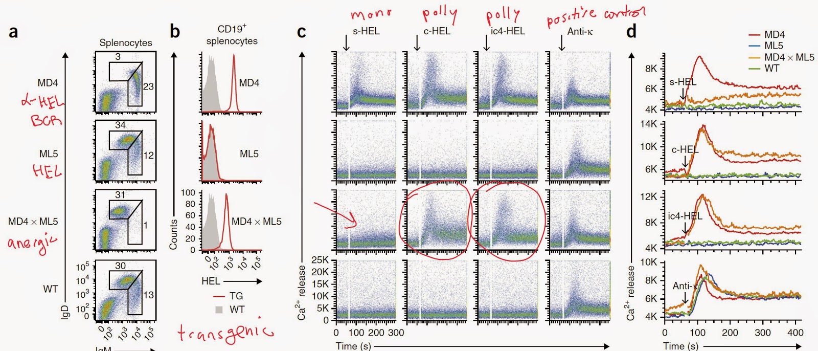
In summary, this study revealed that anergic B cells (IgMlowIgDhigh) can respond to antigen when stimulated with polyvalent antigens. Probably the role of IgD receptor is to prevent improper activation of naive B cells in response to soluble antigens. I wonder what is the (a) phenotype of IgD KO mice or (b) whether auto-antigens targeted by natural antibodies are mono or polyvalent in nature?
David Usharauli