Showing posts with label IgM. Show all posts

Thursday, October 1, 2015

B cell activation by antigen promotes desegregation of IgM and IgD islands

There are two models of B cell activation. In one model, B cell activation is promoted by clustering of individual IgM molecules. Second model, however, argues that B cell activation is promoted by de-clustering of IgM "islands". This second model is mainly based on research by Micheal Reth's lab.


To study B cell receptor architecture on resting or activated B cells, the authors have used 3 different but complementary approaches: (1) two-color direct stochastic optical reconstruction microscopy (dSTORM), (2) two marker transmission electron microscopy (TEM) and (2) proximity ligation assay (PLA). 

First, the authors showed that on resting B cells, both IgM and IgD are segregated into islands containing on average 30 IgM and 48 IgD molecules, respectively.


Next, the authors showed that upon activation, both IgM and IgD islands show reduction of number of receptors per islands, implying that individual IgM or IgD receptors are leaving the islands.  


Interestingly, the authors showed that both IgM and IgD islands could independently respond to the antigen.  


In addition, detection of IgM and IgD (co)-localisation by TEM after antigen exposure confirmed receptors meetup (receptors were detected by secondary antibodies conjugated to two different size gold particles).  


In summary, this study supports the hypothesis that upon B cell activation IgM and IgD receptors leave "home islands" and move close to each other, thus facilitating sharing of downstream signaling molecules. This study is a "biochemical" study that does not incorporate "biological" significance of BCR dissociation, however.   

David Usharauli

Saturday, April 11, 2015

Anergic B cells respond to polyvalent antigens via IgD receptor

Naive B cells express IgM and IgD receptors. Both receptors share the same unique immunoglobulin variable region but differ in immunoglobulin constant regions. IgM is secreted in response to antigenic signaling but not much is known regarding the role of IgD in immune response.

New study in Nature Immunology provided very interesting data about the function of IgD. It turned out that unlike IgM, IgD receptors only respond to polyvalent antigens.

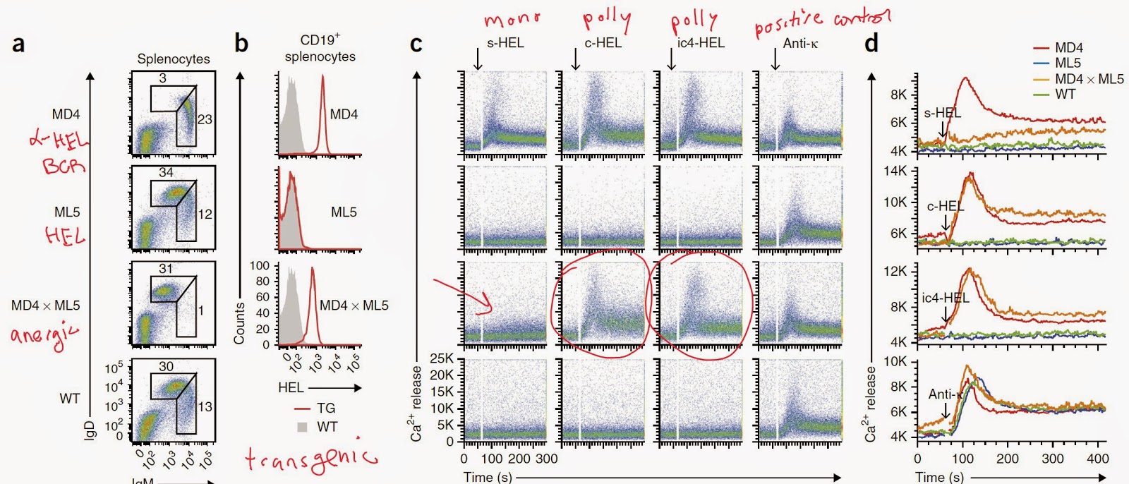
The authors, led by Hassan Jumaa at the Institute of Immunology (Ulm, Germany), first showed that unlike IgM receptor, cell line expressing IgD receptors specific for hapten (NIP) or antigen (HEL) responded only to polyvalent antigenic forms.


Next, the authors find that these difference in response between IgM and IgD was related to the difference in hinge region (that connects variable and constant regions). IgD with no hinge region (IgDΔhinge) responded as if IgM and IgM with IgD hinge region responded as if IgD.


The authors observed that anergic B cells obtained from antigen (HEL)-specific B cell double transgenic mouse expressing soluble HEL (s-HEL) could still respond to polyvalent HEL antigen.


The authors showed that monovalent antigen could competitively inhibit IgD signaling in response to polyvalent antigens.


Next, the authors observed that functionally, absence of IgM receptor could compromise innate B-1 cells scavenging response (natural antibody response) to a soluble auto-antigen phosphatidylcholine (PtC).


Indeed, IgD receptor itself were unable to respond to a soluble phosphatidylcholine (PtC).


Finally, the authors also showed that the absence of IgM signaling compromised IgG scavenging functions against multiple auto-antigens (oxidized LDL, etc) as well.


In summary, this study revealed that anergic B cells (IgMlowIgDhigh) can respond to antigen when stimulated with polyvalent antigens. Probably the role of IgD receptor is to prevent improper activation of naive B cells in response to soluble antigens. I wonder what is the (a) phenotype of IgD KO mice or (b) whether auto-antigens targeted by natural antibodies are mono or polyvalent in nature?

David Usharauli


Tuesday, December 9, 2014

Sweet rejection: sugar-coated malaria parasite

Life is a competition, even for a parasite like a malaria. To infect the host the parasite needs to overcome not just host's defense system but to out-compete the local resident micro-flora as well. 

This new paper in Cell is a thorough research about the initial events of of malaria infection and the role of sugar molecules, called glycans, in the host defense against malaria.

It is well-known that hosts and their parasites may share molecular signature. Blood types, ABO system is one such example. Burnet's clonal selection theory predicts that any such similarity between host-pathogen prevents the host to mount an efficient immune response against shared antigens. As a consequence, based on pathogen burden and evolutionary pressure, the hosts started to loose the capacity to express such shared molecules.

It appears that at some point in their evolution human ancestors lost the capacity to make one type of sugar, alpha-gal, expressed by malaria. This change conferred an improved capacity to defend against malaria. 

This papers shows how exactly such modification provided protection. It turns out to be dependent of natural immunization conferred by gut resident microbes expressing the same exact sugars.

Analysis of serum samples across different age group from children in Mali (malaria endemic region), the authors noticed a gradual increase in anti-alpha-gal IgM level over time. Interestingly, 6-months parasite free condition correlated with higher level of anti-alpha-gal IgM in the serum.
To study this observation in laboratory setting, the authors used mice deficient in the capacity to make alpha-gal (alpha-gal KO). This "human-like" mice can produce anti-alpha-gal antibodies upon colonization with alpha-gal-positive E.coli O86:B7, but not alpha-gal-negative E.coli K12 strain. 

The authors showed that colonization of alpha-gal KO mice with alpha-gal-positive E.coli O86:B7 provided a protection against malaria transmission.

This protection after gut flora colonization was conferred by soluble germ-line, non-mutated IgM.
Immunization of alpha-gal KO mice with  (a) alpha-gal conjugated to BSA (protein carrier) or (b) rabbit RBC (naturally expressing high levels of alpha-gal) conferred protection against malaria transmission.
Deep analyses of mechanism of protection after immunization revealed it was dependent on T cell help and on both IgM and IgG (of note, immunization with rabbit RBC conferred protection even in IgM-deficient mice, unlike protection conferred after colonization with alpha-gal expressing E.coli O86:B7).
The protection could be conferred by passive transfer with IgM, IgG3 and IgG2b, but not IgG1 and IgG2a.
In summary, this study provides evidence how gut flora affects host's defense against parasites by a way of natural immunization. Of course, it is remains to be determined whether immunization against alpha-gal will protect humans as well as it does for "human-like" mice.

David Usharauli