Saturday, February 28, 2015

Specific food additives modify gut microflora and cause metabolic syndrome

Here is another paper that highlights unsuspected health risks of commonly used food emulsifiers, carboxymethylcellulose (CMC) and polysorbate-80 (P80).

Ordinarily, mucus separates gut epithelial cells from the gut microflora. New study in Nature provided evidence suggesting that this separation of gut epithelial cells from the gut microflora is disrupted by CMC and P80, two commonly used food emulsifiers (but not by sodium sulfite, another commonly used food additive).


The authors showed that presence of either CMC or P80 in either drinking water or dry food pellets could promote development of colitis in susceptible mouse strain (IL-10 KO).


In addition, the authors  showed that presence of either CMC or P80 in water or dry pellets could promote development of metabolic syndrome in wild-type as well as in susceptible mouse strain (TLR5 KO).


Interestingly, metabolic syndrome did not develop in germ-free mice treated with CMC or P80.


More importantly, effects of CMC or P80 on animal health were transferable to germ-free mice by fecal transplantation from treated mice, implying direct role of modified gut microflora (note, CMC or P80 did not directly modify mucus thickness in germ-free mice).


In summary, these results support the growing evidence that chemically-processed food carries long-term health risks (development of type II diabetes, metabolic syndrome, inflammatory bowel disease).

David Usharauli

Wednesday, February 25, 2015

Inflammatory signals from the peripheral tissue modulates immune response in lymph nodes

Very unusual results have been published in journal Science. This study came from Facundo Batista's lab at the London Research Institute.

The authors studied lymph node's adaptive re-organization in response to peripheral inflammatory signals, with the focus on CD169-positive macrophages. These specialized macrophages are ordinarily positioning themselves in the subcapsular sinus of the lymph nodes intercepting any incoming antigens from the peripheral tissue for presentation to B cells.

Using different type of inflammatory signals (live or dead bacteria, live or inactivated viruses, or TLR agonists), the authors observed that live bacteria or virus or TLR agonist injected in the mouse footpads caused temporal disruption of CD169+ macrophage layer in the draining lymph node.   


Further experiments showed that disruption of CD169+ macrophage layer in the draining lymph node upon CpG injection was abolished in mice selectively deficient for MyD88 signaling in dendritic cells.


In addition the authors noticed that disruption of CD169+ macrophage layer was also reduced in CCR7-KO mice where DCs could not migrate from the peripheral tissue to the lymph node.  


To understand physiological consequences of CD169+ macrophage layer disruption, the authors injected labeled B cell antigen into footpad following initial CpG or PBS (control) injection (+ 4 / +7 days later). These experiments revealed that fewer CD169+ macrophage and fewer antigen-specific B cells could acquire antigen following CpG injection.   


More importantly, the authors showed that initial CpG injection reduced subsequent anti-viral antibody response.


In summary, these results shows that initial inflammatory signals coming from peripheral tissue disrupt CD169+ macrophage layer and antibody response to subsequently injected antigen. 

This is strange results. No good explanation is given. For one, the authors did not provide results regarding viral titre or tissue pathology in mice injected with primary CpG. 2nd, isn't lymph nodes function to respond to the inflammation in peripheral tissue? How could it be beneficial for the host to shut down immune response following inflammatory stimuli? Could it be that lymph nodes are modified to activate different type of immune response (CTL?)

David Usharauli


Saturday, February 21, 2015

Fasting-induced ketone metabolite, β-hydroxybutyrate suppresses NALP3-mediated inflammasome activity

Inflammasome super-complex represents an ancient innate detection and defense mechanism against exogenous as well as endogenous irritants and toxins. Spontaneous inflammasome activity causes several known human immune-pathologies, like Muckle-Well and familial cold auto-inflammatory syndromes.  

New paper from Nature Medicine provided evidence showing that alternative energy source, β-hydroxybutyrate, elevated during fasting, calorie-restiction or high-intensity exercise, suppresses NALP3-mediated inflammasome activity.

Initially, the authors led by Prof. Vishwa Deep Dixit at the Yale School of Medicine, showed that β-hydroxybutyrate, but not another ketone body acetoacetate or short-chain fatty acids, butyrate or acetate, could inhibit NALP3-mediated inflammasome activity (AIM2- or NLRC4-mediated inflammasome activities were unaffected).


Suppression of NALP3-mediated inflammasome activity by β-hydroxybutyrate was independent of autophagy and required neither G protein-coupled receptor GPR109a signaling via β-hydroxybutyrate nor histone deacetylase inhibition by β-hydroxybutyrate.


The authors found that β-hydroxybutyrate suppressed NALP3-mediated inflammasome activity through its inhibition (by unknown mechanism) of potassium, K+ cation efflux from cytoplasm.


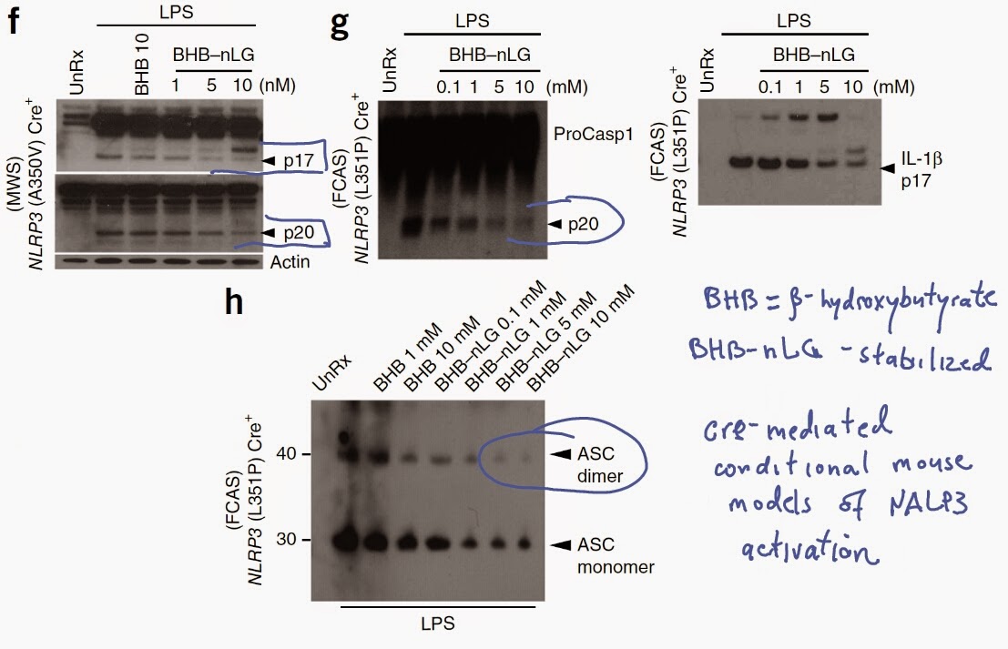
Finally, using cre-mediated mouse conditional models of human Muckle-Well and familial cold auto-inflammatory syndromes, the authors showed that stabilized form of β-hydroxybutyrate could suppress spontaneous NALP3-mediated inflammasome activity and ASC oligomerization characteristics to these gain-of-function NALP3 mutations.


In summary, these results indicate complex interplay between energy metabolism and inflammation. In condition of glucose deficiency, elevated level of β-hydroxybutyrate acts as a new source of ATP in brain and heart and at the same time inhibits NALP3-mediated inflammasome activity.

Leave your comments below and let me know what do you think about this paper.

David Usharauli