Wednesday, December 5, 2018

A minor population of tumor infiltrating CD8+ T cells actually express tumor antigen specific TCR

Tumor immunotherapy with checkpoint inhibitors (anti-CTLA-4, anti-PD1/PDL1 antibodies) showed remarkable therapeutic effect in narrow slice of cancer patients (~20% - 25% of cases). It relies on reactivation of tumor infiltrating T cells called TILs (tumor-infiltrating lymphocytes). Obviously, the more we know about nature of these TILs the better medical approaches could be implemented.

A new study in Nature Medicine by Ton Schumacher's group in Netherlands analyzed single cell sorted TIL TCR specificity derived from 4 treatment-naive patients. Their limited and [technically inadequate, in  my view] analysis revealed that tumor specific T cells represent a minor population among TILs from such cancers as ovarian cancer (OVC) and microsatellite stable colorectal cancer (CRC). 

First, the authors validated their approach with T cells derived from melanoma samples. 88 CD8+ TILs were single cell sorted, TCR sequenced and TCR α/β chain pairs established. 15 such TCR pairs were trasduced into primary T cells and exposed to melanoma samples. 9 different TCR transduced T cells showed IFN-γ expression (60%). It is not clear what tissue they used as non-melanoma negative samples. They mentioned B cells in methods section though it is not obvious why would B cells act as a good negative samples for skin tissue. Also, I find it highly inadequate to limit tumor antigen-specificity detection to intracellular IFN-γ expression when using primary T cells as a TCR transduction carrier. Based on TCR affinity it might engage other types of response.


With these caveats in mind lets examine their results. When they tested single cell sorted TIL TCRs derived from ovarian cancer sample (from a single patient, OVC21), out of 20 TCR transduced T cells only 1 TCR showed IFN-γ response to tumor tissue. Another patient's TCR assay showed zero reactivity. Again, it is not clear whether low reactivity is a biological effect or simply technical deficiency as discussed earlier. We cannot even be sure whether that 1 TCR is actually tumor specific either. It also appears that pairing of TCR α/β chains is not a straightforward either because out of 95 sorted cells only 37 (39%) TCRα/β pairs could be identified.


A slightly more encouraging results were obtained with TCRs from one CRC sample (patient CRC11). Here 5 out 16 TCR tested showed IFN-γ response to cancer organoid tissue. however, similar test on another patient's TCRs showed zero response.


In summary, this study tried to show [but utterly failed in my view] that for some tumors tumor-reactive TCRs among TILs are quite rare. What are then such TILs' TCR specificity are unknown. Why are they recruited in tumor tissue is not known either. The authors have not even formally tested what neoantigens, if any, these tumors from those 4 patients actually expressed. Very unsatisfactory study.

posted by David Usharauli


Thursday, October 25, 2018

Can microbial innate signaling generate peripheral FOXP3+ regulatory T cells?

In the past several years few papers have been published suggesting that some commensal microbiota species acting via their conserved molecules [i.e. polysaccharide A (PSA) from Bacteroides fragilis (Bf)] and metabolic products [i.e. short-chain fatty acids (SCFAs)] could induce peripheral FOXP3+ Tregs. The mechanisms are still controversial due to fact that there is no consensus whether thymus-derived and peripherally generated FOXP3+ Tregs (pTregs) represent two distinct lineages or distinct differentiation phases of unique thymus-derived FOXP3+ Tregs.

In this regard, a new paper in Science Immunology is of interest. In this study the authors tried to produce evidence that microbial cell surface β-glucan/galactan (CSGG) polysaccharides of Bifidobacterium bifidum [B. bifidum], a probiotic strain PRI1, could induce generation of FOXP3+ pTregs specific for diverse set of antigens. Lets review it.

First, the authors showed that monocolonization of germ-free mice with B. bifidum dramatically increased frequency of colonic Tregs compared to control species. Of note, Bacteroides fragilis (Bf) showed no effect thus putting some doubt on original observation.


Second, to nominally distinguish between conversion vs. expansion, the authors transferred naive CD4+Foxp3− T cells sorted from Foxp3GFP reporter mice into germ-free mice and analyzed for GFP expression 3 weeks later. Around 5% of donor T cells up-regulated FOXP3 in colon tissue.


Of note, monocolonization of germ-free mice with B. bifidum induced quite robust IFN-γ response too.


In addition, transfer of OT-II TCR transgenic T cells [on RAG1 KO background] recognizing OVA into OVA-fed B. bifidum treated mice or transfer of CBir TCR transgenic T cells that recognize bacterial flagellin into B. bifidum treated mice on SPF RAG1 KO background could augment their differentiation into FOXP3+ Tregs. Of note, first, OT-II were transferred into germ-free mice that harbor natural, thymus-derived Tregs that could direct naive OT-II conversion into FOXP3+ Tregs in a antigen[epitope]-specific manner (OVA cross-reactivity). Second, there is some inconsistency regarding CBir T cell experiment. In the figure legend it says they were transferred into SPF RAG1 KO hosts but in the supplemental data it says "naïve CBir CD4+CD62LhiCD44loFoxp3GFP- T cells (1x10⁶) were transferred intravenously to the C57BL/6 mice maintained in SPF conditions"These difference could make huge difference in data interpretation. Third, CBir T cells specificity to flagellin has now been questioned in new study.


In vitro co-culture of DCs and T cells pretreated with B. bifidum or its cell surface β-glucan/galactan (CSGG) polysaccharides could induce FOXP3+ Treg phenotype in a TGF-β1 dependent manner. Again, Bacteroides fragilis (Bf) could not do it.


Also, TLR2 appears to be involved in the action of B. bifidum cell surface β-glucan/galactan (CSGG) polysaccharides.


In summary, this study tries to suggest that B. bifidum conserved microbial wall component, β-glucan/galactan (CSGG) polysaccharides could convert naive T cells into FOXP3+ Tregs. It is certainly obvious that β-glucan/galactan (CSGG) polysaccharides could increase frequency of FOXP3+ Tregs but whether it is genuine conversion vs. expansion of pre-committed proto-Tregs via direction of existing thymus-derived Tregs were not cleanly verified. And lets remember that B. bifidum monocolonization increased frequency of IFN-γ producing T cells too. How these data fit to tolerance model? I am also puzzled by the fact that the authors primarily or exclusively showing frequency and not the actual numbers of FOXP3+ Tregs without providing any guidance.

Finally, lets step back and think a little bit more about general premise of such papers. The authors concluded that because B. bifidum and its microbial wall component, β-glucan/galactan (CSGG) polysaccharides could increase frequency of FOXP3+ Tregs it indicates it is a tolerogenic effect. But what's biological function or context that would require increase in frequency of Tregs? Lets assume it is to control inflammation. So, without causing inflammation what is the point of increasing the numbers of Tregs? So, does it mean that B. bifidum is actually 'inflammatory' and Treg expansion is a compensatory mechanism to counteract to such inflammation? Second, expansion and maintenance of Tregs would require increase of those enigmatic cells that supply IL-2 to Tregs. So, by definition, expansion of Tregs cannot happen without concurrent expansion of IL-2 producing cells. So what kind of signaling could drive such concurrent expansion of both Tregs and IL-2 producing cells? 

posted by David Usharauli

Sunday, October 14, 2018

New auto[cross-reactive]-antigen, GDP-l-fucose synthase, identified in MS patients

Multiple sclerosis (MS) is a debilitating autoimmune disease affecting nervous system. Several auto-antigens have been traditionally suspected for MS pathology, mostly derived from neural tissues such myelin. However, more recently the list of relevant auto-antigens expanded to include such member as RAS guanyl releasing protein (RASGRP1–4) family. Similarly, a new paper in Science Translational Medicine presented evidence that yet another auto-antigen, GDP-l-fucose synthase, is a target antigen in a subset of MS patients.

The authors has used modified version of positional scanning epitope library to identify epitope derived from auto-antigen, GDP-L-fucose synthase, as a target epitope for CD4+ T cell clone, TCC21.1, derived from MS patients with DR15 haplotype. Peptides sequences matching GDP-l-fucose synthase was detected in brain tissue.


MS patients whose T cells displayed high response to peptides from GDP-L-fucose synthase in stimulation assay showed high responses to myelin proteins as well.


Curiously, almost all high responders carried DRB3*02:02 allele. 


Finally, the authors speculated that sequence similarity between human and microbiota-derived evolutionary conserved GDP-L-fucose synthase could be a factor that initiates MS pathology in these patients.


In summary, this study has a lot of nice data that support idea that GDP-L-fucose synthase is a  new auto-antigen that could be relevant in molecular diagnosis of MS pathology.

There are several questions about this study. GDP-L-fucose synthase expression is not restricted to brain tissue. So, it should be relevant to understand if other tissues were affected in those MS patients. Second, cross-reactivity per se is not sufficient to explain how and why autoimmune responses are being initiated. Microbiota expressing GDP-L-fucose synthase most likely reside in patients long before MS, maybe even since birth, and T cells are tolerant to them. So, what has to be changed, in a antigen-specific manner, to make T cells less tolerant to initiate specific autoimmune attack on nervous system and not a total autoimmunity targeting all available auto-antigens? 

posted by David Usharauli

Thursday, September 20, 2018

Hypocretin-specific T cells mediate autoimmune sleeping disorder narcolepsy

Narcolepsy is a sleeping disorder characterized by "excessive daytime sleepiness, cataplexy, hypnagogic hallucinations and sleep paralysis". In recent years several studies provided evidence suggested that etiology of narcolepsy could be autoimmune in nature. Such conclusion has become more mainstream especially following observed relationship between 2009 pandemic flu vaccine, Pandemrix (from GSK), vaccination and development of sleeping disorder in a subset of vaccine recipients. In one study, it was found that Pandemrix but not Focetria flu vaccine (they differ in only one amino acid from each other) caused anti-flu antibody production that cross-reacted with hypocretin receptor.


Antigen-specific T cells are rare so to reveal their presence the authors first non-specifically amplify T cells from PBMCs and after considerable expansion tested on hypocretin peptide pool pulsed autologous B cells. Majority of individuals with narcolepsy showed reactivity in this assay.


Single cell analysis showed that hypocretin-specific T cells consist of several clones (up to 30 different clones).       


Interestingly, most of T cells specific for hypocretin were HLA-DR restricted, rather than HLA-DQ as earlier association studies would have predicted.


Also, notably, hypocretin-specific T cells did not reacted with autologous B cells (or monocytes) pulsed with hypocretin protein suggested that this in vitro pulsing assay could not recapitulate in vivo hypocretin protein processing pathway


Of note, the authors showed that hypocretin-specific T cells did not cross-react with pandemic flu peptide pool or vaccine (influvac). However, they did not use Pandemrix vaccine here so these results are not conclusive


In summary, this study showed that narcolepsy could indeed be a bona fide autoimmune disease. As in many other autoimmune diseases exact molecular events that initiate them are yet to be discovered.

posted by David Usharauli 


Tuesday, August 21, 2018

Concurrent cross‐reactivity of microbiota‐derived epitopes to both self and pathogens may underlie the ‘Hygiene hypothesis’

David Usharauli & Tirumalai Kamala

Abstract


The current iteration of the ‘Hygiene hypothesis’ proposes precipitous decline in exposure to conserved microbial products and metabolites in individuals in developed countries undermines innate self‐nonself ‘training’ of immune system leading to allergy and autoimmunity. However, lack of innate ‘training’ alone fails to account for the antigen‐driven nature of these immunopathologies. Here, we advance an alternative, antigen‐specific interpretive framework, SPIRAL (Specific ImmunoRegulatory Algorithm), that predicts ‘loss’ of commensal microbiota‐derived epitopes cross‐reactive to both self and pathogens, rather than conserved microbial moieties or metabolites, underlies the ‘Hygiene hypothesis’. By mechanistically delineating how loss of selective microbiota in predisposed individuals could lead to corresponding ‘holes’ in the epitope‐specific Foxp3+regulatory T cell repertoire and subsequent selective immunopathologies, SPIRAL represents a novel interpretation of cross‐reactivity that could enable targeted discovery of microbiota species and their associated Treg epitopes ‘missing’ in the diseases ‘Hygiene hypothesis’ implicates, and provides a roadmap for a novel unified interpretation of self‐nonself discrimination and T helper phenotype selection.

Wednesday, August 1, 2018

Cross-reactivity between microbial-derived antigens and tumor neoantigens correlates with long-term survival

This is a very interesting paper published in Nature few months back. In this study the authors wanted to uncover immune correlates of long-term (>10 yrs) survival from pancreatic ductal adenocarcinoma that normally account for less than 2% of all patients. 


First they found that "patients with both the highest predicted neoantigen number and either the greatest CD3+CD8+, or polyclonal T-cell repertoire, but neither alone, exhibited the longest survival". It suggested that tumor neoantigen-derived epitope quality rather than simply quantity correlated with survival. 


More importantly, bioinformatics analysis of similarities (mimicry or cross-reactivity or poly-reactivity) and neoantigen fitness modelling between tumor neoatigens and microbial-derived antigens significantly stratified short- and long-term survivors independent of confounding factors and adjuvant chemotherapy. 


If these data will  hold true in other cancer settings, it will append how immunotherapy is applied to treat cancer patients. While this finding is potentially hugely important for immunotherapy, the authors tried not to make too big a statement about it (I would imagine it is a result of a typical reviewers conservatism), writing that "This hypothesis does not assume any associations between pre-existing antimicrobial immunity and survival, but rather aims to develop a strategy to identify candidate neoantigens based on defined immunogenic pathogen-derived epitopes" and in other place "Our results do not indicate causal associations of pre-existing microbial and anti-tumour immunity in LTSs [long-term survivors]. Instead, our data suggest that embedding microbial homology in the context of our neoantigen quality model can help to create an effective surrogate for immunogenic neoantigens." 

posted by David Usharauli


Friday, July 27, 2018

Outcome of TCR/pMHC interactions is presently unpredictable

This week journal Cell published a very important paper from KC Garcia's lab at Stanford. The focus of the study was to understand and determine, if possible, all the parameters that makes a given peptide/MHC and its similar looking variants stimulatory versus non-stimulatory to a given TCR molecule.

First, they compared two TCR molecules specific for class II HLA-DR4-HIV(Gag164–183). While both TCR expressing clones, TCR6 and TCR11, could bind pHLA tetramer, only TCR6 "proliferated, secreted cytokines, and downregulated TCR in response to stimulation with" HLA-DR4-HIV(Gag164–183). Both TCR clones could respond directly or after transducing them into TCR αβ-deficient cell line to non-specific stimulation such as anti-CD3 (OKT3).


Similar observation were made using TCRs specific for class I HLA-B35-HIV(Pol448–456). Here too, both TCR clones designated as TCR589 and TCR55 could bind specific pMHC tetramer but only TCR589 showed dose-dependent secretion of IL-2 in response to peptide-pulsed APCs.


Next, the authors determined TCR/pMHC affinity by surface plasmon resonance. For TCR55 (non-stimulatory) and TCR589 (stimulatory) clones it was 17μM and 4μM KD, respectively. Surprisingly, for TCR6 (stimulatory) and TCR11 (non-stimulatory) clones affinity was 10μM and 1μM KD, respectively.

Analysis of TCR55 (non-stimulatory) and TCR589 (stimulatory) TCR/pMHC interaction on lipid bilayers by total internal reflection fluorescence (TIRF) microscopy showed similar mean dwell times of 10.1 ± 2.0 s and 8.7 ± 2.1 s, respectively. Structural analysis of the crystal structures of TCR589 and TCR55 in complex with HLA-B35-HIV(Pol448–456) also showed "no substantial differences in the conformations of the constant regions that would indicate differential allostery correlating with signaling."

Functional analysis of TCR proximal signaling molecules showed that stimulatory TCR589 but not TCR55 could up-regulate CD69, induce calcium release and recruit ZAP70 in response to HLA-B35-HIV(Pol448–456).


Next, they generated peptide library similar to HIV(Pol448–456) with potencies "ranging from full agonism to partial agonism to no detectable signaling". Importantly, "the peptides that elicited these disparate signaling outcomes were remarkably similar in sequence."


One common feature of agonist peptides such as pep20 and SQL that could signal TCR55 was that such interaction excluded CD45 with varying degrees of efficiency compared to non-stimulatory peptide ligands in "a cell-free system using giant unilamellar vesicles (GUVs) to eliminate effects of signaling in live cells".

In addition, another common feature of stimulatory peptide ligands were so called catch bond formation between TCR and stimulatory pMHC "under conditions of force using a Biomembrane Force Probe (BFP) bond lifetime assay". In contrast, non-stimulatory ligands formed slip bond formations.


In summary, this study indicates that "catch bond formation and CD45 exclusion as principal emergent properties differentiating stimulatory from non-stimulatory TCR-pMHC interactions" but "the same pMHC can be an agonist or non-agonist for different TCRs, although it is difficult to assign causality to either the TCR or the peptide-MHC. The formation of a catch or slip bond is a collective property of the entire TCR-pMHC interface."

My opinion: one drawback of this study is that not many labs can verify its conclusions. I have not even heard of some of the techniques they used here. I could only trust that the authors tried to interpret their data as unbiased as it is rationally possible. Another weakness of the study is a failure to identify parameters that control CD45 exclusion or catch bond formation when TCR interacts with pMHC. Basically, with the exception of few very high affinity ligands (< 2μM), we cannot predict in silico whether a given peptide will be stimulatory or non-stimulatory without actually doing an experiment on it.   

posted by David Usharauli


Saturday, June 16, 2018

First-born advantage of early produced CD8 T cells

During immune response to infection or vaccine antigen-specific T cells differentiate into various categories of effector/memory population. This is a stochastic process that follows some not yet fully understood "rules". New study in Cell suggests that one of those rules is a "date of birth" of individual CD8 T cells that participate in immune response. 

For this study the authors used genetically modified mice where T cells could be permanently color-labeled after injection of tamoxifen (CD4 promoter-driven tamoxifen-inducible CRE mice, CD4cre-ERT2). When compared to each other, CD8 T cells produced early on during neonatal stage (day 1-7) harbored high proportion of cells with innate-like phenotype (CD44+/CD122+) than those CD8 T cells produced after day 28.


Similar phenotype was maintained even in mice where both day 1 and day 28 CD8 T cells were produced in the same mouse (following neonatal RFP+ thymus transplantation into YFP+ adult mice and analyzed at indicated time period post transplantation).


Notably, early-born CD8 T cells were characterized by heightened sensitivity to innate cytokines such IL-18 and IL-12 and rapid initial response to cognate antigen (between day 1-7 post-infection).


The authors opined that various categories of effector/memory T cells observed in other studies should be re-interpreted in light of "layered" CD8 T cells wherein CD8 T cells of different "date of birth" are producing different effector/memory T cell population.

It is certainly interesting observation. But there are several missing opportunities in this study. First, due to their innate-like phenotype, the authors should have looked at the role of microbiota in imprinting these features of early-born CD8 T cells. Second, it is not clear and the authors did not examined if biologically, lack of early-born vs. adult-born CD8 T cells, would have modified the host's response to infection. Third, the authors did not analyze whether early-born versus adult-born CD8 T cells differed in their TCR profile (even using transgenic CD8 T cells on WT background is not proper control). Fourth, different effector/memory categories have been produced by injection of a single CD8 T cells so individual CD8 T cells can indeed produce diverse phenotype of effector/memory T cells. 

posted by David Usharauli


Saturday, June 2, 2018

Monocyte-derived cytokines IL-1 / IL-6 contribute to CAR-T cell-induced cytokine-release syndrome and neurotoxicity

This week Nature Medicine published two papers showing in mouse CAR-T model that cytokine-release syndrome (in both studies) and neurotoxicity (in one study only) were primarily driven by IL-1 and IL-6 cytokines released by monocytes following interaction with infused CAR-T cells. 

In the first study led by Michel Sadelain at Sloan Kettering Institute, New York, immunodeficient SCID-beige mice transplanted intra-peritoneally with human B cell tumor cell line and later infused with CD19 CAR-T cells develop cytokine-release syndrome (CRS) that were reversible by anti-IL-6 antibody,  injection.  


Injection of anti-IL-1 antibody, Anakinra, had similar protective effect against CRS-driven mortality without compromising anti-tumor effectiveness. 


However, due to some limitation of their mouse tumor model where CAR-T cells are of human origin and non-T cells such as monocytes are of mouse origin, the authors acknowledged that not all features of CAR-T cell toxicity could be reproduced here since "None of the reported pathologic findings indicative of neuropathology or associated with neurotoxicity (cortical laminar necrosis, hemorrhages, disseminated intravascular coagulation (DIC), gliosis or vasogenic, neurotoxic or interstitial edema) in human patients were observed in any of the mice examined in the present study".   

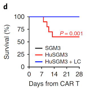
Fortunately, the second study is more extensive and fills much of holes of the first study. Here, the authors led by Attilio Bondanza at San Raffaele Hospital Scientific Institute, Milano, "transplanted human cord blood (CB) hematopoietic stem and progenitor cells (HSPCs) through intrahepatic injection into sublethally irradiated newborn NSG or triple transgenic NSG (SGM3) mice expressing human stem cell factor, granulocytemacrophage colony-stimulating factor (GM-CSF) and IL-3 and initially profiled lymphohematopoietic reconstitution." Basically, this mouse model, referred as newborn humanized SGM3 (nHuSGM3), had both T cell and non-T cell components of hematopoietic system mostly of human origin. In allogeneic tests, human T cells developed in nHuSGM3 mice showed expected functionality. 


Staining of cells in nHuSGM3 challenged with tumor and CAR-T cells that induced CRS showed that monocytes were producing IL-1 early on, followed by IL-6


Depletion of monocytes/macrophages with liposomal clodronate (LC) could rescue mice from CRS.


Importantly, while both anti-IL-1 and anti-IL-6 antibody injection could significantly reduce CRS, only anti-IL-1 antibody were able to reduce neurotoxicity in nHuSGM3 mice. Similar effect were seen with CD44v6 CAR-T cells as well.


In all, these two studies showed that in addition of anti-IL-6 injection, anti-IL-1 antibody therapy could significantly reduce complications of CAR-T cell immunotherapy in humans. They provided a strong evidence to suggest that monocytes were primarily responsible for CRS and neurotoxicity complications of CAR-T cell therapy. 

However, it is not clear why are monocytes getting activated after CAR-T cell infusion in the first place. There is interaction, at certain level, between CAR-T cells and monocytes, but is it antigen-specific via anti-CD19 CAR-T or endogenous TCR, or is it non-specific, is unknown presently. 

posted by David Usharauli 


Wednesday, May 23, 2018

Flagellin-specific T cells induce colitis by recognizing antigen other than flagellin

This is very interesting study from Journal of Immunology (JI). Here, researcher led by Timothy Hand at the University of Pittsburgh Medical School, showed that CBir1 transgenic T cells, thought to cause gut inflammation by recognizing flagellin expressing microbiota, were in fact specific for non-flagellin antigens

CBir1 transgenic mouse have been used for past 10 years to model human Crohn's disease in mice. CBir1 tetramer+ T cells recognize epitope from flagellin, antigen thought to be a target in Crohn's disease. Interestingly, all of those experiments were done using CBir1 T cells on WT background. This could be an issue because it has been known for some time now that transgenic T cells on WT background could use alternative Vα chain to form TCR with new specificity.

To avoid such limitation, here the authors generated CBir1 mouse on RAG KO background that only expressed transgenic Vα:Vβ chains. Surprisingly, unlike CBir1:WT T cells, CBir1:RagKO T cells when transferred in immunodeficient hosts did not induce colitis, and it was not because CBir1:RagKO T cells differentiate into Foxp3+ T cells. It appears that CBir1:RagKO T cells just did not see flagellin epitope in adoptive host.


In vitro tests showed that both CBir1:WT T cells and CBir1:RagKO T cells could respond to DCs pulsed with flagellin epitope [it would have been better and more relevant here to use DCs pulsed with gut flora component].


Other tests showed that CBir1:WT T cells in gut tissue could express alternative Vα chain to form a completely new TCR specificity together with transgenic Vβ chain such as against Ags derived from Helicobacter (HH1713 tetramer). 


In summary, it appears that CBir1 T cells initiate colitis by recognizing non-flagellin antigen from the gut flora through non-CBir1 TCR and only following gut inflammation and gut leakage do flagellin-specific CBir1 transgenic T cells get activated and participate in overall colitis.

So, what is missing from this study? One, it would have been relevant to transfer in vitro activated CBir1:RagKO T cells or activate them in vivo directly to see if then they could initiate colitis. Second, the authors could have tried monocolonization of germ-free mice to see the source of non-flagellin microbiota. Third, there is inconsistency between Fig. 3B and Fig. 6A with regard of proliferation of  CBir1:WT T cells in response to Vanc-treated samples (in vitro it did not proliferate but in vivo it did).

posted by David Usharauli


Saturday, May 5, 2018

IgA protects resident commensal microbiota against competitors

This week journal Science published new study from Sarkis Mazmanian lab at Caltech describing role of IgA in providing strain-specific competitive advantage to certain resident commensal microbiota. 

His lab has been studying immunobiology of Bacteroides fragilis (B. fragilis), a gut commensal. In initial series of experiments they have compared germ-free mice mono-colonized with either wild-type B. fragilis or its mutant variants such as, Δccf, shown to modify biosynthesis of its capsular polysaccharides. They noticed that in co-housing experiments wild-type B. fragilis from one mouse could out compete mutant variant in another mouse in a horizontal transfer assay.   


Since B. fragilis polysaccharides are known to interact with host's immune system, the authors wanted to find out whether host's immune system influenced co-housing experiments. Not surprisingly, the authors found that mutant B. fragilis did not efficiently bind IgA (induced by wild-type B. fragilis) and that it in turn induced IgA repertoire that bound wild-type B. fragilis even less effectively, suggesting some kind of association between IgA and missing antigens on mutant B. fragilis.  


To verify these observations, the authors compared co-housing experiments between germ-free IgA+ and IgA-KO mice (or treated with B cell depleting antibody) mono-colonized with wild-type B. fragilis. Indeed and surprisingly this time, wild-type B. fragilis resident in IgA KO mice were easily overtaken by wild-type B. fragilis from mono-colonized wild-type mice. These results suggested that in absence of IgA wild-type B. fragilis has lost competitive advantage against wild-type B. fragilis resident in IgA+ mice.


What could these results mean in biological context: it appears that certain resident commensal microbiota benefit from interacting with IgA. The authors proposed that "during health, IgA fosters mucosal colonization of microbiota with beneficial properties....while disease states may induce (or be caused by) IgA responses to pathogens or pathobionts that disrupt healthy microbiome equilibria." This is an interpretation that does not provide clear mechanistic explanation as to how IgA response could make such discrimination at the level of antigens between which microbes to keep and which ones to eject from the host. 


posted by David Usharauli

Saturday, April 28, 2018

SUPRA CAR T cell system provides more of cosmetic rather than a real advance

This week journal Cell published a new study from scientists at Boston University describing in their own words "a split, universal, and programmable (SUPRA) CAR system" that supposed to provide several advantages over conventional CAR-T cell system. I reviewed and present here my conclusions on this paper.   

The rationale behind SUPRA CAR T cell design was to develop flexible, "plug-and-play" system to fine tune CAR T cells' activity against tumors without need to redesign it over again. SUPRA consists of two modules: signaling zipCAR construct is artificially expressed by T cells on their surface and soluble zipFv construct expressing tumor antigen specific scFv portion which is injected into system. "Zipper" portions of zipCAR/scFv constructs could interact with each other and by injecting different variants of zipper one can modulate strength of interaction.


What advantage(s) SUPRA CAR T cell design provide? 

1st advantage the authors showed could be to tune signal strength of original SUPRA zipCAR T cells interaction with tumor specific zipFv construct by injecting competitive zipFv constructs that have different affinity to zipCAR module and thus modulate tumor specific zipFv action (to prevent cytokine storm).


2nd advantage is thought to be use of the same zipCAR T cells and inject two different zipFv constructs specific for two different tumor antigens (to prevent tumor escape).


3rd advantage the authors suggested would be to deploy decoy zipFv that could inhibit tumor specific zipFv activity only when decoy scFv interacts with non-tumor specific antigens and thus limiting non-target effects in different tissues (to prevent off-target tissue damage).


Later in the paper the authors went on to present series of experiments that showed comparison of effectiveness of SUPRA CAR T cell construct vs. conventional CAR T cells against two different tumor models in vivo.


On the surface all these experiments look quite impressive. However, close analysis of data shows that advantages are more of cosmetic in nature rather than real ones. First, none of those above mentioned three advantages were actually shown for tumor models in vivo (for some reason the authors did not show how injection of competitive low-affinity zipFv construct could affect tumor protection experiments in vivo or whether double antigen expressing tumors could be efficiently eradicated). Moreover, in vitro experiments showing decoy effect was done in manner that is incompatible for in vivo experiments (one cannot wash away decoy zipFv in vivo before introducing tumor specific zipFv construct and it is likely that free floating decoy zipFv construct could inhibit tumor specific zipFv activity even in absence of decoy tissue antigen).

posted by David Usharauli


Saturday, April 14, 2018

Access to self antigens during germinal center reaction improves self/nonself discrimination against mimicry antigens

This week journal Science published short paper from Chis Goodnow's lab that raises very interesting question about biological significance for existence of anergic self-reactive B cells. Ordinarily, developing B cells when encountering self-antigens undergo deletion, receptor editing or physiological receptor signaling down-regulation that makes such 'anergic' B cells refractory to presence of normal level of self antigens. However, anergic B cells could be re-energized if challenged with high density self antigens or antigens sharing epitope similarity with self antigen.

Now, new study indicates that rather than developing into full blown auto-reactive immune response, anergic B cells when challenged with mimicry antigens mutates its receptors in a such a way, during process of hypermutation, as to achieve a high degree of discrimination between mimicry antigen and actual self antigen.   

The experimental set up itself is quite simple, only complex aspect was to analyze single cell B cell receptor mutation and their binding affinity recovered after antigen challenge. Two type of hosts were used here. Both groups harbor small numbers of self-reactive B cells (CD45.1+ SWHEL B cells)  but only one group also harbored a specific antigen detected by these transgenic SWHEL B cells and expressed "as as an integral membrane protein, mHEL3X, encoded by a transgene with a ubiquitin promoter".


As expected SWHEL B cells in double transgenic hosts were anergic with decreased surface immunoglobulin M (IgM) expression. However, these anergic B cells could be re-activated in germinal centers when challenged with Sheep red blood cells (SRBCs) covalently coupled with self antigen, HEL3X, at high density.


Next set of experiments however showed very unusual results. When challenged with mimicry antigen DEL which slightly differs from self HEL antigen anergic B cell receptors in double transgenic hosts rapidly accumulated mutations that decreased binding affinity to self HEL antigen.


In fact, single cell BCR receptor analysis clearly showed that presence of self antigens dramatically enhanced anergic B cell receptor mutations that allowed up to 5,000-fold better discrimination capacity between self and mimicry antigen (pre vs. post comparison). This is based on assumption that starting affinity to self are the same for both normal and anergic SWHEL B cells population. 


In summary, this study suggests that during germinal center reaction where B cell receptors undergo hypermutation, anergic B cell repertoire, in presence of self antigen, could be salvaged (redeemed) by accelerated accumulation of mutations that modifies their original specificity away from self antigens and allowing more fine discrimination between self and mimicry, cross-reactive nonself antigen. In this scenario, self antigens serve as negative-feedback templates that hypermutating receptors interacts repeatedly in real time to achieve minimal level of binding.

In my view such negative-feedback loop to B cells can only delivered by specialized cell type in germinal center that maintains, keeps memory of host's unadulterated "self antigen collection'' visible to B cells, a task somewhat similar to Foxp3+ Tregs. So, it is possible that new cell type need to be discovered that does it or it is also possible that the same Foxp3+ Tregs localized in germinal centers, referred as follicular Foxp3+ T regs, do it too. 

What are the global implication for such mechanism: It could explain why anergic B cells hang around and how their repertoire could be salvaged without compromising tolerance. The authors also puts forward another intriguing idea that commensal mcrobes and their antigens could serve as negative-feedback loop 'self' templates for anergic B cells that allows them to discriminate between self and mimicking nonself during immune response. 

posted by David Usharauli