Sunday, May 31, 2015

Both non-self and self-specific CD8 T cell clones are found in equal frequency in human PBMCs

Clonal selection model of immune system ontogeny is the most widely accepted concept of immunology. Simply put, it postulates that T cell clones (B cells too) specific for self-antigens (epitopes) are purged from adaptive receptor repertoire during in utero development.

At the time of its formulation, there was no direct proof for this model. It was mainly supported by observations derived from organ transplantation studies. Later, research on TCR transgenic mice confirmed that self-specific T cells were indeed purged from T cell compartment. 

However, more recent studies questioned the validity of results obtain from transgenic mice, mainly due to non-physiological timing of TCR expression in transgenic T cells in the thymus


The authors showed that similar to the frequency of non-specific CD8 T cell clones, healthy human PBMCs contain self-specific CD8 T cell clones with frequency range between 1:104 to 1:10(see below too).


Next, the authors showed that CD8 T cells derived from both female and male donors contained functionally active male antigen, HY-specific CD8 T cells (though, here CD8 T cells were expanded using non-physiological stimuli, such as anti-CD3/anti-CD28 antibodies or PHA).


However, as expected, further experiments with the pool of enriched self-specific CD8 T cell clones revealed that these self-specific CD8 T cells were anergic when stimulated with specific peptide and anti-CD28 antibody (resembling more physiological stimulation, hopefully).


In summary, these results suggest the following: immune system contains lots of self-specific CD8 T cells. These cells are anergic, i.e. non-responders in an ordinary sense. However, they are capable of becoming functionally active when stimulated by non-conventional ways, like PHA or therapeutic antibodies targeting its signaling molecules (CD28, for example) or during the process of autoimmunity?

Why immune system harbors so many self-specific T cells? It was really surprising that on average, PBMCs from male donors still had 1/3 of number of HY-specific CD8 T cells found in PBMCs from female donors. Even though almost every cell in male donors can express HY antigen, the authors still found so many high-affinity HY-specific CD8 T cells in PBMCs from male donors. Staining for mouse HY-specific CD8 T cells showed the same results.

Another interesting observation was the finding that PBMCs from healthy donors contained very high frequency of pre-proinsulin-specific CD8 T cells (almost 1:104) that were significantly increased in donors with T1D. Is it possible that selection of high frequency of pre-proinsulin-specific CD8 T cells has to due with its cross-reactivity for certain evolutionary relevant pathogen-specific epitope?

David Usharauli


Tuesday, May 26, 2015

Leukemia - when RAG genes go Rogue

Receptor diversity of the adaptive immune system (in both B and T cells) is driven by RAG genes. Additionally, B cells also express AID that is responsible for Ig somatic hyper-mutation (SHM) and class switch recombination (CSR). Basically, RAG and AID genes represent naturally occurring gene editing system in mammals.

However, evolution always come at a cost. New paper in Nature Immunology suggests that improperly expressed RAG and AID genes may contribute to the evolution to childhood leukemia.     

By looking at AID expression pattern during human or mouse bone marrow B cell lymphopoiesis, the authors observed that IL-7 signaling played a major role in preventing AID expression prior to small pre-BII cell stage.


In vitro experiments on pre-BII cells from AID-GFP mouse confirmed that withdrawal of IL-7, in combination with LPS (a surrogate for inflammation), induced co-expression of both AID and surface Ig  κ Light-chain (a surrogate marker for RAG gene activity).


Further experiments showed that transduction of mouse pre-BII cells with WT version of pre-leukemic genetic lesion ETV6-RUNX1 induced RAG gene expression.

Finally, adoptive transfer of ETV6-RUNX1 transduced WT pre-BII cells that underwent 5 cycle of in vitro IL-7 withdrawal in combination with LPS, produced leukemia in the NOD.SCID hosts (which lacks its own adaptive immune system). However, ETV6-RUNX1 transduced pre-BII cells from AID-KO or RAG-KO mouse failed to produce leukemia.


In summary, these results indicate that pre-BII cells are vulnerable for improper co-expression of AID and RAG genes due to combination of ETV6-RUNX1 genetic lesion and inflammatory milieu (that may diminish IL-7 signaling). Such condition may arise when the humans are exposed to childhood infections later in their life. It is believed that timely vaccination could reduce incidence of such leukemia in children.     

David Usharauli


Tuesday, May 19, 2015

Tumors repel immune system but attract lytic viruses

Clinically relevant tumors have two characteristics: uncontrolled proliferation (expansion) and immune evasion.

Surprisingly, several recent papers indicated that tumor transformations are associated with inhibition of cell-autonomous anti-viral/anti-modified RNA/anti-modified DNA recognition pathways such as RIG-I or STING.


First, the authors showed that cancer-associated fibroblast (CAF) showed [TGF-β dependent] increase in sensitivity to virus replication compared normal fibroblast harvested from the same cancer patient.


Next, the authors showed that cancer cells too become susceptible to viral infection when co-cultured with cancer associated fibroblast.


This viral titre enhancement was mediated by soluble factor.


Screen for active factors indicated that enhancing factor was fibroblast growth factor-2 (FGF-2). Indeed, inhibition of FGF-2 by RNAi reduced viral titre in tumor-CAF co-culture.


Finally, the authors showed that virus expressing FGF-2 could induce regression of established tumor in mouse model.


In summary, these results suggest that exploiting cancer vulnerability towards lytic viruses may be an alternative path for biological cancer therapy

Note: It is puzzling that virus targets only cancer cells. The authors explained such selective sensitivity of cancer cells based on reduced baseline anti-viral activity in cancer cells. However, we need to consider the fact that cancer patients maybe immune deficient in general, making them generally susceptible to viral infections.

David Usharauli


Saturday, May 16, 2015

Therapeutic antibodies mediate their anti-cancer effect through two different Fcγ receptors

Humanized therapeutic monoclonal antibodies play substantial role in cancer immunotherapy. For example, one of the most recognized member of this class is Rituximab (Rituxan), a humanized α-human CD20 monoclonal antibody used for treatment of B cell-derived malignancies.

It is thought that in humans their anti-cancer effects are mainly driven via human Fcγ receptor IIIA (FcγRIIIA)-mediated ADCC (antibody-dependent cellular cytotoxicity).

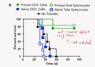
New study in journal Cell from Jeffrey Ravetch lab provided new evidence that α-human CD20 monoclonal antibody have two distinct anti-cancer effects: one, short term, via FcγRIIIA-mediated ADCC, and second, long-term, via FcγRIIA-mediated anti-cancer T cell priming.

Of note, the article has only one first author and one senior author.

Initially, the authors confirmed that presence of Fcγ receptors are necessary for anti-cancer effect of α-human CD20 monoclonal antibody.


Next, the authors showed that wild-type mice treated with α-human CD20 monoclonal antibody and challenged with tumor cells expressing hCD20 antigen became "immune" to secondary challenge of hCD20 antigen expressing cancer cells, implying long-term memory development.


The authors showed that this anti-cancer memory was mediated by T cells generated after primary cancer challenge.


Using mice model with selective expression of human Fcγ receptors, the authors showed that primary anti-cancer effect of anti-human CD20 antibodies was restricted to antibody preferentially binding human Fcγ receptor IIIA, thus activating [macrophage]-mediated ADCC pathway.


The authors confirmed this observation with mice transgenic model expressing only human FcγRIIIA.


Conversely, the authors showed that long-term T cell mediated anti-cancer effect of anti-CD20 treatment was driven by FcγRIIA receptors.


In summary, these results provided new evidence how to improve effectiveness of anti-cancer therapeutic antibodies (it appears that commercial antibodies available on the market today target mainly FcγRIIIA, i.e. ADCC pathway). These results also reinforces the idea that successful cancer protection would require activation of several immune pathways (ADCC and T cell priming).

David Usharauli

Thursday, May 14, 2015

Foxp3+ Tregs depletion activates anti-cancer immunity via eosinophils

Eosinophils are usually associated with atopic/allergic conditions. However, as with all cells involved in type II immune response, eosinophil's role in host's defense is not entirely clear.

This new paper in Nature Immunology provided evidence for eosinophil's anti-tumor function. The paper itself is quite "primitive", observation-type of research article. It belongs more to JEM, if you ask me. Still, it was accepted within 1 month of its submission to Nature Immunology. 

It appears that initial focus of this research was Foxp3+ Tregs. The authors showed that Foxp3Tregs depletion in Foxp3.LuciDTR-4 mice induced B16 melanoma rejection expressing nominal OVA antigen (MO4 tumor cells).

Surprisingly, this rejection of MO4 tumor was accompanied with selective eosinophil infiltration at tumor site.


Indeed, concomitant depletion of eosinphils with Siglec-F antibody significantly reduced anti-cancer effect seen with Foxp3Tregs depletion.

Additional experiments showed that anti-tumor effectiveness of adoptively transferred OVA-specific CD8 T cells were eosinophil dependent, though only in vitro IFN-γ + TNF-α activated, but not resting eosinophils could provide such help to CD8 T cells. Eosinophils alone were not effective.


The authors went on to show that activated eosinophils attracted CD8 T cells to tumor site and promoted normalization of tumor vasculature.

In summary, these results suggests that depletion of Foxp3Tregs activates eosinophils which in turn recruit CD8 T cells into tumor site leading to anti-tumor effect.

Now, why are eosinophils specifically involved in tumor protection in this model is not clear. What attracts eosinophils into tumors?

David Usharauli

Tuesday, May 12, 2015

B cells reduce effectiveness of platinum-based anti-cancer compounds

Some things cannot be explained by conventional thinking. Here is one example. Earlier I reviewed new Nature paper discussing the beneficial role of allo-IgG in cancer therapy. 

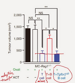
This time, however, another team of scientists reported in the same issue of Nature that B cells and IgA secreting cell in particular, suppress effectiveness of platinum-based anti-cancer compounds, such as oxaliplatin.

The authors, led by Michael Karin at the University of California, San Diego (UCSD), analyzed oxaliplatin's effect in tumor bearing mice. Surprisingly, B cell deficient mice showed significant synergy with oxaliplatin.   


This beneficial effect of B cell ablation on anti-cancer effect of oxaliplatin was CD8 T cell-dependent.


Next, the authors showed that inhibition of oxaliplatin-induced anti-cancer effect was related to TGF-beta signaling in B cells and their differentiation into IgA producing cells.  


Furthermore, the authors showed that B cell-specific expression of IL-10 and PD-L1/2 contributed to cancer resistance towards oxaliplatin.


Finally, the authors showed that adoptive co-transfer of B cells with T cells induced cancer resistance to oxaliplatin's treatment via TGF-beta signaling.


In summary, these results suggest that initial anti-cancer action of oxaliplatin primes B cells to develop into IgA secreting cells and to inhibit CD8 T cell's anti-cancer effector function via IL-10 and PD-L1/2 signalling.

It is not clear whether IgA secretion per se has any inhibitory role. It appears that oxaliplatin activates or amplifies pathways towards IgA differentiation that is inhibitory to CTL functions. Does oxaliplatin action mimics gut immune environment? This fact requires further investigation to understand its mode of action. It could have a separate usefulness for oral vaccine formulations. 

David Usharauli


Thursday, May 7, 2015

Minor antigen-specific allogeneic IgG molecules initiate robust anti-cancer immunity

Cancer cells express two type of antigens relevant for immune system: major and minor antigens. Major antigens are MHC class I, II and related antigens (HLA in humans). All other antigens are classified as minor antigens. Essentially, when scientists talk about cancer associated antigens they mean minor antigens.

In general, we are tolerant towards both our own major and minor antigens both at B cell or T cell level. This is why it is very difficult for the immune system to spot cancerous cells. Cancer cells must accumulate sufficient number of non-synonymous mutations in its minor antigens before body's immune system can sense it.

New paper in journal Nature discusses new way how to accelerate anti-cancer immunity using allogeneic priming. This is how it works.

The authors employed concept of allogeneic priming. Basically, there are B6 mouse strains which differ only in minor antigens but have the same MHC molecules (antigenic differences between humans are frequently located in minor antigens too). 

Depending on number of minor antigenic differences between these MHC-matched allogeneic hosts, immune system of each sub-strain lacks tolerance towards other's minor antigens. For example, B16 melanoma cells are MHC-matched but minor antigenic mismatched in 129S1 substrain of B6 mice and vice versa LMP cancer cells are MHC-matched but minor antigen mismatched in C57 substrain of B6 black mice. Accordingly, each tumor is rejected easily by MHC-matched allogeneic hosts via T cell dependent manner [because of abundance of minor antigenic difference].


Unexpectedly, this rejection was dependent of host B cells too


Next the authors showed that syngeneic hosts injected with syngeneic BmDCs that were pre-incubated with tumor lysate coated with allogeneic IgG (but not IgM) were protected against syngeneic tumors. (syngeneic cells should have identical major and minor antigens)  


Interestingly, tumor associated DCs (TADC) were incapable of providing such protection, nor direct intra-tumoral allo-IgG injection was effective (however, in Fig. 1k, the authors showed that injection of allo-IgG was effective. Don't know what is the difference).


However, priming with allo-IgG in combination with CD40L/TNF-alpha or PolyI:C could provide anti-cancer immunity. In addition, injection of TADC harvested from immunogenic cocktail treated mice could protect naive mouse against cancer.


In summary, these results (and there are lot of figures) suggest that using allo-IgG priming strategy could help overcome host tolerance towards syngeneic tumors. 

How this could apply for human case: one can harvest patient's DCs and incubate with tumor cells coated with allo-IgG. After re-injection, these DCs should then activate patient's T cells and participate in tumor protection

I don't see that direct injection of allo-IgG in combination with CD40L/TNF-alpha or PolyI:C is a feasible approach though.

Strange thing about this study is the fact that pre-absorbing of allo-IgG serum with normal mouse tissue syngeneic to tumor could abolish protection. If immune system detects normal self-antigens and then initiate cancer immunity, how is then autoimmunity prevented? Most likely, self-antigens are somehow linked to mutant antigens in cancer cells and help to deliver them to DCs for presentation to cancer-specific T cells. Since majority of self-specific T cells are tolerant to self-antigens to begin with, it is less likely that this procedure would initiate autoimmunity.

David Usharauli

Sunday, May 3, 2015

exTh17 cells cannot go unnoticed

Interleukin-17 (IL-17) is clinically relevant target molecule. T helper subset called Th17 are thought to produce large amounts of IL-17. Paradoxically, IL-17 is implicated in several human diseases such as psoriasis and inflammatory bowel disease (IBD), as well as autoimmune arthritis and autoimmune encephalitis. So, scientists are naturally very curious to understand Th17 subset's biology.

This new paper in journal Nature provided half-hearted data suggesting that Th17 cells undergo trans-differentiation into "regulatory T cells".    

Initially when I saw the article's title and read its abstract I thought it was about Th17-Foxp3 conversion since the authors used such term such as "regulatory T cells" which ordinarily refers to Foxp3+ CD4 T cells. However, it turned out results had nothing to do with Foxp3 T cells. 

Initially, the authors showed that in both steady state or during α-CD3 induced inflammation, CD4 T cells expressing IL-17 could lose its expression and instead up-regulate IL-10 (called here Tr1exTh17 cells). However, for some reason the authors failed to show IL-10 expression level on CD4 T cells maintaining IL-17 expression (this could have been useful control). In addition, the authors did not discuss or show whether presence of two different types of IL-17A gene in a single cell could affect their individual expression maximum.


Next, the authors showed that Tr1exTh17 cells could prevent colitis development in RAG KO hosts when co-transferred with pathogenic Th17 cells.


The authors also showed that anti-CD3 injection in mice immunized 35 days earlier with brain protein MOG  could induce Tr1exTh17 cells.


These data so far suggest that non-physiological stimulation of T cells with α-CD3 antibody induced down-regulation of IL-17 and up-regulation of IL-10 in a subset of activated CD4 T cells. Whether these Tr1exTh17 cells are derived from bona fide Th17 is not clear.   

Next, the authors used two disease models.  In Th2 dominant disease model of Nippostrongylus Brasiliensis, the authors observed the development of Tr1exTh17 only after 2nd round of re-infection.


In a second Th17 dominant disease model of Staphylococcus Aureus, however, the authors conclusions that there is Tr1exTh17 cell development is premature, in my view. The authors simply did not apply proper flow cytometry gating in this case.


In summary, in my view the correct interpretation of the data in this paper is following:

1.  Tr1exTh17 can develop from activated T cells previously expressing IL-17, however those cells may not be bona fide Th17 cells.  

2. Effect of non-physiological stimuli such as α-CD3 antibody on T cells differentiation may lead to incorrect conclusions.

3. Based on results, in Staphylococcus Aureus model, there is no or very limited trans-differentiation of exIL-17 expressing T cells into Tr1exTh17.   

Shortly, the major flaw in this paper is its heavy focus on gene-modified mice (on technology) rather than on biology.    

David Usharauli