Saturday, February 28, 2015

Specific food additives modify gut microflora and cause metabolic syndrome

Here is another paper that highlights unsuspected health risks of commonly used food emulsifiers, carboxymethylcellulose (CMC) and polysorbate-80 (P80).

Ordinarily, mucus separates gut epithelial cells from the gut microflora. New study in Nature provided evidence suggesting that this separation of gut epithelial cells from the gut microflora is disrupted by CMC and P80, two commonly used food emulsifiers (but not by sodium sulfite, another commonly used food additive).


The authors showed that presence of either CMC or P80 in either drinking water or dry food pellets could promote development of colitis in susceptible mouse strain (IL-10 KO).


In addition, the authors  showed that presence of either CMC or P80 in water or dry pellets could promote development of metabolic syndrome in wild-type as well as in susceptible mouse strain (TLR5 KO).


Interestingly, metabolic syndrome did not develop in germ-free mice treated with CMC or P80.


More importantly, effects of CMC or P80 on animal health were transferable to germ-free mice by fecal transplantation from treated mice, implying direct role of modified gut microflora (note, CMC or P80 did not directly modify mucus thickness in germ-free mice).


In summary, these results support the growing evidence that chemically-processed food carries long-term health risks (development of type II diabetes, metabolic syndrome, inflammatory bowel disease).

David Usharauli

Wednesday, February 25, 2015

Inflammatory signals from the peripheral tissue modulates immune response in lymph nodes

Very unusual results have been published in journal Science. This study came from Facundo Batista's lab at the London Research Institute.

The authors studied lymph node's adaptive re-organization in response to peripheral inflammatory signals, with the focus on CD169-positive macrophages. These specialized macrophages are ordinarily positioning themselves in the subcapsular sinus of the lymph nodes intercepting any incoming antigens from the peripheral tissue for presentation to B cells.

Using different type of inflammatory signals (live or dead bacteria, live or inactivated viruses, or TLR agonists), the authors observed that live bacteria or virus or TLR agonist injected in the mouse footpads caused temporal disruption of CD169+ macrophage layer in the draining lymph node.   


Further experiments showed that disruption of CD169+ macrophage layer in the draining lymph node upon CpG injection was abolished in mice selectively deficient for MyD88 signaling in dendritic cells.


In addition the authors noticed that disruption of CD169+ macrophage layer was also reduced in CCR7-KO mice where DCs could not migrate from the peripheral tissue to the lymph node.  


To understand physiological consequences of CD169+ macrophage layer disruption, the authors injected labeled B cell antigen into footpad following initial CpG or PBS (control) injection (+ 4 / +7 days later). These experiments revealed that fewer CD169+ macrophage and fewer antigen-specific B cells could acquire antigen following CpG injection.   


More importantly, the authors showed that initial CpG injection reduced subsequent anti-viral antibody response.


In summary, these results shows that initial inflammatory signals coming from peripheral tissue disrupt CD169+ macrophage layer and antibody response to subsequently injected antigen. 

This is strange results. No good explanation is given. For one, the authors did not provide results regarding viral titre or tissue pathology in mice injected with primary CpG. 2nd, isn't lymph nodes function to respond to the inflammation in peripheral tissue? How could it be beneficial for the host to shut down immune response following inflammatory stimuli? Could it be that lymph nodes are modified to activate different type of immune response (CTL?)

David Usharauli


Saturday, February 21, 2015

Fasting-induced ketone metabolite, β-hydroxybutyrate suppresses NALP3-mediated inflammasome activity

Inflammasome super-complex represents an ancient innate detection and defense mechanism against exogenous as well as endogenous irritants and toxins. Spontaneous inflammasome activity causes several known human immune-pathologies, like Muckle-Well and familial cold auto-inflammatory syndromes.  

New paper from Nature Medicine provided evidence showing that alternative energy source, β-hydroxybutyrate, elevated during fasting, calorie-restiction or high-intensity exercise, suppresses NALP3-mediated inflammasome activity.

Initially, the authors led by Prof. Vishwa Deep Dixit at the Yale School of Medicine, showed that β-hydroxybutyrate, but not another ketone body acetoacetate or short-chain fatty acids, butyrate or acetate, could inhibit NALP3-mediated inflammasome activity (AIM2- or NLRC4-mediated inflammasome activities were unaffected).


Suppression of NALP3-mediated inflammasome activity by β-hydroxybutyrate was independent of autophagy and required neither G protein-coupled receptor GPR109a signaling via β-hydroxybutyrate nor histone deacetylase inhibition by β-hydroxybutyrate.


The authors found that β-hydroxybutyrate suppressed NALP3-mediated inflammasome activity through its inhibition (by unknown mechanism) of potassium, K+ cation efflux from cytoplasm.


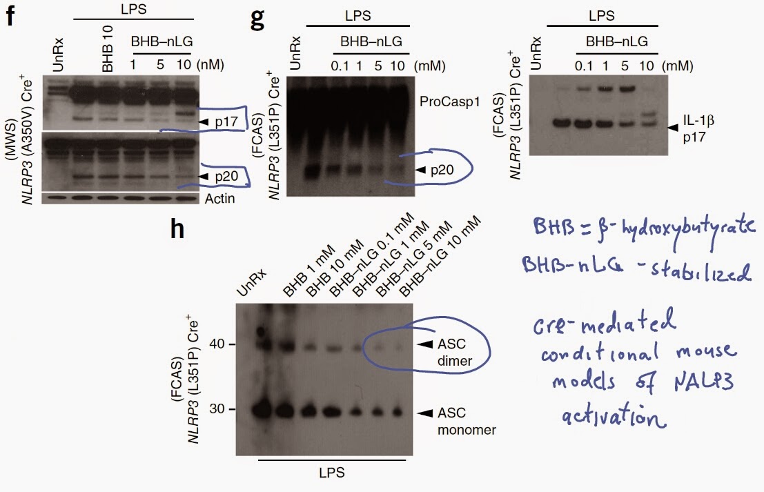
Finally, using cre-mediated mouse conditional models of human Muckle-Well and familial cold auto-inflammatory syndromes, the authors showed that stabilized form of β-hydroxybutyrate could suppress spontaneous NALP3-mediated inflammasome activity and ASC oligomerization characteristics to these gain-of-function NALP3 mutations.


In summary, these results indicate complex interplay between energy metabolism and inflammation. In condition of glucose deficiency, elevated level of β-hydroxybutyrate acts as a new source of ATP in brain and heart and at the same time inhibits NALP3-mediated inflammasome activity.

Leave your comments below and let me know what do you think about this paper.

David Usharauli

Tuesday, February 17, 2015

microbiota influences non-genomic hereditary immune phenotype

The major advances in fundamental immunology for the past 25 years have to do with major advances in artificial manipulations of gene expressions (knock-out, knock-in, conditional knock-out, CRISPR-Cas9, etc).

Until recently very little attention was given to the extra-genomic influence of such gene manipulation. The scientists rarely bothered and still many don't to study gene effects in homozygous offspring derived from heterozygous parents to balance for exrta-genomic influence

For example, it is conceivable that homozygous gene effect are influenced wholly or in part by extra-genomic factors such as microbiota. This is exactly what a new study in journal Nature suggests.

In this study, the authors made initial observation that two colonies of genetically identical B6 mice kept in two different research facilities differed in their gut derived fecal IgA level (IgAhigh and IgAlow). Interestingly two colonies did not differ in serum IgA level. The authors found that fecal IgA phenotype was vertically transmittable from parents to progeny.


Surprisingly, co-housing experiments revealed that IgAlow phenotype was dominant. This dominance of IgAlow phenotype was reversed when fecal materials were passed through 0.45 micron filter before transplantation into IgAhigh hosts.


In addition, the authors found that pre-treatment of IgAlow mice with broad-spectrum antibiotics or ampicillin could reverse IgAlow phenotype as well, indicating that presence of ampicillin sensitive large microbes were responsible for this microbiota effect.


The authors found that IgAlow mice intestine were more sensitive to chemical injury compared to IgAhigh mice.


However, intestinal sensitivity to DSS were independent of microbiota and dependent of intestinal IgA secretion.

To understand the mechanism behind IgAlow phenotype the authors tested components of fecal IgA complexes. These experiments revealed that IgAlow mice had profound deficiency in secretory component of IgA complexes in fecal samples. Secretory component is known to protect IgA from degradation.  
 

Finally, the authors showed that microbial pellets derived from IgAlow mice could degrade secretory component in a protease-dependent manner (use of germ-free host mice would have been beneficial to know exactly what microbe was responsible for this effect).


In summary, these results showed that non-genomic microbiota could influence hereditary immune phenotype. When studying gene-manipulated mice models, it is of critical importance to evaluate the model in proper manner (fecal transplantation, co-housing or heterozygous parents).

David Usharauli


Saturday, February 14, 2015

Cancer-bacterial relationship: how wrong focus can spoil everything

New concepts are only useful if they can explain something or predict something. Sometimes scientific idea is good but using wrong model to test it could produce negative impression.

Here is the one example for a such scientific approach. This paper was published in journal Immunity few days ago. This study identified bacterial component, Fap2, that can protect cancer cells from NK cell cytotocixity through TIGIT interaction. This concept of bacteria protecting tumors against immune system is very interesting, so I decided to analyse it.

Initially the authors showed FITC labeled Fusobacterium nucleatum (F. nucleatum) strain 23726 could bind both hematopoietic and epithelial cancer cell line. Surprisingly, these cell lines pre-incubation of F. nucleatum strain 23726 showed resistance to NK cell natural cytotoxicity (but there was no effect on IFN-γ or TNF-α production). This inhibition of NK natural cytotoxicity was specific for F. nucleatum strains since Escherichia coli lacked this effect.


The authors speculated and then showed that inhibition of NK natural cytotoxicity by F. nucleatum strains could be due to its interaction with inhibitory receptor TIGIT.


Indeed, transfection of TIGIT-negative NK cell line with human TIGIT could reduce its natural cytotoxicity against EBV-transformed B cell line.


Similar reduction of NK cell natural cytotoxicity were observed when primary human NK cells were used, though effect was minimal when using human colorectal carcinoma cell line, RKO.


Next, the authors were able to show the actual clinical isolates of F. nucleatum strain recovered from human colon adenocarcinoma samples could inhibit NK cell cytotoxicity.


Using F. nucleatum transposon-based insertion-inactivation mutant library the authors showed that inhibition of NK cell natural cytotoxicity was reversed with Fap2 mutation.


Additional the authors showed that Fap2 mutant F. nucleatum strains failed to induce IL-2 production from cell line transfected with human TIGIT.


Fap2 mutant F. nucleatum strains also failed to interact with purified human TIGIT.


Finally, the authors showed F. nucleatum strains could (1) inhibit TIL cytoxocity against autologous melanoma cells in a Fap2-dependent manner and (2) inhibit peptide-specific T cell IFN- response.


In summary, these results show that F. nucleatum strains could interact with human TIGIT and this interaction reduces NK cell cytotoxicity in a Fap2-dependent manner but has no effect on NK derived IFN-γ or TNF-α production. Strangely, it appears that Fap2 inhibited both T cell cytoxicity and IFN-γ production.

In my opinion since the authors argue in the beginning that F. nucleatum strains are mostly found in human colon adenocarcinoma tissue, but showed that Fap2 effect on NK cytotoxicity against colorectal carcimona cell line, RKO, is negligent, it puts the significance of this finding into question.

David Usharauli

Wednesday, February 11, 2015

pH sensitive targeting of miR-155 reverses lymphoma oncomiR addiction

Acidity is a general property of tumor environment. The new research in journal Nature described new platform for selective delivery of anti-sense RNAs targeting OncomiR155, a well known target that fuels lymphoma growth.

To this end, the authors attached anti-miR155 RNA sequence to pH sensitive peptide (pHLIP-miR155). In acidic environment, anti-miR155 is cleaved from the peptide and can silence oncomiR155.


In vitro experiments confirmed that pHLIP-miR155 cellular uptake was pH dependent.

The authors showed that pHLIP-miR155 was selectively accumulated in lymphoma tissue and also in kidney. However, the authors did not see any kidney toxicity (see above).

Using spontaneous miR155-driven tumor model, the authors showed that pHLIP-miR155 delivery could improve mice survival, compared to anti-mirR155 alone.


pHLIP-miR155 showed similar anti-tumor effectiveness as mice treated with DOX (in miR155 Tet-Off tumor model) or CHOP (chemotherapy + steroids). Importantly, pHLIP-miR155 did not have toxicity associated with CHOP treatment.


In summary, this approach could be beneficial for treatment of tumors heavily addicted to OncomiRs.

Leave your comments below. Lt me know what do you think about this paper or my analysis.

David Usharauli


Tuesday, February 10, 2015

Mitochondrial DNA abnormality activates cytosolic type I IFN system

Mitochondria is a cell's energy hub. It is of a prokaryotic (simple bacterial) origin. It has its own DNA. However, prokaryotic DNA can activate innate immune system. So, what would happen if our mtDNA undergo misfolding stress?

This is exactly what the new Nature paper tried to uncover. This study led by Gerald Shadel at the Yale School of Medicine, has examined the outcome of mitochondrial DNA (mtDNA) packaging abnormality by studying mouse model heterozygous for mitochondrial transcription factor A (TFAM-/+), a protein responsible for mtDNA higher-order nucleoid organization.

Gene expression profiling revealed that cells in TFAM-/+ mice showed enrichment in IFN-stimulated pathway genes.


The authors showed that this type I IFN response was significantly abrogated in cGAS, STING, IRF3 and TBK1 deficient backgrounds indicating that in TFAM-/+ mice aberrant mtDNA released into cytosol activates cGAS-STING-IRF3 pathway leading to type IFN production.

Additional experiments showed that such heightened type I IFN response in TFAM-/+ mice reduced viral cellular burden.

Finally, the authors showed that viruses from herpesviridae family could induce mtDNA misfolding (by TFAM depletion via viral-encoded UL12 M185 molecule) and mtDNA dependent type I IFN response in wild type mice, mimicking TFAM-/+ mice, suggesting physiological function of mtDNA stress response.


In summary, these results highlight how mitochondrial DNA stress could activate innate immune system and cause inflammation (in human disorders such as systemic lupus erythematosus, cancer, multiple sclerosis, etc).

This study provides additional support for recently described immunological function of mtDNA and its detection by cGAS-STING-IRF3-type I IFN pathway. It is of note that many cellular abnormalities analysed from immunological point of view are converging on type I IFN response.

David Usharauli


Saturday, February 7, 2015

antigen-specific IgG1 isotype protects against type III allergy (hypersensitivity)

IgG antibody family consists of four isotypes: IgG1, IgG2a, IgG2b and IgG3 (IgG1-4, in humans). They differ in their Fc portions. Current knowledge suggests that IgG isotypes have different function depending on a degree of their interactions with complement and FcRs.  

New study in journal Nature, however, provided evidence for complement- and FcR-independent function of IgG1 isotype.

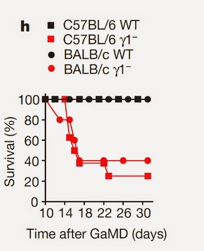
This research led by Fred Finkelman at the University of Cincinnati College of Medicine, have analysed immune response of mice deficient for IgG1 isotype (Igγ1mice). Strangely, immunization with goat anti-mouse IgD anti-serum (GaMD, protein immunogen) led to the death of Igγ1- mice, but not wild-type mice.


The authors showed that this death of Igγ1mice was caused by kidney insufficiency.


Analysis of immune response to GaMD in Igγ1mice revealed selective increase in antigen-specific IgG3 isotype and IgM in serum and their deposition in kidney's glomerular capillaries. Interestingly, kidney damage in Igγ1mice was still present in FcγR1- or FcγR1- and complement C3 double deficient mice.


The authors showed that kidney damage could occur even in wild-type mice by passive injection of antigen (TNP)-specific IgG3 and antigen (TNP-protein). Other IgG isotypes did not induce renal disease.


Importantly, concomitant injection of antigen specific IgG1 and disease-inducing IgG3 could prevent renal disease development independent of complement C3 or inhibitory FcγRIIb.


In summary, these results indicate that induction of antigen-specific IgG1 response during protein immunization prevents the development of type III hypersensitivity. Mechanistically, IgG1 competes for antigen binding with antigen-specific IgG3, thus inhibiting large antigen-antibody complex formations typical for IgG3.

This new knowledge could help to design new treatments for human cryoglobulinemia.

David Usharauli


Thursday, February 5, 2015

Tissue-specific regulatory T cells control asthma development

Foxp3+ CD4 regulatory T cells (T regs) maintain immune tolerance to self. Exactly how do they do it, no one really knows. Studies have described so many different mechanisms of T regs function that one can only wonder whether these cells really belong to one family.

Most likely, T regs may function in a tissue-specific manner as proposed by P. Matzinger and T. Kamala. New study published in Nature Immunology may support this hypothesis.

This study is a combined effort by French and German scientists. Here, the authors have examined the role of protein kinases in T regs function. Initial experiments showed that protein kinase, CK2 (casein kinase 2), was the most up-regulated kinase in activated T regs compared to effector T cells.


Next, the authors generated a mouse model carrying Foxp3-specific deletion of CK2 in T regs by crossing Foxp3-cre mice with CK2 fl/fl mice. 


Interestingly, analysis of different tissues in Foxp3-cre CK2  fl/fl mice revealed that only lung tissue was affected by Foxp3-specific CK2 deletion in T regs.


Further analyses revealed that Foxp3-specific deletion of CK2 in T regs resulted in selective increase in type 2 immune signature in lung draining lymph nodes and in serum.


In fact, spontaneous lung inflammation in naive Foxp3-cre CK2  fl/fl mice was comparable to allergic lung inflammation (asthma) in antigen sensitized control mice.


However, surprisingly, T regs homing into lung tissue was not affected by Foxp3-specific CK2 deletion in T regs. More detailed analysis of T regs from Foxp3-cre CK2  fl/fl mice revealed selective up-regulation of inhibitory receptor ILT3.


The authors showed that ILT3 expression in CK2-deficient T regs could dampen TCR signalling in T regs.


Finally, the authors showed that CK2 deletion in T regs led to selective increase in numbers of type 2 response promoting IRF4+ PD-L2+ dendritic cells (DCs).


In summary, these results indicate that CK2 deletion in T regs induces ILT3 up-regulation and attenuation of TCR signaling in lung-tissue T regs, leading IRF4+ PD-L2+ dendritic cells (DCs) expansion and type II inflammation in the lung

How these results advance our knowledge in Foxp3+ T cells? No data are provided to explain why lung-tissue T regs were so selectively affected by CK2 deletion or how IRF4+ PD-L2+ DCs are amplified. Very interesting and very strange.

Let me know what do you think about this study.

David Usharauli