Wednesday, February 22, 2017

Antigen-specific Tregs maintain immune privileges of male reproductive tract

This week Journal of Clinical Investigation published very good research article that shed light on how tolerance to sequestered self-antigens expressed by immune privileged  tissues are established

Some tissues such as brain, eye, testis or ovaries are thought to be "immune privileged" organs meaning that ordinarily immune system does not see their antigens. However, in this paper, the authors showed that in fact some testes antigens in male mice, for example lactate dehydrogenase 3 (LDH3), are actually secreted and detected by immune system.   

Series of experiments confirmed that WT male mice did not respond to LDH3 immunization, while female and LDH3-null male mice mount detectable immune response to it. This suggested that male mice were physiologically tolerant to LDH3. Notable, both male and female mice could be immunized against another testis antigen, zonadhesin (ZAN), implying absence of tolerance to ZAN in male mice. 


So, how male mice were tolerant to LDH3? To answer it, the authors temporally depleted Tregs and it led to immune response to LDH3 in immunized WT male mice. since no changes were seen in response to ZAN, the authors concluded that tolerance to LDH3 in WT male mice was dependent of presence of FOXP3+ Tregs.


Furthermore, depletion of Tregs even in absence of testes antigen immunization still led to autoimmune pathology in testes in ~40% of male mice ("autoimmune orchitis occurs in autoimmune polyendocrine syndrome 1 (APS1) patients due to mutations of AIRE, possibly associated with impaired thymic deletion of autoreactive T cells and deficient Treg function")


In summary, this study suggests that "immune privilege" is not absolute and self-antigens that naturally leak maintain tolerance by induction of antigen-specific Tregs.

David Usharauli


Thursday, February 16, 2017

IL-33 → MyD88 pathway drives type II immunity in female genital mucosa

Type II immunity underlies several types of immune conditions such as asthma, allergy or response to parasitic helminths. It is not yet fully clear how type II immunity contributes to host's defenses. At this stage scientific inquiry is mostly focused on uncovering cellular and molecular mechanism behind type II immunity.


For these experiments the authors have used papain, a well established experimental type II inducer protease. As reported before, papain required enzymatic activity for its type II response.


Papain application to female vaginal mucosa induced IL-33, a member of IL-1 family implicated in type II immunity.


Indeed, IL-33 deficient mice showed reduced secretion of components of type II immunity (IL-4, IgE).


Interestingly, type II immunity in response to papain in female genital tract depended on IRF4+ CD11c+ dendritic cells but it was independent of basophils and eosinophils.


Furthermore, type II immunity in response to papain application to female vaginal tissue depended on MyD88 signaling.  


In summary, this study confirms the role of IL-33 in type II immunity.

David Usharauli


Thursday, February 9, 2017

Newborns' resistance to pneumonia is driven by acquired microbiota

This week Science Translational Medicine published new study that showed that in newborn mice resistance to pneumonia is driven by neonatally acquired microbiota. It revealed how antibiotic therapy given to mothers near time of delivery could alter and weaken baby's defenses against airway pathogens.

Newborn mice derived from germ-free or from antibiotic-exposed pregnant mice display increased susceptibility to Streptococcus pneumoniae serotype 19A-induced pneumonia that could be reversed by microflora.


Application of epithelial-focused cytokine IL-22 had similar effect on reversing newborn mice susceptibility to pneumonia.


In the lungs of newborn mice, majority of IL-22 is made by RORgt+ group 3 innate lymphoid cells (ILC3).


Antibiotic therapy of pregnant mice reduced IL-22+ ILC3 population in the newborn lungs that could be reversed by microflora.


Moreover, depletion of endogenous ILC3 increased host's susceptibility to pneumonia that could be reversed by adoptive transfer of WT ILC3.


In summary, this study showed that antibiotic therapy of pregnant females at the time of delivery could profoundly affect newborns' ability to mount proper defense against airway pathogens by depleting microflora and disrupting microflora → ILC3 → IL-22 axis.


David Usharauli


Tuesday, February 7, 2017

cGAS puts gas on anti-tumor effect of checkpoint inhibitor

This week PNAS published new article explaining mechanism of action of checkpoint inhibitors, such as anti-PDL1, in tumor immunity. It shows that cytoplasmic DNA sensor cGAS/STING pathway synergize with anti-PDL1 therapy in mouse model of melanoma.

While this study provides noteworthy observation, it is poorly done. For example, when comparing WT and KO mice (cGAS or STING KO mice), the authors did not mention if they used littermate control in these experiments.


In addition, when the authors used exogenous cGAMP (product of cGAS activity detected by STING), they did not use it on cGAS or STING KO mice as controls to verify relationship between cause and effect.   


In summary, the connection between DNA recognition system and checkpoint inhibitors is very interesting. Whether it is a simple generic augmentation of T cell priming or specific stimulation of tumor-specific T cells is to be seen.   

David Usharauli


Sunday, February 5, 2017

New synthetic molecule prevents autoimmunity without compromising anti-pathogen immunity

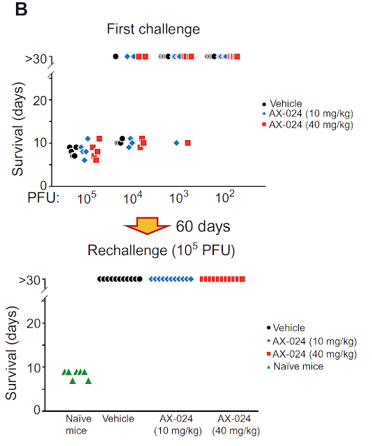
In December of 2016 Science Translational Medicine published new study where the authors introduced new, low-molecular weight compound that specifically inhibited autoimmune/allergic responses while sparing anti-pathogen immune response.


This new molecule, AX-024, specifically targets adaptor protein Nck that is involved in low affinity TCR signaling


It does not affect signaling delivered via IL-2, BCR or PMA/Ion stimulation.


AX-024 inhibited TLR7/8 agonist, imiquimod (IMQ), induced psoriasis skin symptoms in mice.


Also, AX-024 significantly reduced clinical symptoms associated with mouse model of human multiple sclerosis (MS). Such effect of AX-024 was superior to Fingolimod, a sphingosine 1-phosphate receptor modulator approved for the treatment of relapsing-remitting MS.


Notably, AX-024 in therapeutic dosage did not impair anti-pathogen immunity or memory generation.


In summary, by blocking Nck adaptor protein involved in low-affinity TCR signaling, AX-024 inhibited autoreactivity while preserving necessary capacity for anti-pathogen immunity. Such property makes it desirable candidate for clinical trials in humans.

David Usharauli 


Saturday, February 4, 2017

Tregs muscle up in the periphery

There is still a lot of confusion regarding role of peripherally generated Foxp3+ Tregs. Data so far indicate that peripherally induced Tregs play limited [if any] role in maintaining check on auto-reactive T cells. However, extra-thymic Treg population could prevent "excesses" of other T helper class differentiation.


High level of Musculin expression was detected in in vitro induced Foxp3+ Tregs (iTregs).


Musculin deficient mice harbor fewer Tregs at gut mucosal sites.


Musculin deficiency in T cells shifted their differentiation pathway from Foxp3+ Tregs into Th2 cells.


Indeed, removal of canonical Th2 cytokine IL-4 reversed such Th2 shift in Musculin deficient T cells under iTreg differentiation setting.


Musculin deficient iTregs also failed to inhibit house dust mite allergen induced inflammation in lungs upon adoptive transfer.


For me these data confirm that stability of extra-thymic Foxp3+ T regs in the periphery requires coordinated action of several factors, one of which, appears to be MusculinMusculin prevents excessive Th2 differentiation in settings that may favor local Tregs generation. Extra-thymic Treg differentiation pathway does not appear to play a significant role in maintaining peripheral tolerance. Rather, their role is mostly, in my view, consist of absorbing and neutralizing any "excesses" in T helper differentiation, in this case Th2 pathway.

David Usharauli


Friday, February 3, 2017

Regulatory T cells with a single TCR specificity prevent lethal autoimmunity

Foxp3+ regulatory T cells, Tregs, prevent autoimmunity and their total or partial defects can cause lethal or variable autoimmunity. For most part, endogenous ligands recognized by Tregs are not known.

Based on data derived from artificial transgenic models it is suggested that development and maintenance of naturally derived Tregs could be antigen-specific, though it is not known how diverse Treg TCR repertoire should be to keep auto-reactive T cells in check.

To answer this question, Rudensky's research group conducted series of experiment on transgenic mice (G113TgFoxp3YFP-CreTcraFL/FL mice) which harbor normal repertoire of conventional T cells but where "all" naturally developed Treg cells would express single TCR specificity (derived from Vβ6+Vα2+ G113 TCR).

Surprisingly, unlike control mice (which did not survive past 4 wk of age), 50% of G113TgFoxp3YFP-CreTcraFL/FL mice survived to 4 mo of age (the end point of the experiment) though all of them displayed "visible evidence of significant autoimmunity".


So what does this result tell us? These are quite unexpected results. It would be highly unusual to imagine that a single antigen and single Treg specificity could be sufficient to prevent lethal autoimmunity in 50% of mice. We don't even know the source of the endogenous antigen G113 TCR supposed to recognize.

I personally would rather focus on technical aspect of this result: how robust is TCR modification in G113TgFoxp3YFP-CreTcraFL/FL mice? Maybe there is still "leakage" of endogenous TCRalpha chains? I wonder about this because of "50% data". Maybe in those 50% surviving mice Tregs were able to assemble TCR with endogenous TCRalphas?

David Usharauli