Thursday, March 31, 2016

Classical DCs are just that classy for oral tolerance and peripheral Tregs

This week I had a hard time to find any good immunology paper that makes me happy to review. Two days ago I read one new paper from immunity with intriguing title about "Organ-Specific Regulatory T Cells" that was so weak (structurally, conceptually and experimentally) that I have no idea how it ended up in that journal. Yesterday I read another new paper from Nature Immunology about dendritic cells (DCs) that had 8 figures in total but only 3 figures were relevant and conclusive, though even that lacked experimental strength. Let me explain my point.

In this paper the authors wanted to find out which type of DCs were involved in oral tolerance (oral tolerance has been used in clinics, for example, to reduce allergy to food products). To do that, the authors compared mice that lacked either monocyte-macrophage–derived DCs [MMDTR mice] or pre-DC–derived classical DCs [zDCDTR mice or CD11cDTR mice as a positive control]. When these mice were fed with OVA protein (to induce oral tolerance) and later challenged with the same antigen in the skin, only mice lacking cDCs showed loss of oral tolerance induction (no reduction in ear swelling or OVA-specific IgG1/2c levels).


Similar results were obtained in upper respiratory airway and lung allergy model (no reduction of eosinophil recruitment in BAL in zDCDTR mice that lacked classical DCs).


Mechanistically, lack of oral tolerance was "associated" with total lack of OVA-specific peripheral [adoptively transferred OT-II] Treg induction in zDCDTR mice (please note that though MMDTR mice showed dramatic reduction of peripheral OVA-Treg generation, it did not affected oral tolerance induction. See Figure 1).


In summary, this study showed that classical DCs were relevant for oral tolerance and antigen-specific peripheral Treg induction, but these two processes were not necessarily connected (at least no experiments were shown to make this connection). So what's the point of showing all these? I don't have an answer to that and probably neither the authors do.

David Usharauli

Saturday, March 26, 2016

Papain protease activity and mast cells, but not IL-33, are necessary for papain allergic sensitization

Papain is a proteolytic enzyme from papaya. Like other protease allergens such house dust mites (HDM) group 1 allergen, Der p 1, papain is allergen that belongs to family of cysteine proteases. Papain can induce skin contact sensitization or airway hyper-reactivity. 


When injected in mouse ear lobes, intact papain, but not protease-inactive papain (E64-papain), induced skin inflammation and IgE production.


Papain induced IgE production after skin challenge depended on presence of functional mast cells as (a) such response was diminished in mast cell–deficient WBB6F1-W/Wv (W/Wv) mice, and (b) such response could be recovered after transfer of WT mast cells. 


However, unlike mast cells, IL-33 was dispensable for IgE production after papain skin [primary] challenge (though IL-33 played the role in lung eosinophil infiltration later on, upon airway re-challenge with intact papain).


In summary, this study indicate that initial skin sensitization with papain required mast cells but not proto-TH2 innate "primer" cytokine IL-33. This knowledge should be incorporated in therapeutic strategies targeting IL-33 pathway (IL-33 and its receptor ST2).

David Usharauli


Thursday, March 24, 2016

Dying cells release eicosanoid lipid PGE2 to suppress sterile inflammation and immunogenicity

PGE2 is the most abundant eicosanoid lipid in the inflammatory environment and acts via its receptors EP4 and EP2. Earlier this week I wrote about new study that showed the role of PGE2 in suppressing excessive inflammatory response to endogenous microbiota that gained access to internal organs during systemic infection such as sepsis

This time I am reviewing another new paper about PGE2 from PNAS wherein the authors showed that PGE2 released during the process of cell death modulates its immunogenicity.  

Initially, the authors showed that supernatants from cells cultures undergoing various forms of cellular death (freeze-thaw, cisplatin, etoposide or ATP + LPS combination) rather than inducing TNF-α from macrophages it could actually suppress macrophage's response to a canonical inflammatory stimulus such as gram-negative bacterial wall-derived endotoxin, LPS.


Since supernatants treated with DNase I, RNase A, proteinase K or trypsin retained its suppressive activity on the LPS induced production of TNF-α, the authors focus on lipids. Indeed, lipid cellular fraction could reproduce inhibitory effect of the necrotic cell supernatant.


Next, they found that PGE2 was highly enriched in these supernatants and could mediate its suppressive effect.


Synthesis of PGE2 is catalyzed by two cyclooxygenase enzymes, COX-1 and COX-2. Pre-treatment of cells with indomethacin, an inhibitor of COX-1 and COX-2 enzymes, reduced suppressive effect of necrotic cell supernatant.


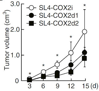
Finally, using CRISPR/Cas9 gene editing technology, the authors constructed COX-2 deficient tumor cell lines and tested their growth pattern in mice. As expected, growth of COX-2 deficient tumor cell  lines were delayed in absence of COX-2 enzyme.


In summary, this study showed that cells undergoing sterile cell death (for example, during excessive tumor growth), release suppressive lipid, PGE2, and this mechanism represents one of the natural anti-inflammatory processes that is hijacked by tumors to evade efficient immune detection. Aspirin's beneficial effect as an anti-cancer therapy could be attributed to its effect on PGE2

David Usharauli


Wednesday, March 23, 2016

Availability of free cytoplasmic cholesterol augments TCR signaling in CD8 T cells


This study focus on Acat1 that encodes cholesterol esterification enzymes that convert free cholesterol to cholesteryl esters for storage. The scientists noticed that chemical inhibition of Acat1 in CD8 T cells could augment their cytolytic effector differentiation (granzyme B ↑).


Similar results were obtained with CD8 T cells from Acat1 conditional knockout mice (Acat1CKO).


Boost of CD8 T cell cytolytic functions with Acat1 inactivation yielded better tumor protection in adoptive transfer experiments as well.


Mechanistically, it appears that availability of excess free cholesterol in Acat1CKO CD8 T cells improved TCR downstream signaling.


Finally, the authors showed that avasimibe, an Acat inhibitor with a good safety profile in humans, could delay tumor progression in mice and even show synergy with checkpoint inhibitor, anti-PD1 antibody.


In summary, this study suggests that commonly used cholesterol modulating drugs could have important role in CD8 T cells effector function and could influence tumor immunotherapy results.

Here I would like to note that one earlier study showed that reduction of cellular free cholesterol triggered universal STING-cGAS mediated anti-viral type I IFN response. This suggests that on one hand free cholesterol could augment CD8 T cell cytolytic function, but on the other hand it could diminish cell-autonomous anti-viral response.  

David Usharauli


Tuesday, March 22, 2016

Pregnancy-associated microbiota modules newborn's group 3 innate lymphoid cells (ILC3)

It is common believe that newborns are born sterile and their immune system undergoes changes at post-natal period in response to their own microbial exposure.

However, new study in Science challenges this concept by showing that mother's microbiota during pregnancy has permanent impact on offspring's postnatal innate immune system

To distinguish effects of maternal vs. neonatal microbiota on neonate's immune system, the authors used a "system in which pregnant dams are transiently colonized with genetically engineered Escherichia coli HA107", a E. coli strain that does not persist in the host and "pregnant dams become germ-free again before term and naturally deliver germ-free pups". Compared to control, microbiota-nil pregnant females, offsprings derived from temporally colonized pregnant females contained more IL-22 producing intestinal ILC3 (there was no effect on T or B cell numbers).


Maternal microbiota effect on newborn's ILC3 numbers could be reproduced by injecting germ-free pregnant females with serum (IgG) derived from HA107 colonized females (though it is not clear whether microbiota-specific IgG or any random IgG could produce the same effect).


Similarly, maternal microbiota effect on newborn's ILC3 numbers was lost in pregnant females lacking B cells (IgH-/-).


In summary, this study suggests that pregnancy-associated microbiota does have an independent effect on maturation of newborn's innate immune system, at least via maternally-derived antibodies.

David Usharauli


Monday, March 21, 2016

IL-23p19 has an inside job in endothelial cells

IL-12 cytokine family is full of surprises. Ordinarily, these cytokines (IL-12, IL-23, IL-27, IL-35) are made of two heterodimeric sub-units. But one after another, each of the sub-units were found to have an independent function.  

Latest in these series is a study from Science Signaling that showed that human endothelial cells express intra-cellular IL-23p19 that signals via gp130 molecules mimicking IL-6.

Initially, the authors showed that endothelial cells from Giant-cell arteritis (GCA) patients express IL-23p19 subunit, but not another IL-23p40 subunit (while I support fully-human studies, in controversial situations such as this, I preferred if the authors have included confirmation staining on IL-23p19 KO cell from KO mice or CRISPR/Cas9 edited IL-23p19 KO human cells). 


Next, in vitro experiments with primary human umbilical vein endothelial cells (HUVECs) and human dermal microvascular endothelial cells (HDMECs) confirmed selective expression of IL-23p19 within endothelial cells in response to pro-inflammatory signaling.


IL-23p19 was detected in endothelial cell lysate but not in supernatants, suggesting that it was intra-cellular protein and wasn't secreted in culture medium.


Mechanistically, IL-23p19 transduced endothelial cells up-regulated adhesion molecules (VCAM-1, ICAM-1, PECAM-1) and induced gp130-dependent STAT3 activation.


The authors hypothesized that IL-23p19 could be similar to viral IL-6 (vIL-6), a viral cytokine product of human herpesvirus 8 (HHV-8, also known as Kaposi’s sarcoma-associated herpesvirus).


In summary, this study provided evidence that in humans IL-23p19 could have an independent biological function in endothelial cells by promoting adhesion and recruitment of inflammatory cells thus contributed to vasculitis.

David Usharauli


Saturday, March 19, 2016

PGE2-IL-22 axis prevents gut microbiota leakage during systemic inflammation


This is very neatly done study. In the initial experiments, the authors showed that pre-treatment of mice with indomethacin (which suppresses PGE2 production) increased their susceptibility to systemic inflammation following endotoxin (LPS) injection.


Interestingly, when mice were also pre-treated with EP4 agonist (PGE2 receptor), it reduced systemic inflammation following LPS injection


Moreover, beneficial effect of EP4 agonist on systemic inflammation following LPS injection was mimicked by antibiotic pre-treatment, suggesting the role of gut barrier in protection mediated by PGE2.


Indeed, EP4 agonist pre-treatment prevented bacterial translocation from gut into sterile internal tissues such as liver.


Finally, the authors showed that PGE2-EP4 signaling in group 3 innate lymphoid cells was necessary for IL-22 production that contributed to maintaining gut barrier function during systemic inflammation (of note, EP4 agonist had (a) no effect in IL-22KO mice and it was still active in (b) RAG KO mice that lack adaptive immune system).


In summary, this study provided additional support for innate cell-derived IL-22 as a cytokine necessary for keeping gut tissue in healthy condition.   

David Usharauli

Tuesday, March 15, 2016

DC vaccines produced with tumor cells undergoing immunogenic cell death

Dendritic cells are the most potent activators of naïve T cells. A lot of effort has been spent to develop DC-based cellular vaccine. We even had some success in mouse models. But, so far no DC based vaccine worked in humans to significantly prolong patients survival. It is not surprising that only company that developed FDA-approved DC vaccine prostate cancer went bankrupt. There are many reasons as to why it is so hard. For one, there are several types of dendritic cells. Secondly, just pulsing [tumor] antigens on dendritic cells were found not to be sufficient.

Few weeks ago, Science Translational Medicine published study wherein the authors had tested next-generation tumor vaccine produced by pulsing DCs with tumor cells undergoing immunogenic cell death.

Basically, the authors had used a high-grade glioma (HGG) cell lines as a source of tumor. To generate immunogenic cell death, tumor cells were exposed to hypericin-based photodynamic therapy (Hyp-PDT). This treatment induces expression/release of several damage-associated molecular patterns (DAMPs) acting as potent danger signals for DCs. When mice were injected with DCs pulsed with Hyp-PDT treated glioma cells, they showed resistance to subsequent live glioma brain challenge.


Mechanistically, tumor protection provided by Hyp-PDT DCs depended on (1) DAMPs, (2) DC-specific MyD88, (3) CD8 T cells (T cells data are shown here).


Importantly, Hyp-PDT DC vaccines improved survival of brain tumor-bearing mice when applied therapeutically and in combination with standard-of-care therapy such as temozolomide (TMZ).


In summary, this study showed that hypericin-based photodynamic therapy could generate immunogenic tumor cells required for proper activation of DC for vaccination purpose. Conceptually, such mode of DCs preparation makes a lot of sense, but there are several technical challenges before this system could be translated into clinic, as discussed by the authors (such as, how to avoid presence of "surviving" live tumor cells within DC vaccine, route of DC vaccine injection [the authors have used intra-peritoneal injection, for some reason], what type of DC is the best for this purpose). 

David Usharauli


Saturday, March 12, 2016

CD47 and PD-L1 work in tandem with MYK to keep T cells at bay

This week Science published a short study examining the role of MYK in tumor immune evasion mechanisms [MYC is a transcription factor that regulates the expression of a multitude of gene products involved in cell proliferation, growth, differentiation, and apoptosis"].The authors found that MYK selectively directs expression of two immune-inhibitory molecules, CD47 and PD-L1, in several mouse and human tumor cells lines.

Using Tet-off transgenic mouse model to control MYK expression in T cell acute lymphoblastic leukemia (MYC T-ALL), the authors showed that both "in vitro or in vivo MYC inactivation resulted in a rapid downregulation of CD47 and PD-L1 [but not other surface markers], both at the mRNA level, as detected by quantitative real-time PCR (qPCR), and at the protein level, as detected by flow cytometry".


Since previous results demonstrated "complete tumor clearance following the inactivation of oncogenes, including MYC", the authors wanted to examine whether MYK effect on tumor regression involved CD47/PD-L1 tandem. Indeed, forced expression of either CD47 or PD-L1 in MYC T-ALL reduced anti-tumor effect of MYK inactivation.


In summary, this study suggests that MYK oncogene provides tumor cells with defenses to repel immune cells. 

David Usharauli

Tuesday, March 8, 2016

Gut-specific eosinophils suppress IL-17 production by antagonizing IL-1 signaling

Prior to discovery of Toll-like receptors (TLR) in late 90s, immunology was dominated by concepts involving adaptive immune cells. However, for past 15 years it has become evident that innate immune cells of "all shapes and forms that exist out there" are equal partners with T and B cells and frequently provide instructions and actually control the magnitude and directions of adaptive immune response. For example, development of successful, next generation vaccines would require incorporation of these new concepts. 

It seems innate immune system is even more complex and unpredictable than T or B cells are. For instance, this week Journal of Experimental Medicine published new study wherein the authors reported that eosinophils, a cell population usually implicated in allergy and anti-helminth response, suppressed Th17 cells by antagonizing IL-1 signaling.

First, the authors observed that Th17 cells in ΔdblGATA-1 mice [which lack eosinophil-lineage cells] were significantly increased in the small intestine, but not in the spleen or mesenteric LNs (MLNs).


In vitro generation of Th17 cells were inhibited by presence of small intestinal eosinophils. 


Functional profiling revealed that small intestinal eosinophils secreted high amount of IL-1 receptor antagonist (IL-1Ra), which competes with IL-1β for receptor binding (IL-1 signaling is involved in Th17 development).


Interestingly, eosinophils were the major producers of IL-1Ra in small intestine, since no IL-1Ra was detected in small intestinal tissue from ΔdblGATA-1 mice. Of note, eosinophils from blood or bone marrow produced little or no IL-1Ra. 


In addition, the authors showed that small intestinal eosinophils derived from MyD88-KO or Germ Free (GF) mice produced WT-level IL-1Ra, suggesting its independence from gut flora or major TLR signaling.


Role of IL-1Ra in eosinophil mediated Th17 suppression in small intestine was confirmed in experiments with eosinophils derived from IL-1Ra-KO mice.   


In summary, this study showed that small intestine harbors specialized eosinophils secreting high amount of IL-1Ra and suppressing steady-state development of Th17. 

David Usharauli


Saturday, March 5, 2016

Sensing of RNA:DNA heteroduplexes by cGAS/STING pathway mirrors human autoimmune Aicardi–Goutières syndrome

Autoimmune diseases occur when bodies immune system continues to respond to self-antigens. Ordinarily, such [mis]directed responses are ascribed to adaptive immune system which possesses myriads of randomly generated receptors. So it is quite reasonable to assume that some of those receptor equipped clones (T or B cells) could go haywire and attack their own body's particular antigen or set of antigens. There are few autoimmune diseases known thus far that fall in this category (e.g. in Graves' disease, thyroid stimulating immunoglobulin [over]stimulates the same receptor that normally responds to only thyroid stimulating hormone). This is what is called lack or break of tolerance [mostly due to cross-reactivity]. 

However, much larger category of autoimmune diseases are due to genetic defects of different kind. These conditions harbor mutations in "housekeeping" molecules that are required for proper cellular homeostasis. Such mutations leads to immunopathologies that target broad spectrum of antigens or could even be restricted solely to the activation of innate immune system.    

For example, mutations in enzymes that ordinarily degrades self-DNA could lead to systemic immunopathologies due to activation of DNA sensing molecules cGAS or STING.

One such genetic mutation in ribonuclease (RNase) H2B (a enzyme that degrades cellular RNA:DNA heteroduplexes) leading to pathological cGAS/STING activation was recently described in the EMBO Journal.


This study revealed that mutation in RNase H2B that renders it less active led to up-regulation of interferon‐stimulated gene (ISG) transcripts. 


Follow-up experiments confirmed that interferon "signature" in RNase H2B KO cells was driven by cGAS (deletion via CRISPR/Cas9 genome editing) and STING activation (STING KO).


Exactly what type of self-DNA is detected that leads to cGAS and STING activation in cells with RNase H2B mutation is not yet clear. It could be (a) cytosolic RNA:DNA heteroduplexes accumulated as a consequence of reduced RNA:DNA degradation, or it could be (b) cytoplasmic DNA with embedded ribonucleotides that may accumulate due to impaired ribonucleotide excision repair (RER).

With the advance of human genomic testing, it is becoming evident that large portion of autoimmune diseases / immunopathologies may have a clear genetic basis and could be specifically targeted for therapeutic purpose.

David Usharauli
    

Wednesday, March 2, 2016

Microbiota-specific serum IgG protects against systemic bacterial dissemination

This week journal Immunity published a study from Gabriel Núñez lab describing the role of serum IgG in protecting the body against systemic dissemination of gut commensal bacteria.

Initially, the authors showed that WT mouse serum contained IgG specific for fecal bacteria and its production depended on (a) gut microflora and (b) T cells.


These data indicated that gut bacteria [or its parts] "circulate systemically in spite of intact intestinal barriers." Indeed, the presence of bacterial 16S rRNA gene was confirmed in the spleens, for example.


However, the authors showed that such "access" was not a "random" translocation. Only certain gut bacteria (for example, Enterobacteriaceae and Moraxellaceae, but not Porphyromonadaceae and Prevotellaceae) were able to disseminate internally.


Correspondingly, immunoblotting analysis of serum IgG against fecal bacterial antigens confirmed "selective" IgG response to gut flora gram-negative Escherichia coli (EC) or Klebsiella pneumoniae (KP), but not to gram-positive Enterococcus faecalis (EF) or Clostridium bifermentans (CB).


In parallel experiments, the authors showed that serum IgG from WT mice, but not from quasi-monoclonal (QM) mice which have limited B cell repertoire, could protect B cell deficient mice from gram-negative E.coli infection.


Since gram-negative bacteria are recognized by TLR4 the authors tested mice deficient TLR4 signaling pathway. Indeed, absence of TLR4 specifically on B cells selectively reduced serum IgG against fecal bacteria (single TLR4-KO mice showed the same effect, while single TLR2-KO had no effect).


Similar to B cell-deficient mice, mice with TLR4-deficient B cells were highly susceptible to E.coli infection and could be rescued only by IgG from WT mice, but not by IgG from quasi-monoclonal (QM) mice.  


Analysis of gram-negative WT or mutant bacteria showed that serum IgG was mostly directed against murein lipoprotein (MLP), a highly conserved outer membrane protein of ~7 kD expressed abundantly in gram-negative enterobacteria.


Finally, the authors showed that monoclonal anti-MLP IgG injection could protect B cell deficient mice from E.coli infection almost as efficiently as total WT IgG injection.


In summary, this study suggests that normal serum IgG protects the body in situations when gram-negative bacteria from gut gains access to systemic circulation (for example, in "leaky gut" or during a sepsis). Generation of such protective IgG requires presence of T cells and normal B cell receptor diversity.

It is not clear what role IgA plays in this process. The authors have not tested IgA-KO mice to determine its exact contribution in preventing gut flora translocation. No data are provided regarding presence and [amount] of gut bacteria in systemic circulation in different KO mice shown in this study either. This would have given some valuable information.

David Usharauli