Thursday, October 30, 2014

Did mythical Hydra got it right?

In Greek mythology Hydra from lake Lerna was immortal. Interestingly, Hydra, a metozoan, is indeed an immortal multi-cellular organism.

I want to recommend for a reading a new article by Thomas Bosch in Trends Immunology. This is a short review article. It has a very good thought provoking idea. Not sure whether it is an original idea but still good to see that such paradigm shifting ideas are welcomed for publication.  

In this paper, the author opines that the role for innate immune system is to select and keep beneficial microbes rather than fight off pathogens. The author made such conclusion based on experiments on Hydra polyps, where compared to control polyps, Hydra with specific deficiency in anti-microbial peptide, arminin, has a different microbial flora, suggesting that innate immune sensing were driving microbiota selection. The author also speculated that gene responsible for Hydra's immortality, FoxO, affected Hydra's innate immune system as well.  

Why is this idea so important? We were thought to believe the immune system is directed to defend against virulent pathogens. But what if immune system keeps "friendly" microbes as proxies to defend against pathogenic ones. This can be a mutually beneficial arrangement. The host will provide nutrition and living space and microbiota (not just in the gut lumen, but on the lung epithelial surface, on the skin surface, etc) will defend against uninvited "guests".

It is well known that good proportion of individuals in any given population is naturally immune against pathogens, known or new ones. We think it has to do with good immune system. But what does good immune system really mean? Maybe it has to do with microbiota diversity in such individuals? Microbiota diversity will be invariably linked with specific sets of immune genes that make microbiota diversity and their accommodation possible in the host. Mechanisms of accommodation of endogenous microbiota will be linked to mechanism of disease tolerance, a recently introduced new concept of host defense. It is much more easy to imagine that "disease tolerance" against virulent pathogens in individuals is a byproduct of microbiota accommodation by the host, a learned, adaptive process during the host ontogenesis.

"Selfish" view of immune system could explain why antibiotic treatment has such negative effect on the host, sometimes leading to development of allergic and autoimmune disease.

David Usharauli  


Monday, October 27, 2014

Control of EBV is so elemental

Genetic mutations provide a window into inner workings of the biological systems. 

Here is one example. This paper, Magnesium and NKG2D, was published in journal Science in summer of 2013. It came from Michael Lenardo's lab at NIAID.

I have noticed that recently his lab's focus shifted in more clinical direction, like understanding molecular mechanisms behind natural mutations affecting immune system in humans.

In this paper, his lab provided further analysis of patients (total of 7 patients) with mutation in Magnesium transporter 1, called XMEN disease. These patient show inability to control EBV infection and can develop lymphomas.


Initial study, (Second messenger role for Mg2+), published in 2011 in journal Nature showed that deficiency in Magnesium transporter 1 (MagT1) affected T cell activation. It was quite new idea since we were thought that Ca2+ was necessary for T cell activation.

Here, the authors made observation that cytotoxic activity of CD8 T cells and NK cells from XMEN patients were reduced.


Interestingly, supplementation of culture medium with magnesium could partially (for CD8 T cells) or fully (for NK) restore cytotoxic activity against target cells. 


Since activation of CD8 T cells was not restored by magnesium supplementation, increase in cytotoxicity was probably related to increase in efficiency in releasing cytotoxic granules from already differentiated effector T cells. 

Luckily for the authors, they were able to pinpoint surface molecule that was involved in Magnesium-dependent improvement in cytotoxicity. It turned out to be NKG2D, a well known molecule in NK cell activity. XMEN patients had dramatically reduced level of surface NKG2D expression on NK cells. The authors showed that increase in cytotoxicity with magnesium supplementation was correlated with up-regulation of NKG2D (on both NK and CTL), since this effect was abolished in presence of blocking NKG2D-Fc molecule in CTL assay.


Finally, in vivo supplementation of XMEN patients with magnesium could improve their ability to control EBV.      

This proof of concept (POC) studies are the most straightforward to understand and easy to incorporate into patient's treatment guides. In this situation the authors could perform well established assays to verify the hypothesis. However, mutations can happen in proteins with no known functions and no known functional assays. In there any way to rapidly identify protein function and design the assay?

David Usharauli


Saturday, October 25, 2014

Microbiota needs sleep too

We all heard over and over that sleep deficiency could lead to obesity. One may wonder why it is the case. What sleep has to do with insulin insensitivity and metabolic syndrome?

Surprisingly, new study from Weizmann Institute of Science, Israel, suggests that alteration in sleep-wake rhythmicity leads to obesity through modification of gut microbiota.

This study, diurnal rhythmicity and a microbiome-dependent mechanism for common metabolic disturbances in humans, from Cell, examined how disruption of food intake rhythmicity due to time zone travel (jet-lag) affected development of metabolic syndrome.

First, the authors observed that there was rhythmic variation of gut microbiota composition wild-type mice due to dark-light cycle, that was lost in Per1/2−/− mice, which are deficient in a functional host clock genes. Interestingly, artificially modulating food availability could alleviate circadian clock deficiency in Per1/2−/− mice.


To mimic natural dark-light cycle disruption, the authors created experimental jet-lag condition for mice. Within 6 weeks, jet-lagged mice showed weight gain and glucose intolerance, hallmark of metabolic syndrome. Interestingly, antibiotic treatment of jet-lagged mice reversed signs of metabolic syndrome.


Finally, the authors examined gut microbiota composition from individuals undergoing jet-lag. Interestingly, during jet-lag phase there was an increase in Firmicutes which were linked to obesity in earlier studies. Moreover, gut flora taken collected during jet-lag phase but not before or after, could promote metabolic syndrome in germ-free mice.


In summary, this study showed that human circadian rhythm control gut microbiota composition which in turn influence development of metabolic syndrome (weight gain, type II diabetes). It shows that some of the effects of dark-light cycle disruption could be minimized by antibiotic treatment. Of course, in this case antibiotic treatment must be directed to specific gut microbiota species. In addition, this study indicates that modulating or restricting access to food to particular time periods could minimize effect of dark-light cycle disruption in humans.

David Usharauli


Thursday, October 23, 2014

niaIDEAlist's Report

If interested, please check my new blog niaIDEAlist's Report. I have just started it using free wordpress.com platform.    

As you may have noticed, titles of both my blogs are wordplay. While NIHilist's Immunology is focused on research articles and represents hybrid approach in evaluating newly published studies, niaIDEAlist's Report will have more diverse focus. Its topics will cover science news and its interpretation, my experience as a research scientist and my opinions about science matter.  

David Usharauli


Tuesday, October 21, 2014

Ebola virus is no match for feminine power

These days everyone is talking about Ebola. I remember reading news article in early 2014 discussing spread of Ebola in West African countries. It was very puzzling since such highly virulent infectious agents are usually self-limiting. Something is certainly different this time. It appears it now developed a mutation allowing prolonged incubation time.

It is obvious Ebola is new virus for human immune system and most likely there is no natural (genetic) immunity against it. It means that adaptive immune system goes into overdrive because innate system lacks genetic memory of Ebola and is unable to properly instruct effector T and B cells. In this situation, survival depends on presence antibody or T cells cross-reactive to environmental microbes or host commensals.

However, sometimes protection against virulent infections can come from unexpected corner. Here is such example for Ebola. While researching Ebola publications prior to 2014 outbreak, I came across two research articles about Ebola that immediately caught my attention.

These papers were published in April-June 2013 by two independent groups. One was published in Plos One (A Systematic Screen of FDA-Approved Drugs for Inhibitors of Biological Threat Agents), and another in Sci Transl Med (FDA-Approved Selective Estrogen Receptor Modulators Inhibit Ebola Virus Infection). Only similarity was that some of the authors (but not the same authors) from each group were from U.S. Army Medical Research Institute of Infectious Diseases (USAMRIID)

Basic idea of the studies were to screen FDA-approved drugs to see if any of the drugs would protect against category A biological threat agents, like Ebola. This is called drug re-purposing and has many advantages considering the fact that lengthy human safety studies have already been conducted on such medications.


Using both in vitro cell culture assay or in vivo mouse models, both studies independently showed that FDA-Approved Selective Estrogen Receptor Modulatorstoremifene or clomiphene, blocked Ebola virus host cell entry capacity by 97% and protected >50% (90% protection in clomiphene group) of infected mice (100% death in untreated group).  


Sci Transl Med 19 June 2013
Fig. 4


Interestingly, the authors think this protection is estrogen-receptor independent, basically meaning that response appeared to be an off-target effect, completely unexpected. 

It maybe time for FDA to have a quick look for such data. It may indeed provide quick and safe alternatives (to vaccines) in times of uncertainty.

David Usharauli


Thursday, October 16, 2014

Eosinophil's cooling effect


Obesity develops when there is an imbalance between energy intake and expenditure. A brown adipose tissue (BAT) is a specialized adipose tissue involved in thermogenesis and energy dissipation in response to cold. 

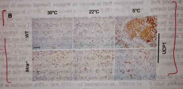
This new study (1) published in Cell indicates, that strangely, BAT response to cold is mediated via cytokine IL-4.

The authors in this study wanted to understand how type 2 immunity (Th2 response dominated by IL-4 and IL-13) affects cold-induced brown fat remodeling. 

To this end, wild-type or IL-4 /IL-13 double KO mice were exposed to thermoneutral (30C), sub-neutral (22C) and cold temperature (5C) and subcutaneous white adipose tissue response were examined. Unlike wild-type mice, IL-4 /IL-13 double KO mice did not show response to cold.


Similarly, IL-4ra KO mice did not show response to cold.


Examination of subcutaneous white adipose tissue (scWAT) in STAT6 KO mice confirmed that IL-4 signaling was crucial for thermogenic response to cold.

To understand what cell type was producing IL-4, the authors examined 4get mouse that express GFP protein from endogenous IL-4 locus. Expression pattern indicated that IL-4 producers were eosinophils. Indeed, using 4get mice deficient in eosinophils, the authors could abrogate cold response in scWAT.


In addition, the authors found that mice deficient for CCR2 also lacked scWAT response to cold. CCR2 is required for recruitment of macrophages into tissue.

To authors speculated that IL-4 driven macrophage-specific tyrosine hydroxylase (TH) expression was responsible for scWAT response to cold. To test this hypothesis, they have generated mice with specific deletion of tyrosine hydroxylase in macrophages. Indeed, this TH f/f Lyz2-cre mice lacked scWAT browning response to cold.


Finally, to examine the role of exogenous IL-4 on browning of scWAT, the authors injected mice, fed with high-fat diet, with IL-4/anti-IL-4 complexes to mimic efficient IL-4 signaling. Such treatment mimicked cold-response in scWAT with corresponding body fat and mass reduction and increase in insulin sensitivity.


In summary, the authors showed that long-held idea that cold response is neuronal driven response is outdated. It appears that type 2 immune response, specifically, eosinophil-derived IL-4 stimulates local macrophage-specific tyrosine hydroxylase activity and catecholamine production leading to local browning of scWAT and thermogenic response.

Of course, it is very interesting to know how eosinophils are sensing cold or suboptimal temperature. I think it will be a local commensial microbiota-induced effect.

The idea that immune system can sense a temperature gradient is quite surprising in itself. Though almost everyone can affirm common observation that when children or even adults happened to fall asleep in the couch, others are trying to cover them with blanket to prevent them from getting or catching a “cold”. Does such empirical observation imply that cold-induced type 2 response has its undesirable effects?


David Usharauli


Sunday, October 5, 2014

High Five to inactivated Flu vaccine


Here is another Immunity paper about microbiota with interesting twist. This paper came from Bali Pulendran's lab in Emory. His lab focuses on systems biology approach to immunology. The systems biology is a basically an algorithm-driven science with minimal involvement of human experience and intuition.

In this paper (1), the authors made initial observation TLR5-deficient mice immunized with FDA-approved inactivated Flu vaccine, TIV, mounted impaired antibody response compared to wild-type mice. 


Reduced antibody response was more apparent at early stage, at day 7, but not at day 28. Correspondingly, number of TIV-specific antibody-secreting cells in draining lymph nodes was reduced at day 7. However, interestingly, serum level of TIV-specific IgG at day 84 in TLR5-deficient mice was again lower compared to wild-type mice suggesting that both early and long-term antibody production were affected by absence of TLR5.


Since TIV itself does not interact with TLR5 directly, 


the authors tried to understand how absence of TLR5 impacted response to TIV. Interestingly, the authors found that germ-free mice or mice treated with broad-spectrum antibiotics showed similar impaired response to TIV, similar to TLR5-deficient mice. 

Germ-free mice inoculated with gut microbiota showed restored response to TIV.


In addition, the authors showed that supplementation of TIV with flagellin (TLR5 agonist) improved TIV-specific antibody response in antibiotic-treated mice.


Further experiments showed that TLR5 signaling in hematopoietic cells (specifically, in macrophages) were critical to TIV antibody response.


Finally, the authors showed that dependency on TLR5 for TIV-specific antibody response was not unique for TIV. IPOL, another inactivated vaccine consisting of polio virus subunits, also required TLR5 for full response. However, live-attenuated vaccine or alum-adjuvanted vaccines did not require TLR5 for full response.


In summary, this study indicates that even vaccine efficacy is depended on gut microbiota.

Why specifically, TLR5 plays such important role is not clear. Earlier research showed that TLR5 had a protective role against development of metabolic syndrome and radiation-induced toxicity. If those mechanisms also responsible for improved antibody response is to be seen. Or is it possible that an absence of TLR5 affects the composition of gut microbiota in such manner to impact the magnitude of TIV response?

David