Showing posts with label inflammasome. Show all posts

Saturday, April 23, 2016

New mouse model to mimic immune response to Flu virus in elderly

This week Science published several research papers in immunology. One of these papers is the study from Yale School of Medicine showing that Tlr7–/– × Mavs–/– double KO mice expressing human anti-viral molecule Mx1 display susceptibility to influenza A virus in a caspase 1/11-dependent manner.

It is known that elderly individuals show susceptibility to influenza A virus. One reason for this susceptibility is a reduced ability of elderly immune system to produce type I IFNs in response to influenza A virus.


In contrast, aging mice do not show such susceptibility to influenza A virus. Mouse does not express Mx1, a dynamin-like guanosine triphosphatase that blocks primary transcription of influenza in humans. To make mouse model "usable" for  studying human response influenza A virus, the authors created Mx1+ mice. Mx1+ mice were resistant to experimental influenza A virus infection and this resistance was dependent on Tlr7 and Mavs (both molecules are involved in anti-viral response).


Interestingly, while Mx1+ mice double deficient for Tlr7–/– × Mavs–/– were susceptible to influenza A virus infection,  Mx1+ mice triple deficient for Tlr7–/– × Mavs–/–× Casp1/11–/– were resistant to influenza A virus infection even though both showed similar viral burden (Of note, Tlr7–/– × Mavs–/–× Casp1/11–/– mice were eventually cleared the virus by 30 days after infection).


Moreover, Mx1+ mice triple deficient for Tlr7–/– × Mavs–/–× Casp1/11–/– were resistant to influenza A virus infection even though they too showed similar secondary bacterial "bloom" in their airways.


These data suggested that caspase 1/11 signaling reduced "tissue tolerance" [rather than increased anti-viral response] to influenza A virus infection and secondary bacterial "bloom" in Mx1+ mice deficient for anti-viral innate signaling via Tlr7–/– × Mavs–/–.

In summary, this study showed that intact caspase 1/11 signaling compromises tissue tolerance to acute influenza A virus infection and secondary bacterial "bloom" in host with a weakened anti-viral signaling.

David Usharauli

Friday, April 22, 2016

PAMPs and DAMPs cooperate to drive vigorous adaptive immune response

This week Science published interesting article related to basic question of dendritic cell (DCs) activation and initiation of productive adaptive immune response. It showed that endogenously generated oxidized phospholipids (oxPAPC) cooperate with bacterial-derived LPS in a caspase 1/11 dependent manner to enhance viability and activation status of DCs enabling a better adaptive immune response.  

I would like to note here that title of this paper does not exactly captures the main idea behind this study. In fact, only reading the final portion of the paper one can get the sense and larger meaning of this study. I will try to explain it.   

This paper's main idea is to understand the difference between PAMPs and DAMPs with the regard of their impact on DCs. Every scientists in immunology is familiar with pathogen-associated molecular patterns (PAMPs, such as LPS) and damage-associated molecular patterns (DAMPs, such as ATP). Nonetheless, it is still unclear what role(s), for example, DAMPs play within immune system.

To understand it, the authors focused on one particular type of DAPMs, oxidized phospholipids (oxPAPCs). oxPAPCs are found in inflammatory milieu and can reach concentrations of 10-100 μM in damaged tissues. Initially, the authors showed that unlike LPS, oxPAPCs do not signal via TLR4.


However, similar to other DAMP molecules, oxPAPCs could induce IL-1β release from LPS-primed DCs. As expected, oxPAPC-induced IL-1β release from LPS-primed DCs required inflammasome activation, since such effect were absent in DCs from ASC knockout (KO), caspase-1 KO, caspase-1/caspase-11 double KO or NLRP3 KO mice, each of which are defective for inflammasome functions.


Pyroptosis is an inflammasome-dependent cell death characterized by loss of plasma membrane integrity. Interestingly, unlike LPS or LPS+ATP combination, oxPAPC-induced inflammasomes did not promote pyroptosis. Thus oxPAPC promoted IL-1β release from living DCs.


Finally, the authors showed that mice immunization with antigen in combination with oxPAPC+LPS could improve priming of adaptive T cells in a caspase 11-dependent manner.


In summary, this study showed that DAMP molecule, oxPAPC, promotes DC viability and IL-1β release when combined with PAMP molecule, LPS. Such combination of DAMP and PAMP promoted more productive adaptive immune response.

David Usharauli


Saturday, February 21, 2015

Fasting-induced ketone metabolite, β-hydroxybutyrate suppresses NALP3-mediated inflammasome activity

Inflammasome super-complex represents an ancient innate detection and defense mechanism against exogenous as well as endogenous irritants and toxins. Spontaneous inflammasome activity causes several known human immune-pathologies, like Muckle-Well and familial cold auto-inflammatory syndromes.  

New paper from Nature Medicine provided evidence showing that alternative energy source, β-hydroxybutyrate, elevated during fasting, calorie-restiction or high-intensity exercise, suppresses NALP3-mediated inflammasome activity.

Initially, the authors led by Prof. Vishwa Deep Dixit at the Yale School of Medicine, showed that β-hydroxybutyrate, but not another ketone body acetoacetate or short-chain fatty acids, butyrate or acetate, could inhibit NALP3-mediated inflammasome activity (AIM2- or NLRC4-mediated inflammasome activities were unaffected).


Suppression of NALP3-mediated inflammasome activity by β-hydroxybutyrate was independent of autophagy and required neither G protein-coupled receptor GPR109a signaling via β-hydroxybutyrate nor histone deacetylase inhibition by β-hydroxybutyrate.


The authors found that β-hydroxybutyrate suppressed NALP3-mediated inflammasome activity through its inhibition (by unknown mechanism) of potassium, K+ cation efflux from cytoplasm.


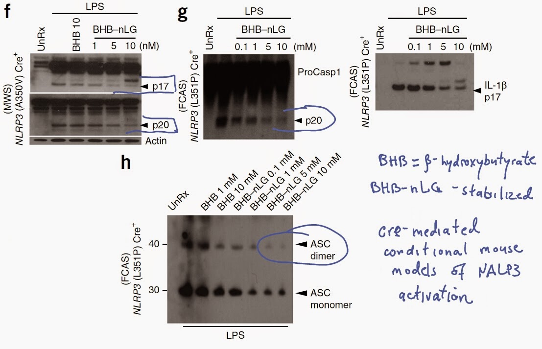
Finally, using cre-mediated mouse conditional models of human Muckle-Well and familial cold auto-inflammatory syndromes, the authors showed that stabilized form of β-hydroxybutyrate could suppress spontaneous NALP3-mediated inflammasome activity and ASC oligomerization characteristics to these gain-of-function NALP3 mutations.


In summary, these results indicate complex interplay between energy metabolism and inflammation. In condition of glucose deficiency, elevated level of β-hydroxybutyrate acts as a new source of ATP in brain and heart and at the same time inhibits NALP3-mediated inflammasome activity.

Leave your comments below and let me know what do you think about this paper.

David Usharauli