Showing posts with label dendritic cells. Show all posts

Friday, October 28, 2016

A minor subset within CD11c+ dendritic cells is primarily responsible for peripheral Treg expansion

FOXP3+ Tregs play a major role in tolerance maintenance in the periphery. Most of FOXP3+ Tregs are thymus derived. Thymus is a specialized lymphoid tissue that generates novel T cells from their bone marrow precursors. The question whether naive T cells could convert into FOXP3+ Tregs in the periphery has not been settled. 

It is believed that a "steady-state" condition favors FOXP3+ Tregs conversion in the periphery. However, a concept of "steady-state" is an arbitrary one, defined as absence of deliberate immunization or experimentally observed infection. In fact, whether "steady-state" truly exist is an open question as well.

Why this matters? Almost everyone agrees that in absence of so called "steady-state" naive T cells would convert into effector T cells rather than into FOXP3+ Tregs following antigen recognition. For example, if one wants to generate new FOXP3+ Tregs specific for particular antigen to treat autoimmune diseases, this task would be almost impossible to achieve if condition of "steady-state" does not actually exist in the body [from T cells' "point of view"]. 

Also, what cell types are responsible for that supposed FOXP3+ Tregs conversion? A new study in Immunity clarified this question somewhat. It showed that even in "steady-state" condition only minor subset of DCs within CD11c+ population defined by DEC205/CD8 expression were responsible for FOXP3+ Tregs "conversion" in T cell-replete mice [which harbor endogenous FOXP3+ Tregs].

For this study, the authors have used chimeric anti-DEC205 Ab [or anti-CD11c chimeric Ab as a control] that incorporate antigen of interest [MOG or OVA]. When injected into mice chimeric anti-DEC205 Ab, but not control, could "convert" naive MOG or OVA-specific T cells into FOXP3+ Tregs.

It appeared that DEC205+ CD11c+ DCs were also primarily BTLA+ and its expression were required for FOXP3+ Tregs induction.


Interestingly, the authors proposed that BTLA to HVEM signaling in naive T cells up-regulated CD5 and permitted FOXP3+ Tregs conversion even in presence of inflammatory cytokines such as IL-4 and IL-6.  

However, in my view, such mechanism of FOXP3+ Tregs conversion even in presence of inflammation sounds counter-intuitive. Wouldn't it also induce FOXP3+ Tregs conversion from naive T cells specific for nonself antigen derived from pathogens during inflammation? Otherwise, how can system make sure that only self antigens are presented by DEC205+ DCs? The authors could only admit that this tolerance mechanism somehow only affects "self and tolerizing antigens". Also, what about endogenous FOXP3+ Tregs in these mice? Is it possible that endogenous thymus FOXP3+ Tregs are involved in assisting in FOXP3+ Tregs conversion, rather than DEC205+ DCs doing it alone from scratch? If so, implications are very different.  

David Usharauli


Monday, November 30, 2015

Tregs new motto: united we stand


Tregs are a major players within immune system but Tregs-mediated "negative" regulation of antigen-specific immune response has a fundamental problem at the theoretical, mechanistic level. No major, widely accepted immune theories such as Burnet's clonal selection theory, 2-signal model, Janeway-Medzhitov's PAMP model, or Matzinger's Danger theory could satisfactorily explain the role of Tregs within immune system. 

Actually, all these major immune theories excludes or simply prohibit the existence of any antigen-specific T cells that are of inhibitory nature. Antigen cannot be the basis of inhibition, according to these models, because T cell has no way of "knowing" the difference between self or nonself antigens. Moreover, at the innate immune system level, simply eliminating "irritants" that caused initiation of immune response in first place would automatically return immune system into quiet state without need of any negative regulation.

So why Tregs exist and how they regulate immune response? So this new paper tried to unlock the first door for this theoretical labyrinth. The authors showed that fraction of Tregs within lymph nodes are constantly active and constantly inhibiting proto-effector T cells. This suggests that Tregs function is more complex that typically assumed "off"/"on" model.

Initially, the authors led by Ron Germain at NIH, showed that lymph nodes contains functionally active Tregs clusters responding to local IL-2 in real time (phospho-STAT5).
Interestingly, the authors found no difference between active Treg clusters in wild-type and germ-free mice, suggesting that commensal bacteria derived non-self antigens were not responsible for Treg cluster formation (i.e. clusters were formed in response to self-antigens).
Next, the authors showed that when TCR signaling was interrupted in Tregs, these Treg clusters became small or disbanded.

Actual quantification of active, pSTAT5+ Tregs revealed both Treg cluster number and density of T regs within pSTAT5+ clusters were reduced in absence of TCR signaling, implying active role of antigen recognition for Treg cluster formation. Interestingly, total number of pSTAT5+ Tregs was not affected in absence of TCR signalling suggesting wider availability of free IL-2 secreted by proto-effector T cells in absence of suppressive Treg clusters (this could be compensatory increase in functionally impotent single Tregs).


This conclusion was supported by the fact that when IL-2 was blocked by anti-IL-2 antibody (that indirectly would have "inactivated" Tregs within clusters), there was an increase in IL-2 production by proto-effector CD4 T cells.


Finally, using more refined antigen-specific adoptive transfer experiment, the authors showed that when proto-effector CD4 T cells lacked IL-2 (and hence could not signal Tregs in clusters, these IL-2-deficient proto-effector CD4 T cells were able to form long-lasting interaction with dendritic cells, potentially increasing their chance for effector differentiation.     


In summary, this study suggests that Tregs are constantly scanning, responding and provide feedback to self-reactive proto-effector T cells in "steady state" within specialized "suppressor" clusters. But what happens to clusters during typical immune response?

In general the model proposed here is more like a network and I clearly see its basic similarity with Niels Jerne's Network model, a theory that was very popular during 80s but later was abandoned. Who knows, may be this paper is a new beginning for Network theory.


David Usharauli          

Wednesday, September 16, 2015

Pain sensing neurons alert dendritic cells for presence of skin fungal parasites

IL-23/IL-17 axis plays important role in the host's defense against fungal parasites. Recent studies show that skin CD11bdendritic cells and skin γδ T cells contribute for anti-fungal protection.


Using mouse model of candida albicans fungal infection, the authors first re-confirmed that cytokine IL-17 showed anti-fungal activity.

Next, the authors showed that the source of this protective IL-17 were skin γδ T cells (and not conventional αβ T cells or non-conventional dendritic epidermal T cells (DETCs).


Next, the authors re-confirmed that IL-23 was upstream of IL-17 in host's protection against candida albicans fungal infection.


Next, the authors showed that skin langerhans cells (LCs) and Batf3 + CD103+ DCs (LCΔ Batf3 Δ) were dispensable for anti-fungal protection.


However, the authors found that IL-23 derived from C-type lectin positive dermal DCs (using Mgl2-DTR + IL-23KO BM chimera) were necessary for this protection.


Afterwards, the authors found that ablation of skin nociceptors, TRPV1, decreased anti-fungal protection.


Finally, the authors found that upon detection of candida albicans, CGRP (calcitonin gene related peptide) secreted by TRPV1 neurons acted on Mgl2+ dermal DCs to induce IL-23 secretion that in turn induce protective IL-17 from local skin γδ T cells (strangely, however, Fig. 6H and 6I do not match the authors text in the results. Here, -DT+CGRPα samples should have significantly lower CFU compared to +DT+CGRPα samples, since DT depletes Mgl2+ DCs).

In summary, these results provide additional support for neuro-immune network affecting host's protection against infection. This does not mean that neurons are important for ultimate clearance of the pathogen. Here, the authors only showed day 3 of infection when there is a maximum burden of fungal pathogen.

However, there is also a broader implications. Mainly, how CNS can influence or even imitate local immune response and produce or augment skin inflammation such as in psoriasis, dermatitis, eczema, urticaria and skin allergies.  

David Usharauli


Thursday, May 7, 2015

Minor antigen-specific allogeneic IgG molecules initiate robust anti-cancer immunity

Cancer cells express two type of antigens relevant for immune system: major and minor antigens. Major antigens are MHC class I, II and related antigens (HLA in humans). All other antigens are classified as minor antigens. Essentially, when scientists talk about cancer associated antigens they mean minor antigens.

In general, we are tolerant towards both our own major and minor antigens both at B cell or T cell level. This is why it is very difficult for the immune system to spot cancerous cells. Cancer cells must accumulate sufficient number of non-synonymous mutations in its minor antigens before body's immune system can sense it.

New paper in journal Nature discusses new way how to accelerate anti-cancer immunity using allogeneic priming. This is how it works.

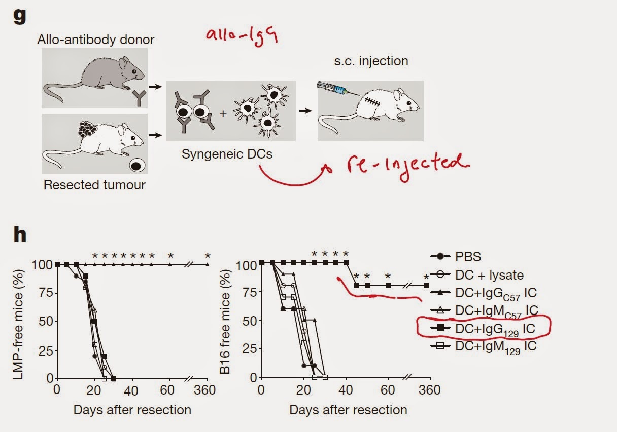
The authors employed concept of allogeneic priming. Basically, there are B6 mouse strains which differ only in minor antigens but have the same MHC molecules (antigenic differences between humans are frequently located in minor antigens too). 

Depending on number of minor antigenic differences between these MHC-matched allogeneic hosts, immune system of each sub-strain lacks tolerance towards other's minor antigens. For example, B16 melanoma cells are MHC-matched but minor antigenic mismatched in 129S1 substrain of B6 mice and vice versa LMP cancer cells are MHC-matched but minor antigen mismatched in C57 substrain of B6 black mice. Accordingly, each tumor is rejected easily by MHC-matched allogeneic hosts via T cell dependent manner [because of abundance of minor antigenic difference].


Unexpectedly, this rejection was dependent of host B cells too


Next the authors showed that syngeneic hosts injected with syngeneic BmDCs that were pre-incubated with tumor lysate coated with allogeneic IgG (but not IgM) were protected against syngeneic tumors. (syngeneic cells should have identical major and minor antigens)  


Interestingly, tumor associated DCs (TADC) were incapable of providing such protection, nor direct intra-tumoral allo-IgG injection was effective (however, in Fig. 1k, the authors showed that injection of allo-IgG was effective. Don't know what is the difference).


However, priming with allo-IgG in combination with CD40L/TNF-alpha or PolyI:C could provide anti-cancer immunity. In addition, injection of TADC harvested from immunogenic cocktail treated mice could protect naive mouse against cancer.


In summary, these results (and there are lot of figures) suggest that using allo-IgG priming strategy could help overcome host tolerance towards syngeneic tumors. 

How this could apply for human case: one can harvest patient's DCs and incubate with tumor cells coated with allo-IgG. After re-injection, these DCs should then activate patient's T cells and participate in tumor protection

I don't see that direct injection of allo-IgG in combination with CD40L/TNF-alpha or PolyI:C is a feasible approach though.

Strange thing about this study is the fact that pre-absorbing of allo-IgG serum with normal mouse tissue syngeneic to tumor could abolish protection. If immune system detects normal self-antigens and then initiate cancer immunity, how is then autoimmunity prevented? Most likely, self-antigens are somehow linked to mutant antigens in cancer cells and help to deliver them to DCs for presentation to cancer-specific T cells. Since majority of self-specific T cells are tolerant to self-antigens to begin with, it is less likely that this procedure would initiate autoimmunity.

David Usharauli

Friday, March 20, 2015

CCL3 and memory CD4 T cells improve tumor antigen RNA loaded dendritic cell vaccine performance

Dendritic cells represent the most potent antigen-presenting cells capable of priming naive T cells against infectious or tumor antigens. However, so far, clinical application of antigen-loaded autologous (patient's own) dendritic cell vaccines produced few encouraging results.

Unlike regular vaccines that contain protein conjugates emulsified in adjuvants, dendritic cell vaccine is a biological vaccine containing live cells pulsed with target antigen. The rules that govern efficacy of live cell vaccines would depend on several conditions such as immunological condition of vaccine injection site, migration and viability of injected live vaccine cells and etc. 


The authors have tested their model both in glioblastoma cancer patients and in mouse model of skin cancer. I would like to point out the this paper was under Nature's review for more than a year, implying that it's data are not as strong as the authors would like to argue.

For one thing, clinical protocol the authors have used is not entirely clear. Initially, the authors showed that immunization of patients with tetanus toxoid (Td vaccine) prior to DC vaccine injection extended glioblastoma patients survival almost two-fold compared to un-pulsed DCs. However, I do not understand why Td was compared to un-pulsed DCs. Even after careful reading of methods section, I still was not able to clearly understand the authors reasoning.


Next, using mouse model, the authors showed that presence of activated memory CD4 T cells contributed to the enhanced migration of antigen-loaded DCs to the local lymph nodes.


Interestingly, Td- or memory CD4 T cell-mediated enhancement of DCs migration was abolished in CCL3-KO host.


In addition, exogenous CCL3 was effective only in presence of activated memory CD4 T cells, implying that both memory CD4 T cells and CCL3 are required for enhancement of DCs migration to local lymph nodes.


In summary, these results revealed that locally Td-activated memory CD4 T cells creates a favorable environment for antigen-loaded DCs migration to local lymph node in a CCL3-dependent manner. Source of Td-driven CCL3 from the injection site is not clear.

If you ask me, only reason this paper ended up in Nature has to do with the fact that somehow the authors' protocol (Td + DC pulsed with pp65 antigen RNA) was able to substantially extend glioblastoma patients' survival. However, it is not clear why would Td-activated skin-resident memory CD4 T cells improve migration of DCs loaded with non-related antigen, CMV pp65? If we consider the fact that glioblastoma patients should already have CMV pp65-specific memory CD4 T cells capable of doing the same function as Td did, then I don't see how Td could have provided any additional benefits over simple CMV pp65 RNA-loaded DCs? This paper raises more questions than provides answers.

David Usharauli